
Papagona papoosa Ball, 1935
|
publication ID |
https://doi.org/ 10.11646/zootaxa.5023.1.6 |
|
publication LSID |
lsid:zoobank.org:pub:F107135D-07EA-48E0-9311-A0BFF06A0D93 |
|
persistent identifier |
https://treatment.plazi.org/id/038D87B4-FFDD-E372-FF5C-FDE3FE027677 |
|
treatment provided by |
Plazi |
|
scientific name |
Papagona papoosa Ball, 1935 |
| status |
|
Papagona papoosa Ball, 1935 View in CoL
( Fig. 5 View FIGURE 5 )
Papagona papoosa View in CoL — Ball 1935: 41 (Original description)
Type locality. Arizona, Santa Cruz River, near Tubac (according to Ball 1935) .
Amended description. Body length. Male, 2.6 mm ( Ball 1935).
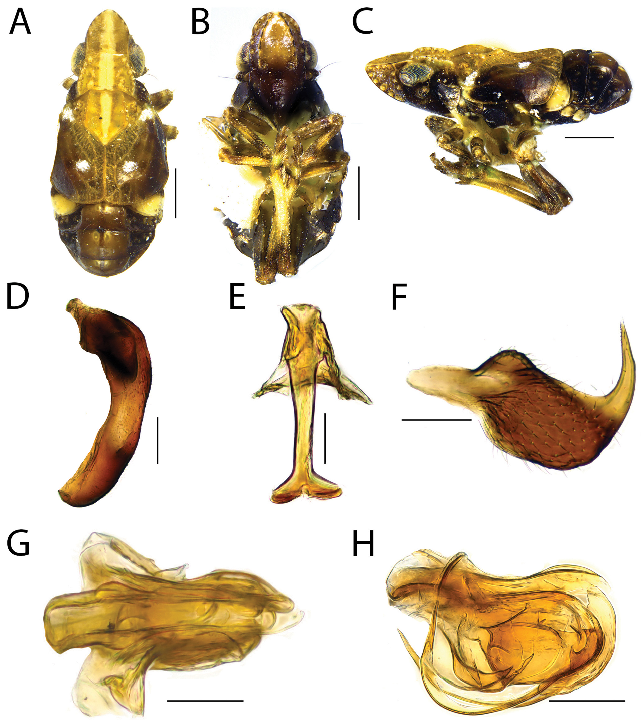
Color. Males ( Fig. 5A–C View FIGURE 5 ). Body mainly black with some regions yellowish-brown. Vertex, pronotum, and mesonotum yellowish-brown with broad median longitudinal white stripe crossing these structures ( Fig. 5A View FIGURE 5 ). Forewing with corium mostly black with apex light brown and two small circular pale maculae in between corium and clavus, one at base and another near midlength of claval suture; clavus brown ( Fig. 5A View FIGURE 5 ). Legs mostly yellow; metafemur black ( Fig. 5B, C View FIGURE 5 ). Abdomen with tergite III with lateral white macula ( Fig. 5A, C View FIGURE 5 ); each segment of abdomen with dorsal median pair of small and few conspicuous yellow maculae ( Fig. 5A View FIGURE 5 ); in lateral view, sternites with lateral white macula ( Fig. 5A View FIGURE 5 ).
Structure. Head and thorax. Vertex ( Fig. 5A View FIGURE 5 ) with anterior margin rounded; as long as basal width at midline; subequal to pronotum length at midline. Frons ( Fig. 5B View FIGURE 5 ) without median carina; in lateral view ( Fig. 5C View FIGURE 5 ) each side with row of seven sensory pits bordering sublateral carina, four pits bordering frontogenal carina, and a pair of pits bordering fastigium (linking the other two rows providing a triangle-like arrangement—with two isolated pits within, aligned diagonally).
Pronotum ( Fig. 5A View FIGURE 5 ) with six sensory pits bordering lateral margins of disc and a group of fiver inner ones at posterior half. Mesonotum ( Fig. 5A View FIGURE 5 ) without median carina; region outerad of lateral carina with eight to nine sensory pits.
Abdomen. Tergite III ( Fig. 5C View FIGURE 5 ), in lateral view, with pair of sensory pits. Tergite IV ( Fig. 5C View FIGURE 5 ), in lateral view, with pair of sensory pits followed by an isolated ventral pair. Tergite V ( Fig. 5C View FIGURE 5 ), in lateral view, with one row of three sensory pits followed by an isolated ventral pair. Tergite VI ( Fig. 5C View FIGURE 5 ), in lateral view, with pair of sensory pits followed by an isolated ventral pair. Tergite VII ( Fig. 5C View FIGURE 5 ), in lateral view, with one row of three sensory pits followed by an isolated ventral pair. Tergite VIII, in lateral view, with pair of sensory pits.
Male terminalia. Pygofer ( Fig. 5D View FIGURE 5 ) with anterior margin concave; with posterior margin convex. Connective ( Fig. 5E View FIGURE 5 ) with tectiform structure bearing tectiductus; ventral support inverted Y-shaped. Gonostylus ( Fig. 5F View FIGURE 5 ) hook-like; anterior portion pointed; caudal portion curved anterodorsally; dorsal margin follows almost straight with a rounded protuberance in between anterior and median third; ventral margin mostly rounded; median third longer than high, setose. Endosoma ( Fig. 5G, H View FIGURE 5 ) enclosing almost all phallobase and aedeagus lengths laterally and ventrally; asymmetrical, with two different sides linked ventrally: one side is longer and with apex curved ventrally, comma-like; and other side is shorter and apically truncated, bearing sub-triangular expansion ventrally directed to the longest side of endosoma at aedeagus midlength ( Fig. 5G View FIGURE 5 ). Phallobase membranous, shorter than endosoma; enclosing aedeagus half-length laterally and ventrally; slightly visible in lateral view, apically and dorsally. Aedeagus ( Fig. 5G, H View FIGURE 5 ) opened dorsally; apically, narrowing and with pair of aedeagal hooks ( Fig. 5H View FIGURE 5 ), both with same length, longer and thinner than aedeagus, and curved anterodorsally, reaching the base of phallus. Suspensorium V-shaped ( Fig. 5G View FIGURE 5 ). Anal tube as long as wide; posterior margin rounded; setose.
Taxonomic notes. Although Ball (1935) states in the generic description that the vertex of Papagona is longer than pronotum length (see Introduction), in the holotype of P. papoosa it appears only slightly longer than pronotum length ( Fig. 1A View FIGURE 1 ). However, this could be an intraspecific variation or artefact the specimen position when photographed. See taxonomic notes of P. dietrichi sp. nov. above for comparative notes.
Distribution. United States: Arizona ( Ball 1935).
Plant associations. Muhlenbergia porteri Scribn. ex Beal (muhly grass, Poaceae ) ( Ball 1935).
Studied material. Holotype: male (dissected herein), USA, Arizona, Santa Cruz River , 6 Aug. 1932, E. D. Ball ( USNM ENT 01513540 View Materials ).

| V |
Royal British Columbia Museum - Herbarium |
| VI |
Mykotektet, National Veterinary Institute |
| USNM |
Smithsonian Institution, National Museum of Natural History |
No known copyright restrictions apply. See Agosti, D., Egloff, W., 2009. Taxonomic information exchange and copyright: the Plazi approach. BMC Research Notes 2009, 2:53 for further explanation.
|
Kingdom |
|
|
Phylum |
|
|
Class |
|
|
Order |
|
|
Family |
|
|
Genus |
Papagona papoosa Ball, 1935
| De Freitas, Abner S., Zahniser, James N. & Takiya, Daniela M. 2021 |
Papagona papoosa
| Ball, E. D. 1935: 41 |