
Opisthosyllis leslieharrisae, Aguado, M. Teresa, Martín, Guillermo San & Nygren, Arne, 2005
|
publication ID |
https://doi.org/ 10.5281/zenodo.170244 |
|
DOI |
https://doi.org/10.5281/zenodo.6269045 |
|
persistent identifier |
https://treatment.plazi.org/id/03FF8780-0F0F-DB35-FE9C-FCD9FD33F91A |
|
treatment provided by |
Plazi |
|
scientific name |
Opisthosyllis leslieharrisae |
| status |
sp. nov. |
Opisthosyllis leslieharrisae View in CoL sp. nov.
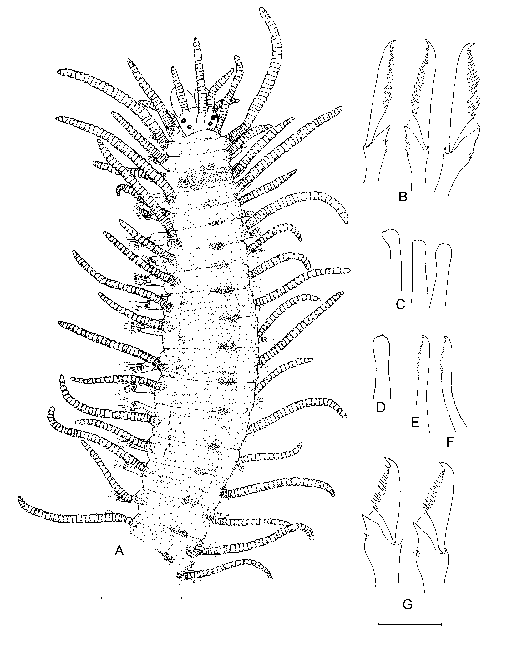
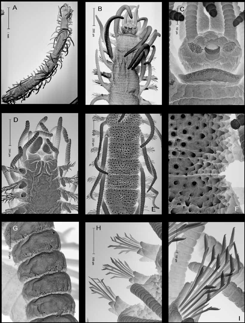
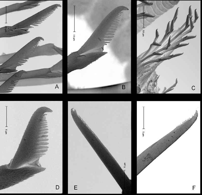
( Figures 1–4 View FIGURE 1 View FIGURE 2 View FIGURE 3 View FIGURE 4 )
Material examined. USA: holotype [ MNCN 16.01/10264], 3 paratypes [ MNCN 16.01/ 10265 (1), MNCN 16.01/10266 (2)], 3 spms., Santa Catalina Island, Wrighley Marine Science center: 33° 26.7 N, 118° 29.1W; 1–4 m, Corallina, Sargussum , red algae, hydroids, bryozoans and sponges, 15 Jan. 2001. Coll. A. Nygren, J. Toth.
Comparative material examined.
Opisthosyllis papillata HartmannSchröder, 1960 . Paratype [P14718HZM].
Opisthosyllis viridis Langerhans, 1879 View in CoL . Several specimens. Cabo Verde Islands [Polychaetes collection, UAM].
Opisthosyllis australis Augener, 1913 View in CoL . Type material [V7947HZM].
Diagnosis. Opisthosyllis with dorsum densely covered by papillae in two sizes, tooth located in the third quarter of the pharynx, long spines on blades of compound chaetae, and a distinct colour pattern consisting of white spots (live specimens) and dark red areas distributed over dorsum (maintained in preserved specimens).
Description.The holotype is 7.6 mm long, 0.48 mm wide, with 73 chaetigers, adult specimen. Paratypes are 9.5 mm long, 0.7 mm wide, 54 chaetigers (MNCN16.01/10265); 6.4 mm long, 0.5 mm wide, 32 chaetigers and 2.1 mm long, 0.5 mm wide, 30 chaetigers (anterior fragment) (MNCN 16.01/10266). Body shape, excluding parapodia, circular in section, venter flattened; body width fairly constant with tapering end. Body in outline long and slender, posteriorly broken, with signs of regeneration in all specimens ( Fig. 1 View FIGURE 1 B, E). Live specimens with distinct colour pattern ( Figs. 1 View FIGURE 1 A–E): white area in chaetigers 1 and 2; chaetiger 3 dorsally pigmented in red, following segments with a reddish oval area at midline of body; tentacular cirri and dorsal cirri of all segments with an oval to circular area of reddish pigment on and around the cirrophores. Colour markings more distinct on the cirri pointing up than on cirri pointing down. White spots in transversal rows on each segment, and white fibrilar material in cirral articles. The red colour pattern is preserved in formalin fixed specimens ( Fig. 2 View FIGURE 2 A). Dorsal surface covered with triangular papillae in two sizes ( Fig. 3 View FIGURE 3 E, F), more distinct posteriorly to proventricle ( Figs. 1 View FIGURE 1 E, 2A). Prostomium wider than long, rectangular to oval, with two pairs of red eyes with lenses, in trapezoidal arrangement, anterior pair larger ( Fig. 1 View FIGURE 1 A); eye spots absent. Palps broad, fused at base, with visible, central groove; palps slightly longer than prostomium. Median antenna inserted medially on prostomium, longer than prostomium and palps together, with 23 articles. Lateral antennae inserted on anterior margin of prostomium ( Figs. 2 View FIGURE 2 A, 3B) with 14–15 articles, approximately half the length of median antenna. Ceratophores present ( Fig. 3 View FIGURE 3 C). Two ciliated areas present between base of median antenna and bases of both lateral antennae ( Fig. 3 View FIGURE 3 C). Two ciliated nuchal organs present lateral and behind the prostomium ( Fig. 3 View FIGURE 3 B, C). Peristomium shorter than subsequent segments, anterior margin of peristomium ciliated and prolonged, partially covering the prostomium ( Fig. 3 View FIGURE 3 C). Dorsal tentacular cirri with 33 articles, ventral pair shorter, with 25 articles. Dorsal cirri of chaetiger one shorter than tentacular cirri, with 22 articles, second dorsal cirri similar in length to first dorsal cirri, with 20 articles, third and fourth dorsal cirri longer, with 34 and 32 articles ( Figs. 1 View FIGURE 1 C, 2A, 3B). Subsequent cirri alternating in length with 20–35 articles, longer ones pointing up and shorter ones pointing down ( Fig. 1 View FIGURE 1 C, E). Alternation in direction of cirri starting from chaetiger 1, where D=cirri pointing down and U=cirri pointing up, with the following formula UDDUDUDDU followed by DUgroups to the posterior end. Distinct alternation in median chaetigers ( Fig. 1 View FIGURE 1 D). Cirrophores well developed. Antennae, tentacular and dorsal cirri with minute ciliation on articles ( Fig. 3 View FIGURE 3 G). Ventral cirri oval and short, proximally inserted and not extending beyond tips of parapodia ( Fig. 3 View FIGURE 3 D). Pre and postchaetal as well as dorsal lobes, all similar in length, present on all parapodia ( Fig. 3 View FIGURE 3 H). Chaetal fascicle with 10–12 heterogomph compounds in anterior chaetigers, 3–6 in median and posterior; distal part of shafts provided with spines. Compound chaetae with bidentate blades, distal tooth longer and broader than proximal one, blade edge with long spines, blades dorsoventrally gradiated in length ( Figs. 2 View FIGURE 2 B, G, 3I, 4A–D). Length of dorsalmost chaetal blades c. 35 µm in anterior parapodia ( Fig. 2 View FIGURE 2 B), and c. 28 µm in median parapodia ( Fig. 2 View FIGURE 2 G). Dorsal and ventral simple chaetae distally bifid (indistinct in ventral ones) with short subdistal spines on margin ( Figs. 2 View FIGURE 2 E, 4E). Ventral simple chaetae only observed in last segment of holotype ( Figs. 2 View FIGURE 2 F, 4F). Three aciculae in anterior parapodia ( Fig. 2 View FIGURE 2 C), one in median and posterior ( Fig. 2 View FIGURE 2 D), all distally blunt. Pygidium regenerating, no anal cirri, median papilla absent. Paratype MNCN 16.01/ 10265 with two anal cirri, pygidium also regenerating ( Fig. 1 View FIGURE 1 B). Pharynx shorter than proventricle, almost as broad as proventricle; large conical tooth located in the third quarter of the pharynx ( Fig. 1 View FIGURE 1 A, 2A). Proventricle shape cylindrical, through segment 10 to 18, with 40–50 cellrows ( Fig. 1 View FIGURE 1 D, 2A).

| MNCN |
Museo Nacional de Ciencias Naturales |
No known copyright restrictions apply. See Agosti, D., Egloff, W., 2009. Taxonomic information exchange and copyright: the Plazi approach. BMC Research Notes 2009, 2:53 for further explanation.
|
Kingdom |
|
|
Phylum |
|
|
Class |
|
|
Order |
|
|
Family |
|
|
Genus |
Opisthosyllis leslieharrisae
| Aguado, M. Teresa, Martín, Guillermo San & Nygren, Arne 2005 |
Opisthosyllis papillata HartmannSchröder, 1960
| Hartmann-Schroder 1960 |
Opisthosyllis australis
| Augener 1913 |
Opisthosyllis viridis
| Langerhans 1879 |