
Onchidella steindachneri ( Semper, 1885 )
|
publication ID |
https://doi.org/ 10.1080/00222933.2010.545486 |
|
persistent identifier |
https://treatment.plazi.org/id/03D887F6-FFCD-FFDC-FE4B-9CDAFD8BB21B |
|
treatment provided by |
Felipe |
|
scientific name |
Onchidella steindachneri ( Semper, 1885 ) |
| status |
|
Onchidella steindachneri ( Semper, 1885) View in CoL
( Figures 1 View Figure 1 , 13–18 View Figure 13 View Figure 14 View Figure 15 View Figure 16 View Figure 17 View Figure 18 )
Onchidium steindachneri Semper 1885: 280 View in CoL , plate XIX, figs 7–8, plate XXI, fig. 15, plate XXIII, fig. 14.
Onchidella steindachneri View in CoL . – Stearns 1894: 384, plate LI, figs 4–5. — Watson 1925: 304. — Hoffmann 1928: 38–44, 70, 94–95, plate 2, figs 3, 5–6, plate 3, figs 4, 7. — Labbé 1934: 241.
Type material
One syntype ( ZMB 39.038 View Materials ): one specimen ∼ 25/ 10 mm preserved [leg. and collecting date unknown]. Type locality: Galapagos Inseln. Type material condition: the original description was based on two syntypes, but there seems to be only one of them held at the ZMB, which was dissected and cut in pieces prior to the present study (so the size indicated above is tentative) .
Additional material examined and dissected
A total of 95 non-type specimens were examined, 29 of which were dissected to study their internal anatomy. Examined specimens were collected from 12 localities on six of the Galapagos Islands, four islands being new records (see Distribution). Ecuador , Galapagos Islands, Isla Cristóbal [Chatham Island], 11 May 1852, 21 specimens 28/20 to 20/ 12 mm preserved [leg. Eugenie Expedition, station 838], identified as Onchidella steindachneri [unknown identifier], ( SMNH 103166 View Materials ) [one specimen dissected here: 25/10 (#1); the jar contains pieces of possibly four additional specimens dissected prior to the present study; those specimens are the vouchers of Hoffmann’s (1928) description of steindachneri ]; Ecuador , Galapagos Islands, Santa Cruz, Academy Bay , 1934, three specimens 23/18 to 20/16 (#1) mm preserved [leg. Blomberg], identified as Onchidella steindachneri [unknown identifier], ( SMNH 103167 View Materials ); Ecuador , Galapagos Islands, Santa Cruz, Academy Bay, 1934, two specimens 24/17 and 16/15 (#1) mm preserved [leg. Blomberg], identified as Onchidella steindachneri [unknown identifier], ( SMNH 103168 View Materials ); south Pacific coast, Ecuador , Galapagos Islands, Sulivan Bay, James Island, 24 July 1938, one specimen 17/ 13 mm preserved [leg. unknown], identified as Onchidiidae [unknown identifier], ( NMNH 574530 View Materials ) [dissected]; south Pacific coast, Ecuador , Galapagos Islands, Elizabeth Bay, Albemarle Island , 26 July 1938, two specimens 25/10 (#1) and 25/ 20 mm preserved [leg. unknown], identified as Onchidiidae [unknown identifier], ( NMNH 574379 View Materials ) ; Galapagos Islands, W coast of Isla Santa Cruz, opposite Guy Fawkes Island , “Venice” cove, 19 February 1964, three specimens 13/7 to 10/9 (#1) mm preserved [leg. D.P. Abbott and J.W. Durham], identified as Onchidella steindachneri by B. Roth ( CASIZ 017922 ) ; Galapagos Islands, Isla Santa Cruz, Academy Bay , February 1964, nine specimens 45/25 to 15/ 11 mm preserved [leg. A.G. Smith], identified as Onchidella steindachneri by A.G. Smith ( CASIZ 078745 ) [four specimens dissected: 45/25 (#2), 30/24 (#5), 20/17 (#4), 18/16 (#3) and 15/11 (#1)] ; Galapagos Islands, Isla Pinta [Abingdon Island], SW coast, 25 May 1964, three specimens 16/14 (#1) to 13/11 (#2) mm preserved [leg. D.Q. Cavagnaro], identified as Onchidella steindachneri by A.G. Smith ( CASIZ 078743 ) ; Galapagos Islands, Isla Pinzon [Duncan Island], 7 June 1932, two specimens 12/11 (#1) and 13/ 10 mm preserved [leg. T. Crocker and F. Taiga], identified as Onchidiidae by L. Borock ( CASIZ 077057 ) ; Galapagos Islands, Isla San Salvadore [James Island], N end James Bay , 2 February 1967, seven specimens 18/15 to 11/ 10 mm preserved [leg. I. Wiggins], identified as Onchidiidae [unknown identifier], ( CASIZ 079303 ) [four specimens dissected: 18/15 (#1), 16/12 (#4), 12/10 (#2) and 11/10 (#3)] ; Galapagos Islands, Isla Santa Cruz, Academy Bay , 18 July 1906, four specimens 18/23 (#1) to 18/15 (#2) mm preserved [leg. W.H. Oschner], identified as Onchidella steindachneri by A.G. Smith, ( CASIZ 078749 ) ; Galapagos Islands, Isla Santa Cruz [Indefatigable Island], Dove Roads , 1905–1906, 37 specimens 26/22 to 12/ 14 mm preserved [leg. W.H. Oschner], identified as Onchidella [unknown identifier], ( CASIZ 078354 ) [10 specimens dissected: 26/22 (#1), 20/23 (#7), 20/21 (#8), 20/18 (#10), 19/24 (#4), 18/15 (#2), 16/21 (#5), 13/15 (#6), 13/10 (#3), and 12/14 (#9)] .
Distribution
Endemic to the Galapagos Islands: San Cristobal ( Hoffmann 1928; present study); Santa Maria (= Charles Island; Stearns 1894); Isabella (= Albemarle; Stearns 1894; present study); San Salvadore (present study); Pinzon (present study); Santa Cruz (present study); Pinta (present study). Semper (1885) recorded O. steindachneri from the Galapagos Islands, without citing any specific island. Watson (1925) and Labbé (1934) only mentioned O. steindachneri from the Galapagos, without new material.
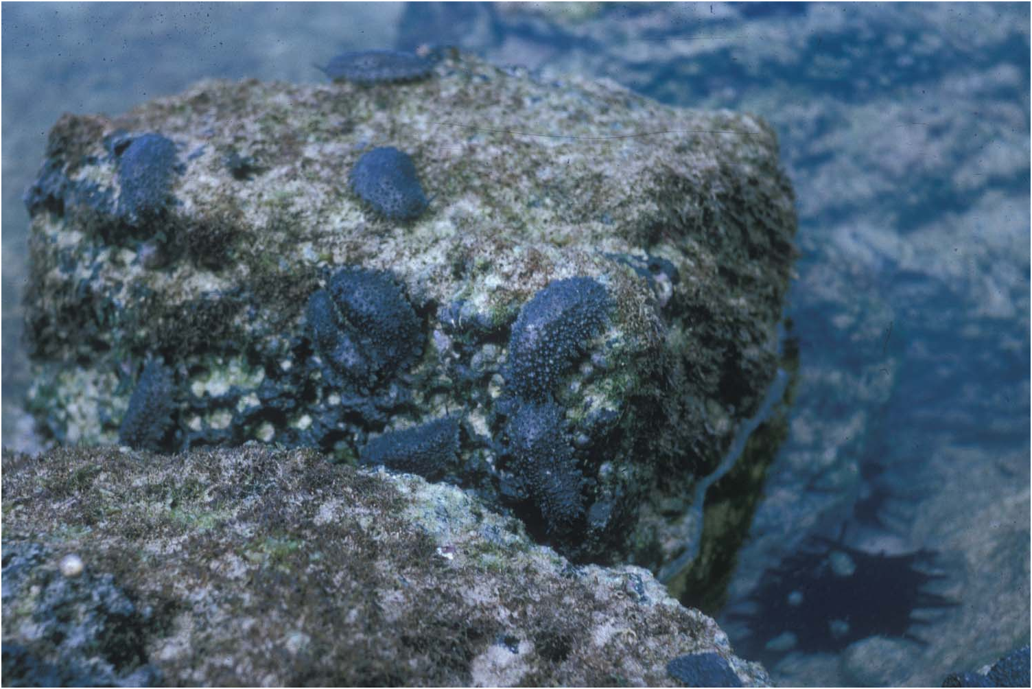
Habitat
Intertidal. On rocks.
Remarks on the original description
The dorsal colour of the two syntypes was described as blackish-greenish, and the ventral surface as yellowish. Semper also described some of the internal anatomy, especially the reproductive system: the insertion of the retractor muscle is at the very end of the visceral cavity, near the anus; also, the vas deferens was described as highly convoluted; finally, there is no penial accessory gland. The drawings of the rachidian and lateral teeth are poorly informative.
Description of new specimens
Several anatomical systems of O. steindachneri are similar to O. binneyi (nervous system, pallial organs, digestive system except for the radular formulae, and the posterior genital organs). Their written description is not provided here again, although some organs, which have not – or only poorly – been illustrated, are illustrated here. Given that the only syntype available has been completely destroyed, the description below is exclusively based on non-type specimens.
External Morphology ( Figures 1 View Figure 1 and 13 View Figure 13 ). Dorsal notum of live animals blackish. In preserved animals, dorsal colour from dark grey or bluish to dark brownish; centre of notum light tan in most individuals; hyponotum and pedal sole whitish.
Size from 45/25 ( CASIZ 078745) to 10/ 9 mm ( CASIZ 017922). Body high, not flattened. Hyponotum horizontal. Dorsal notum oval, longer than wide. In most individuals, entire surface of dorsal notum granular with papillae of various sizes (usually <2 mm high). However, notum of some individuals with smooth centre (e.g. CASIZ 078745, CASIZ 078749, SMNH 103166). Margin of notum is not smooth: characterized by digitiform warts of various sizes up to 2 mm long. In large individuals, many marginal warts much larger (up to 4 mm long) and subdivided (one main, central wart with several, smaller digitiform warts). Papillae with dorsal eyes absent. Dorsal gills absent.
In most individuals, total width of hyponotum (left and right sides added) superior to width of pedal sole (H> S): left side of hyponotum, pedal sole, and right side of the hyponotum of equal width. However, exceptionally, pedal sole wider than left and right hyponotum added (e.g., left hyponotum/pedal sole/right hyponotum as 5/10/4 and 4/10/ 3 mm, CASIZ 078745). On hyponotum, hyponotal line around pedal sole separating hyponotum into inner area (close to foot) and outer area. Distance between hyponotal line and pedal sole varies from one fifth to one third of width of
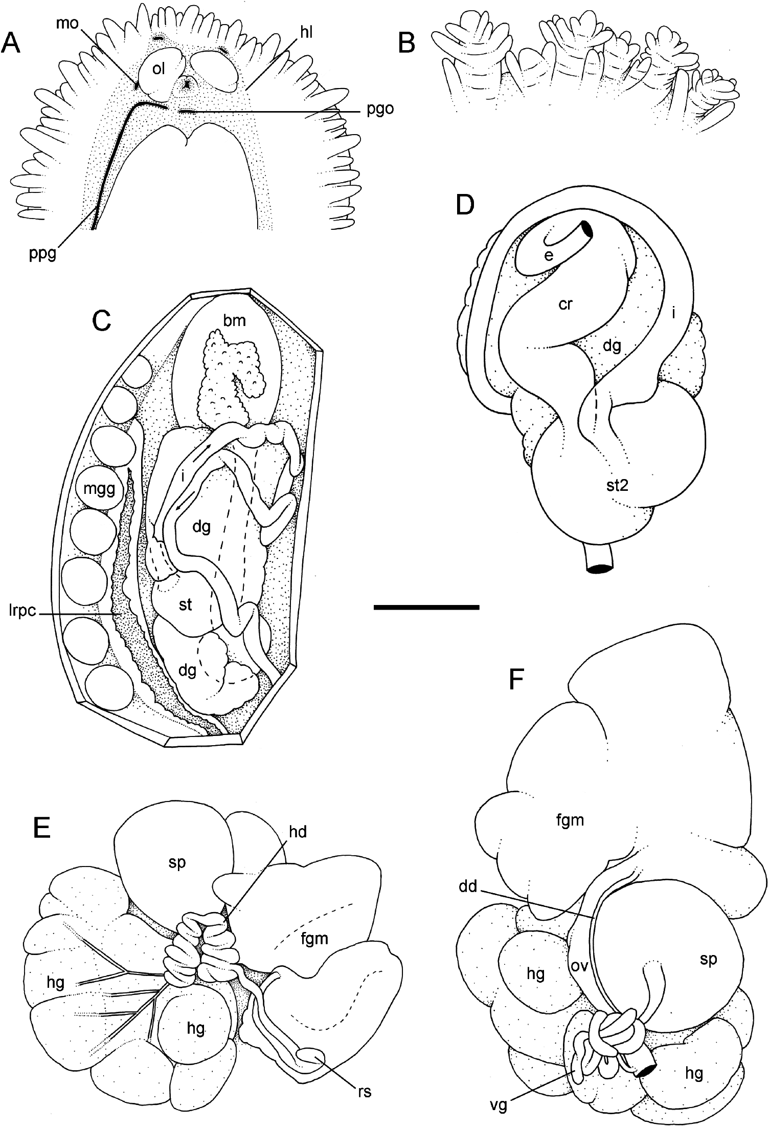
ventral view, SMNH 103167 #1; (E) posterior (female) genital organs, SMNH 103167 #1; (F) posterior (female) genital organs, SMNH 103167 #1. Scale bar: A, 5 mm; B, 4 mm; C, 3.3 mm; D, 2 mm; E, 2 mm; F, 1.7 mm. Abbreviations: bm, buccal mass; cr, crop; dd, deferent duct; dg, digestive gland; e, oesophagus; fgm, female gland mass; hd, hermaphroditic duct; hg, hermaphroditic gland; hl, hyponotal line; i, intestine; lrpc, left reno-pulmonary complex; mgg, marginal gland; mo, male opening; ol, oral lobe; ov, oviduct; pgo, pedal gland opening; ppg, peripodal groove; rs, receptaculum seminis; sp, spermatheca; st, stomach; st2, stomach chamber #2; vg, vaginal gland.
hyponotum (hyponotal line closer to pedal sole than to hyponotum margin). Openings (pneumostome, male opening and eye tentacles) are within smooth area delimited by hyponotal line.
Pedal sole surrounded by two grooves on left and right sides. Left groove shallow. Peripodial groove present on right side, from buccal area (pedal gland opening) to posterior openings (anus and female opening). Anus posterior, median, close to pedal sole. Exceptionally, anus slightly on right side ( CASIZ 078749 #2 and #9). Posterior female genital opening is very close to anus. Position of pneumostome on the hyponotum relative to notum margin and lateral side of the pedal sole varies among and within lots. Pneumostome can be: at an equal distance between pedal sole and hyponotal margin (e.g. CASIZ 078745 #2 and #3); one third closer to sole than hyponotal margin (e.g., CASIZ 078749 #2 and #9) or very close to anus, almost touching it (e.g. CASIZ 078354, two spms). Pneumostome generally close to median line, but slightly to right, occasionally median (e.g. SMNH 103166, three spms; NMNH 574379, two spms). Head bears pair of retractile, ocular tentacles, with eyes at tip. In all preserved specimens, ocular tentacles deeply retracted inside body. Left and right oral lobes distinct, not fused medially, superior to mouth but inferior to ocular tentacles. Opening of pedal gland median, inferior to mouth. Male genital opening on the right lateral side of right oral lobe.
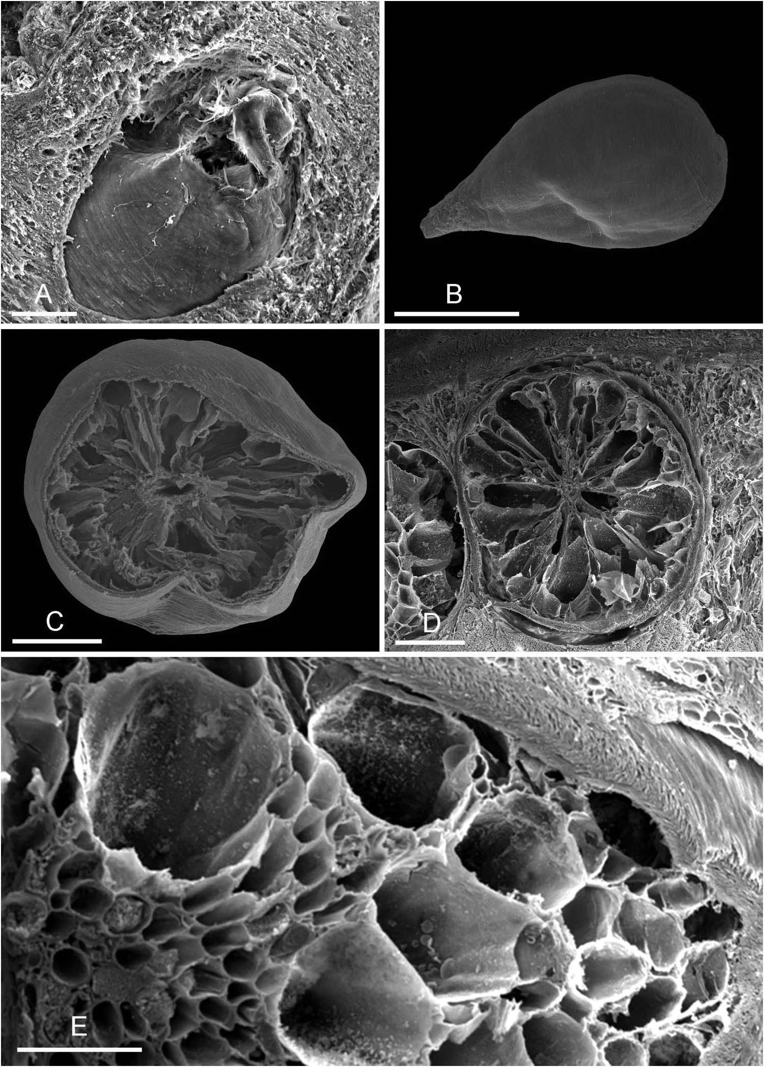
Marginal glands ( Figures 13 View Figure 13 and 14 View Figure 14 ). Marginal glands are similar to those found in O. binneyi . However, between eight and fourteen glands on each side. Glands about 1 mm in diameter, although some smaller glands occasionally found in between larger glands.
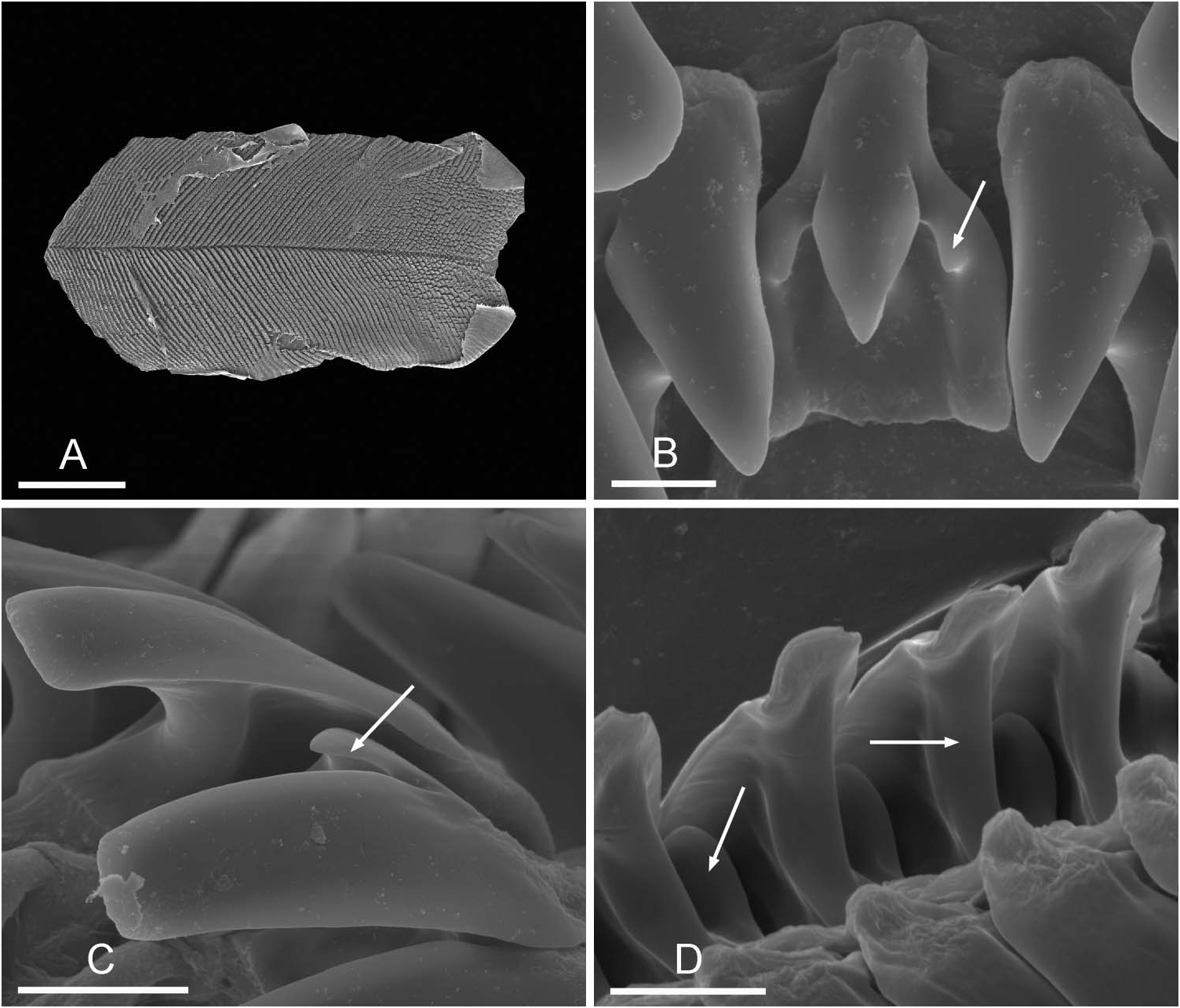
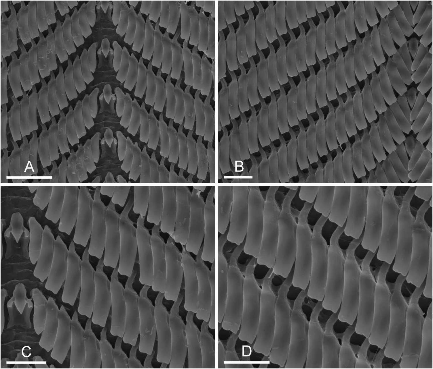
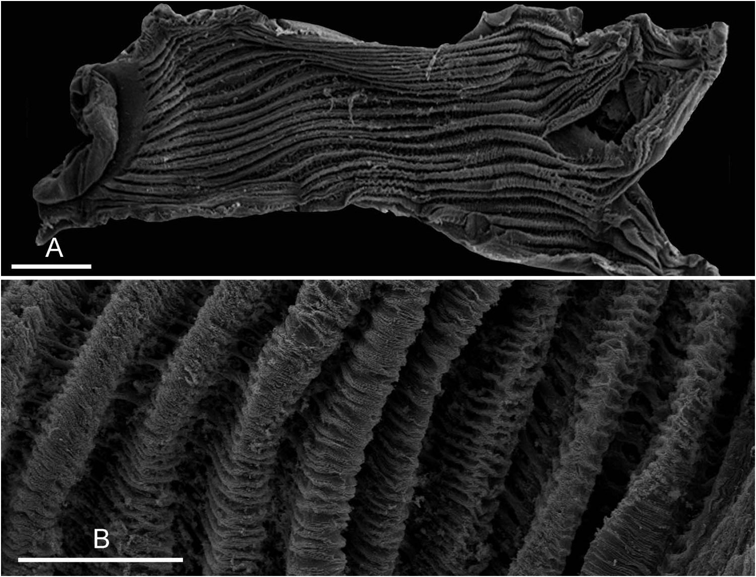
Digestive system ( Figures 13 View Figure 13 , 15 View Figure 15 and 16 View Figure 16 ). Radular maximum size 10/ 4 mm, when flattened. Radular formulae vary among individuals ( Table 1). From 66 to 121 rows, and from 63 to 114 teeth per half row.
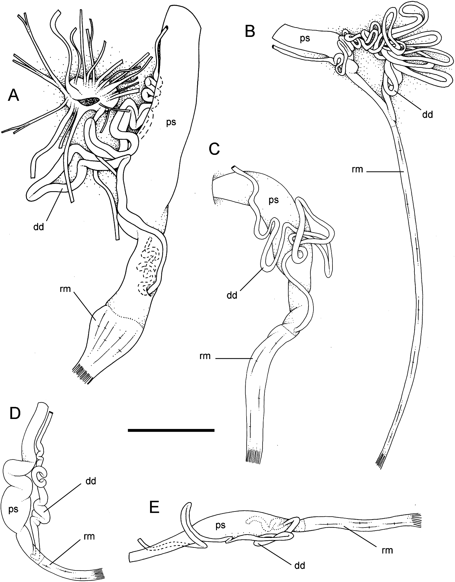
Reproductive system ( Figs 13 View Figure 13 , 17 View Figure 17 and 18 View Figure 18 ). No accessory penial gland. No distinct penis inside penial sheath. No distinct, conspicuous papilla at distal end of deferens duct. Penial sheath covered internally by thick folds. Length of penial sheath up to 15 mm in large specimens. No accessory caecum filled with white concretions opening into penial sheath. Deferent duct highly convoluted and coiled. In most specimens, many loops that can hardly be counted. Number of loops varies among individuals: young, small specimens generally with deferent duct with fewer loops; deferent duct never straight and short. Diameter of deferent duct more or less constant (∼ 300 µm maximum). Retractor muscle anchors where deferent duct enters penial sheath and runs to the end of visceral cavity to insert near posterior openings (female opening and anus). Length of retractor muscle depends on size of penial sheath (long if short penial sheath and short if long penial sheath). However, retractor muscle inserts at posterior end of visceral cavity in all specimens dissected.
Discussion
Prior to the present study, only six specimens of O. steindachneri had been dissected: two by Semper (1885) and four by Hoffmann (1928); Stearns (1894) only described the external morphology. The present study, based on 29 specimens dissected (for 95 specimens examined in total) from 12 localities from six of the Galapagos Islands, provides us with the first opportunity to address the intra-specific variation of some of the characters. In particular, the variation of the male copulatory organs, which clearly distinguish O. steindachneri from other species in the north-eastern Pacific, is described here for the first time: the deferent duct is highly coiled, and, although the number of loops may vary among individuals, the deferent duct is never straight. Also, the variation of the radular formula is now better known: 66/121 × 63/114-1-63/114 (neither Semper nor Hoffmann gave specific radular formulae).
Overall, the taxonomy of O. steindachneri is not problematic. It is a species that clearly differs from other Onchidella from the north-eastern Pacific and for which only one name has ever been proposed in the literature. As for its supra-specific relationships, it clearly does not belong to Onchidium , as originally proposed by Semper, who actually only used one generic name for all onchidiids, with the exception of one species which he classified in Onchidina .
No known copyright restrictions apply. See Agosti, D., Egloff, W., 2009. Taxonomic information exchange and copyright: the Plazi approach. BMC Research Notes 2009, 2:53 for further explanation.
|
Kingdom |
|
|
Phylum |
|
|
Class |
|
|
Order |
|
|
Family |
|
|
Genus |
Onchidella steindachneri ( Semper, 1885 )
| Dayrat, Benoît, Zimmermann, Sara & Raposa, Melissa 2011 |
Onchidella steindachneri
| Labbe A 1934: 241 |
| Hoffmann K 1928: 38 |
| Watson H 1925: 304 |
| Stearns REC 1894: 384 |
Onchidium steindachneri
| Semper C 1885: 280 |