
Neoempheria dilatata, Sueyoshi, Masahiro, 2014
|
publication ID |
https://doi.org/ 10.11646/zootaxa.3790.1.6 |
|
publication LSID |
lsid:zoobank.org:pub:87AB27EC-DC05-48F3-8AB7-5C317B275AF5 |
|
DOI |
https://doi.org/10.5281/zenodo.6138800 |
|
persistent identifier |
https://treatment.plazi.org/id/49168794-FFF1-5655-37AD-FB1D23B43556 |
|
treatment provided by |
Plazi |
|
scientific name |
Neoempheria dilatata |
| status |
sp. nov. |
Neoempheria dilatata View in CoL sp. n.
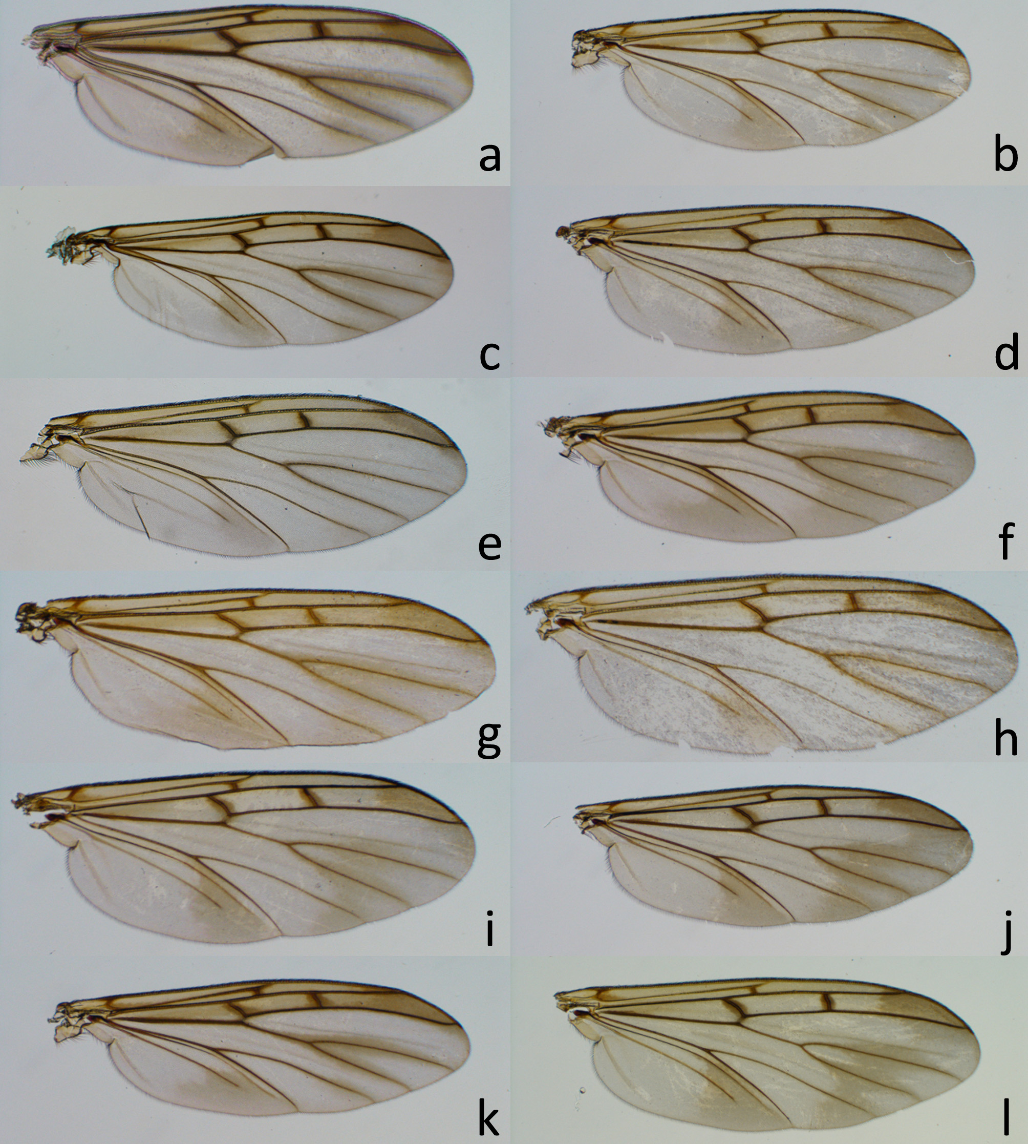
[Japanese name: fukura-nagamado-kinoko-bae] ( Figs. 2 View FIGURE 2 h, 3h, 5, 15a–e)
Neoempheria ferruginea: Okada 1938: 137 View in CoL .
Description. Body length: 4.7 mm (4.4–5.1 mm, n= 5) in male, 4.5 mm (3.8–5.4 mm, n= 7). Wing length: 4.5 mm (3.8–5.0 mm, n= 10) in male, 4.7 mm (3.8–6.2 mm, n= 9) in female. Wing ( Fig. 3 View FIGURE 3 h): vein sc-r ending basal 1/4 of anterior margin of cell r1. Vein Rs longer than distance between basal end of vein Rs and apical end of vein sc-r. Male: genitalia yellow in ground color, gonocoxal projection dark brown, apex of gonocoxal lobe brown to black, surrounding parts brown, basal portion of aedeagus yellow to dark brown. S9 narrow, less than 1/10 as wide as long, visible as seam between gonocoxites 9, without sternal projections. Gonocoxite ( Fig. 15 View FIGURE 15 a, b: gc) with gonocoxal lobe round ( Fig. 15 View FIGURE 15 a: gl) or angled at outer apical margin. Gonostylus ( Fig. 15 View FIGURE 15 a: gs) slender, apical 1/4 less than 1.5 times as wide as base. Aedeagus ( Fig. 15 View FIGURE 15 a, b) dilated at apical half, without projections. Sclerotized part of aedeagus ( Fig. 15 View FIGURE 15 a: sa) tapered to apex, with slender, angled lateral extension. Female: S7 with uniform setae on posterior margin, posterior margin with deep incision at middle. S8 triangular in ventral view. Gonocoxite 8 ( Fig. 15 View FIGURE 15 c: gc8) small, recognizable as bump at sublateral portion in posterior margin of S8. Gonapophysis 8 ( Fig. 15 View FIGURE 15 c: gp8) dilated in apical half, with acute apex. Gonocoxite 9 ( Fig. 15 View FIGURE 15 d, e: gc9) yellow, without projections. Gonapophysis 9 ( Fig. 15 View FIGURE 15 d, e: gp9) dark brown to black, with a single ventromedial ridge ( Fig. 15 View FIGURE 15 d: vr), gradually tapered to apex with gonopore.
Specimens examined. Holotype. Male. “KU-352” [underside: KU-352], “Manchoukuo/ I. Okada” [underside: “ 9/IX-1937 / Andon [written in Chinese character], “ Neoempheria / ferruginea / (Brunetti)/ det. I. Okada”, “RV-4”, green disk label, “Ne.2012” (HUM). Paratypes. CHINA [LIAONING] same data as holotype (6♂, 3♀: Ne. 2006–2011, 2016–2018; HUM); Tieling, 5.ix.1937, I Okada leg. (5♂, 2♀: Ne. 2001–2005, 2013, 2014; HUM). JAPAN [KYUSHU] Kumamoto P: Mt. Tatsuta, Kumamoto C, 23.vii.1977, Z Kuranaga leg. (1♂, Ne.1009; FFPRI); same locality, 16.vii.2008, MS (1♂, 2♀: Ne.1006, 1007, 1042; BLKU); same locality, 29.vi.2006, MS (1♂: Ne.1008; BLKU). [RYUKYUS] Okinawa P: Sueyoshi Park, Naha C, 21.iii.2000, H Nakayama leg. (1♂: Ne.1011; BLKU); Yona, Kunigami V, 25.iii.2000, H Nakayama leg. (1♀: Ne.1012; BLKU).
Specimens collected in indoor facilities. JAPAN [HONSHU] Nara P: Yamato-kohriyama C, 8.ix.2011 (16♂, 10♀: Ne. 1707–1732). [RYUKYUS] Okinawa P: Higashi V, iii.2009 (2♂, 4♀: Ne. 1017–1021, 1041); Nago C, v.2012 (1♂, 1♀: Ne.1733, 1734). All specimens are deposited in FFPRI.
Etymology. The specific epithet is derived from Latin and refers to the gonocoxal lobe, which is dilated (dilatatus) ventrally ( Fig. 15 View FIGURE 15 a).
Distribution. China (Liaoning) and Japan (Honshu, Kyushu, and Ryukyus) ( Fig. 5 View FIGURE 5 ).
Remarks. This species is distinguished from other species similar in general appearances by a combination of the following characters: in the male the gonocoxal lobe is dilated at apex; and sternite 9 is narrow and without any sternal projections, and in the female by the acute apex of the gonapophysis 8 ( Fig. 15 View FIGURE 15 c) and dark brown or black gonapophysis 9 with a single ventromedial ridge ( Fig 15 View FIGURE 15 e). Coloration and shape of the apex of the gonocoxal lobe is variable among the specimens from different localities. The 9th gonapophysis of the females are stable in the coloration and shape among different localities. I consider that the differences we can recognize in the shape and coloration of the gonocoxal lobe are quantitative and continuous among the localities. The male (Ne.1009) was collected at a light trap.


No known copyright restrictions apply. See Agosti, D., Egloff, W., 2009. Taxonomic information exchange and copyright: the Plazi approach. BMC Research Notes 2009, 2:53 for further explanation.
|
Kingdom |
|
|
Phylum |
|
|
Class |
|
|
Order |
|
|
Family |
|
|
Genus |
Neoempheria dilatata
| Sueyoshi, Masahiro 2014 |
Neoempheria ferruginea:
| Okada 1938: 137 |