
Molgula cooperi (Huntsman, 1912)
|
publication ID |
https://doi.org/ 10.11646/zootaxa.1579.1.3 |
|
publication LSID |
lsid:zoobank.org:pub:9DA6758A-01F9-4E81-BB7E-A1152F2658E5 |
|
persistent identifier |
https://treatment.plazi.org/id/03B987E8-FFD2-FFD6-E7B6-CDDC60D2D702 |
|
treatment provided by |
Felipe |
|
scientific name |
Molgula cooperi (Huntsman, 1912) |
| status |
|
Molgula cooperi (Huntsman, 1912)
( Figure 4 View FIGURE 4 )
Caesira cooperi Huntsman, 1912b: 134 View in CoL ; 1912a: 127.
Molgula cooperi: Van Name, 1945: 416 .
Molgula regularis: Sanamyan and Sanamyan, 1998: 214 . Not Ritter, 1907: 8.
Material examined: RV Gefest , 52°47.6N, 158°52.4'E – 52°48.7'N, 158°46.5'E, 300–500 m, two specimens GoogleMaps , KBPIG 663 /2; 58°17.1'N, 163°57.0'E – 58°13.6N, 163°58.3'E, 280 – 720 m, one specimen GoogleMaps , KBPIG 657 /1 ; RV Rubinovyi , 52°56'N, 160°06'E – 52°56'E, 160°12'E, 138–218 m, one specimen GoogleMaps , KBPIG 1049 /3; 52°56'N, 160°10'E – 52°57'N, 160°04'E, 185– 119 m GoogleMaps , KBPIG 1050 /4, three specimens ; from the same region but without exact locality, KBPIG 1051 /5, two specimens. Collector B.Sheiko.
Previous records: NE Pacific, Departure Bay, British Columbia ( Huntsman 1912b) and Newport, Oregon ( Van Name 1945). NW Pacific, E Kamchatka ( Sanamyan and Sanamyan 1998).
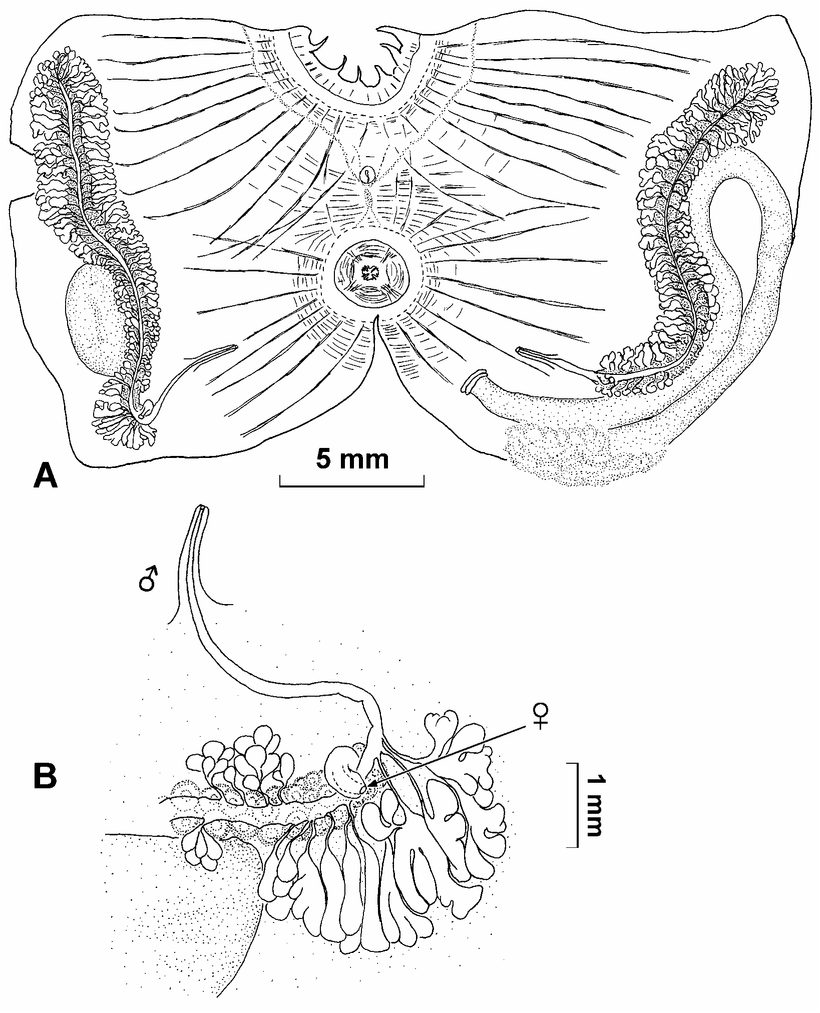
Description. Oval or almost globular specimens are from 15 to 45mm in greatest diameter. The two smallest specimens (KBPIG 663/2) are covered by fine sediment and sand grains and have sparse, short but discernible hair-like processes on the test. Other specimens are covered by a thick crust of rather large gravel and epibionts (Hydrozoa and Bryozoa) and hair-like processes were not detected. The largest specimen (KBPIG 1049/3) is attached to a large stone by a wide area; all other specimens also have a more or less definite area of attachment and apparently were attached to hard objects in life. Siphons are indiscernible on intact specimens and are inconspicuous on the body removed from the test. Body muscles are better developed in the anterior half of the body where crowded circular muscles surround the apertures, thick well-spaced muscle bands radiate from the apertures and a diffuse field of transverse muscles cover the intersiphonal area. In addition the whole body wall is covered by a loose network of thin muscle fibers. The tentacles are much branched, twenty or more larger and numerous smaller ones can be counted. The dorsal lamina has a plain margin. A robust branchial sac has six high equally developed folds, each with seven–13 longitudinal vessels. Vessels are not present between the folds or between the endostyle and most ventral fold. The stigmata are short and almost straight between the folds and form deep broad infundibula with twice subdivided apices in the folds. The narrow gently curved gut loop is open only at the pole. The pyloric region has well developed oblique glandular pouches. Gonads are very long and sinuously curved. The left gonad is in the secondary gut loop and runs in close contact with its descending limb; its posterior end extends above the pole of the gut loop. The right gonad extends along almost the whole extent of the ventral border bending around the dorsal side of the renal sac (which is much smaller than the gonad). Each gonad consists of a central tubular ovary surrounded on the sides and posterior end by numerous rather long male follicles which become well branched in larger specimens. There is one wide common sperm duct running along the mesial surface of the whole ovary and continuing beyond it to form a long male papilla on the body wall halfway between the distal (posterior) end of the ovary and the atrial orifice. The female opening, in contrast, is at the end of the ovary on a short inconspicuous papilla curved ventrally ( Fig. 4B View FIGURE 4 ).
Tailed larvae are found in the peribranchial cavity in the largest specimen (KBPIG 1049/3).
Remarks. The original description of this species is based on numerous small specimens (7–15 mm) collected in British Columbia at about 10– 30 m. These specimens (like the present ones) were directly attached by a large area to hard substrata, rather than by hairlike processes of the test. The long sinuously curved gonads are characteristic, but the most peculiar and significant distinguishing feature of this species is the structure of male and female ducts. Huntsman (1912b: 135) describes the ducts as "Ducts directed upward from the posterior end of each gonad: a long and narrow anterior one, the vas deferens, and a short and broad posterior one, the oviduct". Van Name (1945: 417) completely misinterpreted this description and stated that "Huntsman found a single common sperm duct for each gonad that opened near the end of the oviduct". Actually the description of Huntsman clearly corresponds to that found in the present specimens (in which com- mon sperm duct continues far beyond the end of the oviduct, the distal end projecting from the body wall between the end of the ovary and the atrial aperture, see Fig. 4B View FIGURE 4 ). The other features, including the presence of only six branchial folds (without an additional rudimentary fold near the endostyle) and viviparous development are also in agreement with the original description although many of the present specimens are significantly larger and come from a greater depth.
Van Name (1945) suggested that M. cooperi may be conspecific with M. regularis (described from California) and claimed that the only feature that might separate these species is a viviparous development of M. cooperi . However, the position of the left gonad in M. regularis is different: Ritter (1907) says that it is in front of the intestinal loop and in his figure (plate 1, Fig. 7) the gonad is in contact only with the pole of gut loop, while in all present specimens it follows the descending limb of the gut loop and is in close contact with the intestine. The structure and position of the ducts of M. regularis are not known. There are other minor differences, e.g. the specimens appear not to be attached to hard objects and live freely in sand.
| RV |
Collection of Leptospira Strains |
No known copyright restrictions apply. See Agosti, D., Egloff, W., 2009. Taxonomic information exchange and copyright: the Plazi approach. BMC Research Notes 2009, 2:53 for further explanation.
|
Kingdom |
|
|
Phylum |
|
|
Class |
|
|
Order |
|
|
Family |
|
|
Genus |
Molgula cooperi (Huntsman, 1912)
| Sanamyan, Karen & Sanamyan, Nadya 2007 |
Molgula regularis: Sanamyan and Sanamyan, 1998: 214
| Sanamyan, K. & Sanamyan, N. 1998: 214 |
| Ritter, W. E. 1907: 8 |
Molgula cooperi: Van Name, 1945: 416
| Van Name, W. G. 1945: 416 |
Caesira cooperi
| Huntsman, A. G. 1912: 134 |
| Huntsman, A. G. 1912: 127 |