
Micropora plana, Arakawa, 2016
|
publication ID |
https://doi.org/ 10.12782/sd.21.1.009 |
|
publication LSID |
lsid:zoobank.org:pub:61308999-8455-4892-8464-423FFBACF0A1 |
|
DOI |
https://doi.org/10.5281/zenodo.5526913 |
|
persistent identifier |
https://treatment.plazi.org/id/A388FA38-E989-4014-A3C9-34DECC39129B |
|
taxon LSID |
lsid:zoobank.org:act:A388FA38-E989-4014-A3C9-34DECC39129B |
|
treatment provided by |
Felipe |
|
scientific name |
Micropora plana |
| status |
sp. nov. |
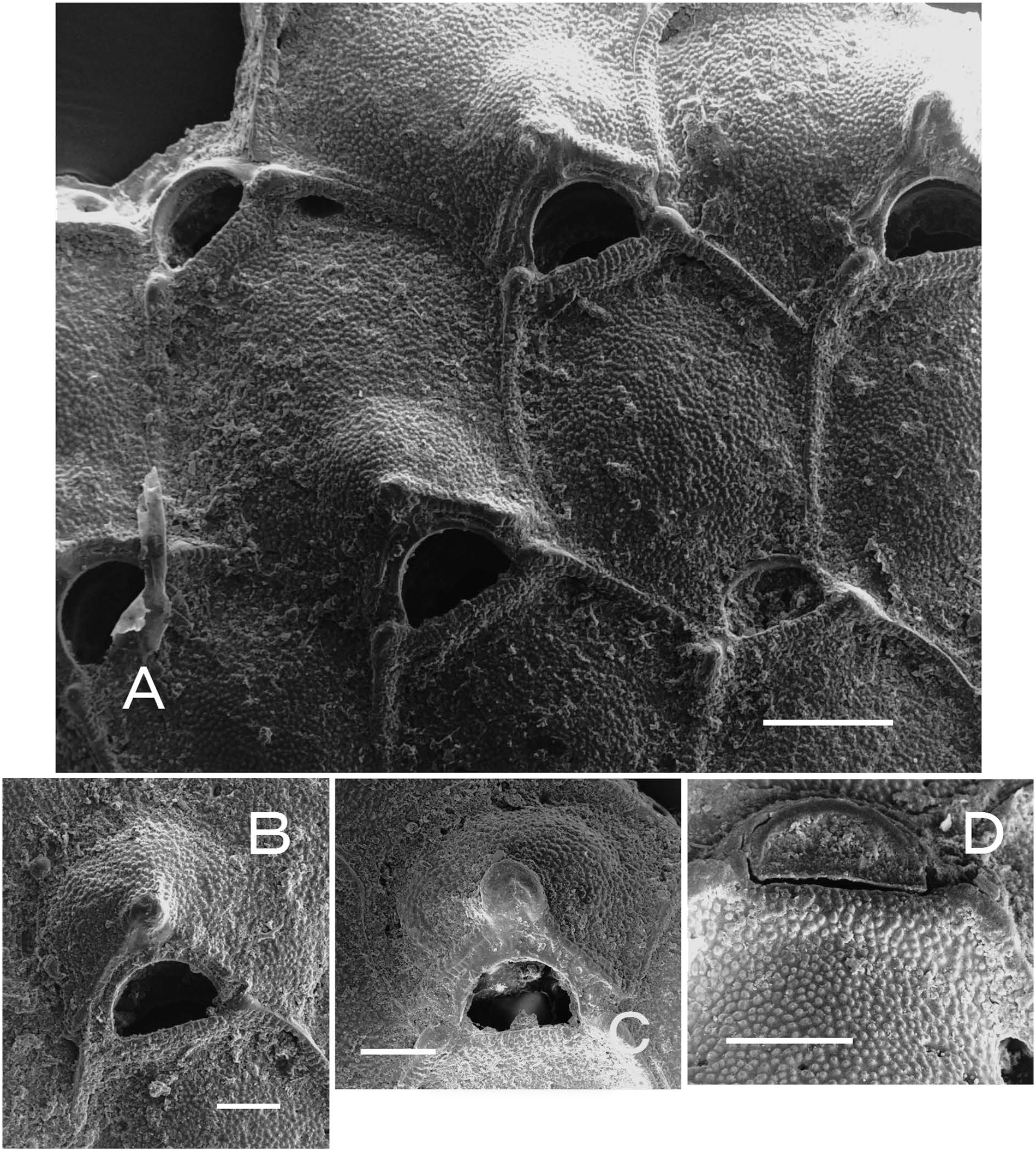
Micropora plana sp. nov.
( Fig. 4 View Fig )
Micropora coriacea (not of Esper in Johnston, 1847): Arakawa 1999: 56 (in part).
Miropora sp.: Arakawa 2014: 22, fig. 4D.
Material examined. Holotype: NMNS PA 16833 View Materials (seven fragments), Station 1739, Hakurei-Maru cruise GH80-2 . Paratype: NMNS PA 16834 (on siltstone, not coated with metal), Station 1733, Hakurei-Maru cruise GH80-2. Other material examined: NMNS PA 16835 View Materials (on bioclast), and 16836 (on mollusc shell), Station 1734, Hakurei-Maru cruise GH80-2; NMNS PA 16837 View Materials (on pebble), Station 1683, Hakurei-Maru cruise GH80-2 . See Table 1 View Table 1 for coordinates and depths of cruise samples.
Diagnosis. Frontal shield nearly flat, finely granulated, with minute pores. Mural rim low and narrow. Small laterooral knobs present. Orifice dimorphic. Spines and avicularia lacking. Ovicell with granular surface and thickened proximal margin, sometimes bearing small umbo.
Etymology. The specific name comes from the Latin planus (flat), referring to the flattened cryptocyst and low mural rim.
Measurements (in milimetres). NMNS PA 16833, 16835, and 16837. Autozooids (n =149, 9): ZL, 0.41–1.04 (0.693±0.099); ZW, 0.35–1.05 (0.563±0.093); OrL, 0.05– 0.12 (0.086±0.013); OrW, 0.12–0.24 (0.164±0.016). Ovicellate zooids (n =38, 9): ZL, 0.56–0.98 (0.716±0.081); ZW, 0.48–0.86 (0.580±0.079); OrL, 0.07–0.14 (0.104±0.014); OrW, 0.12–0.25 (0.185±0.020). Ovicells (n =38, 3): OvL, 0.23–0.45 (0.288±0.040); OvW, 0.29–0.53 (0.355±0.054).
Description. Colony encrusting, unilaminar, multiserial, forming lobate sheet; maximum observed size in largest dimension 7.3 mm (NMNS PA 16835). Zooids roughly subhexagonal to elliptical in outline. Frontal shield cryptocystal, nearly flat, with finely granulated surface, perforated by minute pores, surrounded by relatively low and narrow mural rim ( Fig. 4A View Fig ). Proximal gymnocyst lacking. Laterooral knobs small. Opesiules small, elongate. Orifice dimorphic; autozooidal orifice wider than long, broadly D-shaped; orifice of ovicellate zooids larger, semicircular. Surface of operculum granular, rimmed distally ( Fig. 4D View Fig ). Oral spines lacking. Ovicell globose, raised; surface granular like frontal shield, sometimes surrounded by slightly raised margin and suture line; proximal margin thickened, chevron-shaped, oπen with small, rounded-conical umbo at apex ( Fig. 4 View Fig A–C). Avicularia lacking. Two basal pore chambers in each distolateral wall; pair of widely spaced smaller pore chambers in transverse wall.
Distribution. This species was found from the continental shelf east of the Boso Peninsula at depths from 125 to 196 m, and the Danjo Islands, Nagasaki Prefecture, Japan ( Arakawa 2014).
Remarks. Micropora plana resembles M. mawatarii in showing dimorphism of the orifice and in lacking avicularia. However, this species differs from the latter in the following characters: zooids are larger (average length 0.693 mm, average width 0.563 mm) than those of M. mawatarii (average length 0.574 mm, average width 0.437 mm); the frontal shield is flatter, with smaller pores and finer granulation; the mural rim is lower and thinner; both latero-oral knobs and an umbo on the ovicell are less pronounced; a suture line is sometimes evident around the ovicell; and there are two pore chambers in the distal transverse wall.
Among the species of Micropora in the Pacific Ocean and adjoining seas ( Table 2 View Table 2 ), Micropora inexpectata Moyano, 2002 from Chile is similar to M. plana in having a flattened and finely granulated frontal shield with small pores, and ovicells with a raised rim and suture line, but it differs in having avicularia ( Moyano 2002). Micropora santacruzana Soule, Soule, and Chaney, 1995 from California also has an ovicell surrounded by a marginal rim, but it has avicularia and large frontal pores ( Soule et al. 1995).
| NMNS |
National Museum of Natural Science |
No known copyright restrictions apply. See Agosti, D., Egloff, W., 2009. Taxonomic information exchange and copyright: the Plazi approach. BMC Research Notes 2009, 2:53 for further explanation.