
Microchaetus sophieae, Plisko, 2002
|
publication ID |
https://doi.org/ 10.5281/zenodo.7909864 |
|
DOI |
https://doi.org/10.5281/zenodo.7910145 |
|
persistent identifier |
https://treatment.plazi.org/id/197C4F49-732B-FFC7-DAD6-0055FE81FE9A |
|
treatment provided by |
Felipe |
|
scientific name |
Microchaetus sophieae |
| status |
sp. nov. |
Microchaetus sophieae sp. n.
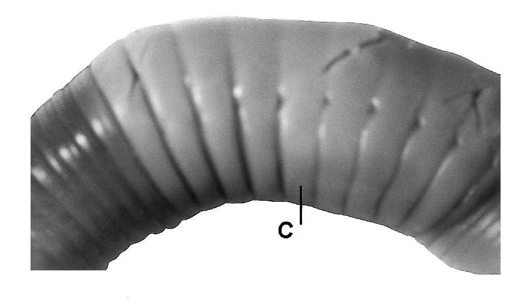
( Fig. 3 View Fig )
Etymology: Named for Dr Sophie Reinecke of the Zoology Department, University of Stellenbosch, who supervised the collection of the type material and donated it to the Natal Museum Oligochaeta Collection.
Material examined: SOUTH AFRICA: Western Cape: Holotype NMSA /Olig.03548, Nieuwoudtville (31º22'S: 19º06'E) from arable field under fungicide experiment, 17 July 1998, M. Maboeta leg. Paratypes: NMSA / Olig. 03549, 3 clitellate collected with holotype GoogleMaps .
Description based on holotype and paratypes.
External characters:
General: Body cylindrical, firm. Colour: Alcohol-preserved: prostomium and segments 1–2 violet all around; other segments dorsally violet, ventrally grey. Dimensions: Preserved and contracted holotype 112 mm, 5 mm wide at segment 10, 7 at tubercula pubertatis; paratypes 100–130 mm long, 4–5 mm wide at segment 10, 6–7 mm at tubercula pubertatis. Segment number: 300–342. Prostomium: Prolobous, large. Segmentation: Secondary annulation present on preclitellar segments; segments 1 and 2 fused, with irregular longitudinal grooves; segment 3 simple, as long as first ringlet of segment 4; 4–9 with 2 ringlets, first longer than second; 10 and 11 short, simple, irregularly annulated; 12 and clitellar segments simple, smooth; postclitellar much shorter than preclitellar, simple, randomly annulated. Setae: Minute, closely paired; postclitellarly ab = cd, aa <bc> dd <1/2u; first pairs on segment 3. Nephridial pores: Much ventral to cd setal lines; on clitellar segments conspicuous, postclitellarly difficult to trace. Female pores: In 14 between bc setae. Male pores: Large openings on 18 in area of tubercula pubertatis. Spermathecal pores: Clearly visible in intersegmental furrows13/14 14/15 15/16 close to nephridial pores.
Clitellar region ( Fig. 3 View Fig ): Clitellum: Extends over segments 13–22, saddle-shaped, with simple, smooth segments; clearly bordered anteriorly and posteriorly, with ventral edges above ab setal lines. Tubercula pubertatis: Oblong, flat pads, overlapping clitellum on 17–1/n20; dorsal edges below nephridial pores, ventral edges slightly above b setal lines at ventral edges of clitellum. Papillae: Paired, minute swellings, various shapes and sizes, associated with ab setae; on segments 10–23.
Internal characters:
Septa: 4/5 thickened only a little, 5/6 7/8 8/9 thickened moderately, firm, not muscular; 6/7 thinner than 4/5; 8/9 slightly thickened, however, thinner than 4/5; other septa thin, firm. Variation in thickness of septa observed. Gizzard: In 7, bell-shaped, firm, widened and softened posteriorly. Calciferous glands: In 10, paired, small glands, lateral, clearly separated dorsally and ventrally. Intestine: Commences in 12. Typhlosole: In paratype with 300 segments, commences immediately with intestine as narrow tube, gradually changing into V-shaped structure; terminates in segment 188. Dorsal blood vessel: Undivided, simple over its whole length; slender vessel in 4–7, enlarged in 8, in 9 thick organ; posteriorly simple, moderate in size. Paired dorsoventral vessels: In 4–7 simple thin vessels, in 8–11 enlarged, moniliform. Nephridia: Meganephridia ; in clitellar segments thin, coiled loops with J-shaped slender tubes.
Reproductive organs: Spermiductal funnels: Holandric arrangement (in 10 and 11); large, free funnels, iridescent, containing sperm. Vasa deferentia : Two pairs of ducts commence from lateral sides of spermiductal funnels, extending transversely in 10 and 11 before curving posteriorly to run backward parallel to axis of body; two ducts being initially separated in segment 10 and 11, in the following segments become very close one to another and run to segment 18, where they enter body wall in posterior part of segment, and into male pores. It is not clear if double ducts fuse before entering male pores. Seminal vesicles: Two pairs of sacs commencing at posterior parts of septa 10/11 and 11/12 respectively; each pair of different size and appearance; an anterior pair being much smaller than the posterior pair curving dorsally and confined to segment 11; posterior pair commencing at septa 11/12 and forming bulged, dissimilar pouches extending backwards differently on left and right side; in holotype the left vesicle extends to segment 16, the right vesicle to segment 18; in paratypes left vesicle extends to segments 16–18, right one to 16 or 17 or 19. Variation in shapes, sizes and extension of posterior pairs were noted in dissected specimens. Spermathecae: In holotype in segments 13 14 16, in paratypes in 13 14 15; paired, globular, large iridescent ampullae with long, slender ducts entering body wall at intersegmental furrows 13/14 14/15 15/16; in one paratype were two spermathecae at left side of segment 13. Iridescence confirming presence of sperm observed in all spermathecae. Ovaries: Not observed. Genital glands: Single or paired, small to minute oval bladders, in segments 10–23. Some of the glands contain genital setae.
Distribution: Known only from the type locality in southwestern South Africa, in the Namaqualand escarpment mountains of the Western Cape Province.
Biological notes: Found in a cultivated field undergoing fungicide treatment. It is not know if the species survived this treatment. Presence of iridescent sperm in the spermiductal funnels and in all spermathecae of dissected specimens indicates sexual activity in winter, when temperatures can be very low, sometimes reaching 0 ºC.
Discussion: Belongs to the group of species with seminal vesicles extended backwards over more than two segments. Most similar to M. ljungströmi Pickford 1975 , having paired spermathecae in three segments, and spermathecal pores in intersegmental furrows 13/14 14/15 15/16. Both species are characterised by extension of the posterior pair of seminal vesicles. However, in ljungströmi seminal vesicles are located in segments 10 and 11, whereas in sophieae they are in 11 and 12. These two species differ also in the shape of the nephridia, position and shape of the tubercula pubertatis, and thickness of anterior septa.
| NMSA |
KwaZulu-Natal Museum |
No known copyright restrictions apply. See Agosti, D., Egloff, W., 2009. Taxonomic information exchange and copyright: the Plazi approach. BMC Research Notes 2009, 2:53 for further explanation.