
Mesomyzostoma katoi Okada, 1933
|
publication ID |
https://doi.org/ 10.1080/00222933.2015.1056266 |
|
DOI |
https://doi.org/10.5281/zenodo.5672664 |
|
persistent identifier |
https://treatment.plazi.org/id/2F2FE66B-940C-FFD1-548C-BBEC18F8FA0C |
|
treatment provided by |
Plazi |
|
scientific name |
Mesomyzostoma katoi Okada, 1933 |
| status |
|
Mesomyzostoma katoi Okada, 1933
( Figures 1 View Figure 1 , 2 View Figure 2 )
Mesomyzostoma katoi in Lanterbecq et al. (2006)
Mesomyzostoma cf. katoi in Helm et al. (2014)
Mesomyzostoma cf. katoi in Summers and Rouse (2014)
Material examined
Sagami Bay ( Japan) 35°09.46ʹ N, 139°36.72ʹ E; 10 – 15 m deep; ten specimens in Anneissia japonica ( Müller 1841) (Comatulidae) . Collector: Greg Rouse, 13 – 28 May 1998. Between one and five individuals in each host. Neotype (SIO-BIC A4070) fixed in formalin, preserved in 70% ethanol; Other specimens: five (SAM-E3407) in 70% ethanol after formalin fixation; one (SIO-BIC A4072) on SEM stub used for SEM observations. Kagoshima, off the slope of Mt Sakurajima (Japan) 31°35.855ʹ N, 130° 36.031ʹ E; 10 – 15 m deep; two specimens (SIO-BIC A4071) from several A. japonica , both fixed in glutaraldehyde and osmium, then embedded in resin for histological studies. Collector: Greg Rouse, 8 May 1998. One specimen from lot SAM-E3407 dissolved in bleach for observation of parapodial hook apparatus and another three specimens digested for DNA extraction and molecular phylogenetic analyses (not catalogued). Other material: Madang Lagoon (Papua New Guinea), 5.136° S, 145.81° E, 3 m deep. Six specimens (SIO-BIC A3689, A3691, A3711) in 70% ethanol after formalin fixation, or in 95% ethanol. Host = Anneissia bennetti ( Müller 1841) (Comatulidae) . Collector: Greg Rouse and Mindi Summers, November – December 2012.
Diagnosis
Mesomyzostoma with elongated, thin, ribbon-like body; no introvert or cirri. Five chaetigers, with very low parapodia located close to body margin. Emergent hooks small with thin shaft, tip curving 90° with respect to shaft. No replacement hooks. Aciculae as long as emergent hooks, much thicker at base. Manubria club-shaped, developed on one side. Tens of low, slit-like lateral organs arranged along body margin. Mouth at anterior tip of body, cloaca (exit for digestive tract and uterus) at posterior tip. Body translucent. Two pairs of digestive diverticula. Simultaneous hermaphrodites. One pair of male seminal vesicles opening at third chaetiger. No penes. Parasitic, living in crinoid coelom.
Description
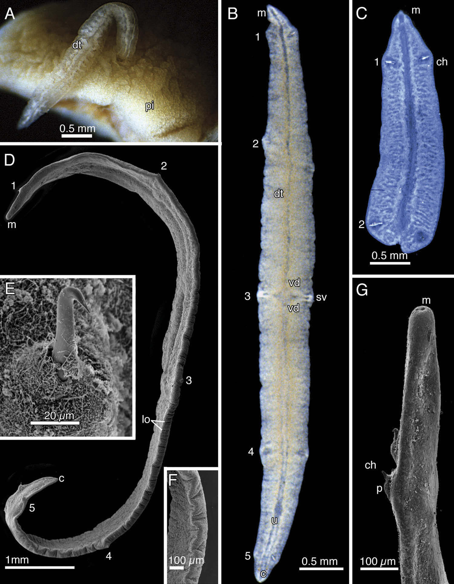
Live individuals observed in coelom of pinnules close to gonads and in coelom of arms ( Figure 1 View Figure 1 A). Neotype ( Figure 1 View Figure 1 B) with very thin, flat body 6.5 mm long, 0.2 mm thick. In life translucent with pale orange gut; ovary and oocytes orange, seminal vesicles white. Width irregular, 0.5 mm maximum, at mid-level of third chaetiger. Preserved body white, curled dorsally with anterior and posterior margins close to each other. Mouth at anterior tip, a small circular opening of less than 50 µm in diameter. Five pairs of low ventral parapodia, with hooks, very close to body edge ( Figures 1 View Figure 1 B, D – F, 2A, B). First to fifth pairs of chaetae located at 0.2, 1.3, 2.5, 3.7 and 4.9 mm from mouth, respectively. Each emergent hook with acicula, but no replacement hook ( Figure 2 View Figure 2 D). Emergent hook up to 0.25 mm long, shaft moderately thick, with distal fifth bowed slightly outward, tip short and sharp curving 90° ( Figures 1 View Figure 1 E, 2D). Acicula of same length as emergent hook, but thicker at base. Manubrium developed on one side of hook, with small ridges along outer margin ( Figure 2 View Figure 2 D).
Body margin irregularly notched, lacking cirri ( Figure 1 View Figure 1 B). Lateral organs evident on body margin as small dorsoventral slits, only visible by SEM ( Figure 1 View Figure 1 D). Cloaca at extreme posterior end ( Figure 1 View Figure 1 B). Penes absent; pair of seminal vesicles near third pair of parapodia ( Figure 1 View Figure 1 B). Spermatogonia/spermatids found dorsally and laterally at least in midbody region ( Figure 2 View Figure 2 A). Vas deferens, filled with sperm, situated in vicinity of third chaetiger ( Figures 1 View Figure 1 B, 2A, B) and leading into seminal vesicles lying level with third chaetiger ( Figure 1 View Figure 1 B). Uterus full of oocytes ( Figure 1 View Figure 1 B). Digestive system a straight central tube with lateral diverticula. Central gut tube including very small pharynx ( Figure 1 View Figure 1 B). Digestive diverticula start from each side of central gut, dichotomizing at regular intervals to give rise to secondary branches that stop near body margin ( Figures 1 View Figure 1 A – C, 2A).
Other specimens range from 5 to 13 mm long, 0.5 to 1 mm wide and 0.1 mm thick ( Figure 1 View Figure 1 A). One specimen incomplete, 2.5 mm long ( Figure 1 View Figure 1 C). Number of parapodia ranging from two to five pairs ( Figure 1 View Figure 1 A, C). Larvae raised from oocytes released by several individuals upon dissection from host, ellipsoidal with 50-µm long axis and bundle of chaetae on each side of mid-body ( Figure 2 View Figure 2 C).
Remarks
Mesomyzostoma katoi was originally described from four specimens observed in the gonads of Comanthus japonicus ( Müller 1841) [a species subsequently placed in Oxycomanthus and now Anneissia (see Summers, Messing, et al. 2014)]. The host crinoids were collected by Kôjiro Kato in shallow waters near the Misaki Marine Biological Station, Sagami Bay ( Okada 1933). Okada (1933) did not mention where the types were deposited. No specimens of M. katoi are at the University Museum of the University of Tokyo ( Nishi and Tanaka 2011). During his career Dr Okada worked at the University of Tsukuba (Shimoda Marine Research Centre), Mie University (Faculty of Fisheries) and at Tokai University (the likely repository there being the Marine Science Museum at Orido in the Shimizu section of Shizuoka city) ( Watanabe 1981; M. Grygier, pers. comm.; current names used here). There are no samples from the time of the description held at any of these institutions (E. Nishi, pers. comm.). It therefore seems reasonable to infer that Okada did not deposit any types for M. katoi , or that they have been lost.
The fact that there are two species of Mesomyzostoma present in A. japonica (see below), from the type locality of Sagami Bay, makes the designation of a neotype necessary. Our specimens from the type locality, Sagami Bay, seemingly differ from those described by Okada by (1) the presence of numerous slit-like lateral organs, (2) the presence of five chaetigers and (3) the greater length of the specimens. Okada (1933) mentioned in the diagnosis of M. katoi that it does not have any ‘ tentacles ’ (i.e. cirri) on the periphery and that ‘ suckers ’ (i.e. lateral organs) were absent. However, Okada (1933) apparently missed these small organs, which, depending on their state of contraction, were not always visible on the specimens we observed. Okada ’ s (1933) four specimens ranged from 2.3 to 4.5 mm long and 0.4 to 0.8 mm wide, with three to four pairs of chaetae and digestive diverticulae at the posterior end of the central gut lumen. These three characteristics strongly suggest that the specimens described by Okada were incomplete. We also observed incomplete specimens with two to three pairs of chaetae, matching Okada ’ s (1933) description ( Figure 1 View Figure 1 C). It is easy to break Mesomyzostoma specimens while trying to extract them from the crinoid host (Rouse pers. obs.), and we strongly suspect that this is what happened with Okada ’ s (1933) material. Although M. katoi is hosted by the same crinoid species as another new Mesomyzostoma species described here (see below), the smooth aspect of the trunk margin in M. katoi distinguishes it easily, as do molecular sequence data. Several specimens of M. katoi were also arguably collected from Papua New Guinea (see Helm et al. 2014) from a different species of Anneissia , A. bennetti . Unfortunately no COI data from Japanese specimens could be obtained to compare the two populations more closely. The 18S sequences for the Papua New Guinea versus Japan specimens differed by only two bases ( Figure 8 View Figure 8 ), once the erroneous part of the sequence from the Japanese specimen was removed.


No known copyright restrictions apply. See Agosti, D., Egloff, W., 2009. Taxonomic information exchange and copyright: the Plazi approach. BMC Research Notes 2009, 2:53 for further explanation.
|
Kingdom |
|
|
Phylum |
|
|
Class |
|
|
Order |
|
|
Family |
|
|
Genus |
Mesomyzostoma katoi Okada, 1933
| Greg W. Rousea, Deborah Lanterbecq, Mindi M. Summersa & Igor Eeckhaut 2016 |
Mesomyzostoma katoi in Lanterbecq et al. (2006 )
| Lanterbecq et al. 2006: 5 |
Mesomyzostoma cf. katoi in Helm et al. (2014)
| Helm et al. (2014) |
Mesomyzostoma cf. katoi in Summers and Rouse (2014)
| Summers and Rouse (2014) |