
Mesocyclops brasilianus Kiefer, 1933
|
publication ID |
https://doi.org/ 10.1080/00222930600761837 |
|
persistent identifier |
https://treatment.plazi.org/id/03C887FC-FF86-BC36-E6C9-EC82FB450BE8 |
|
treatment provided by |
Felipe |
|
scientific name |
Mesocyclops brasilianus Kiefer, 1933 |
| status |
|
Mesocyclops brasilianus Kiefer, 1933
( Figures 1 View Figure 1 , 2 View Figure 2 )
M. varius Dussart, 1987 , junior synonym.
M. venezolanus Dussart, 1987 , junior synonym.
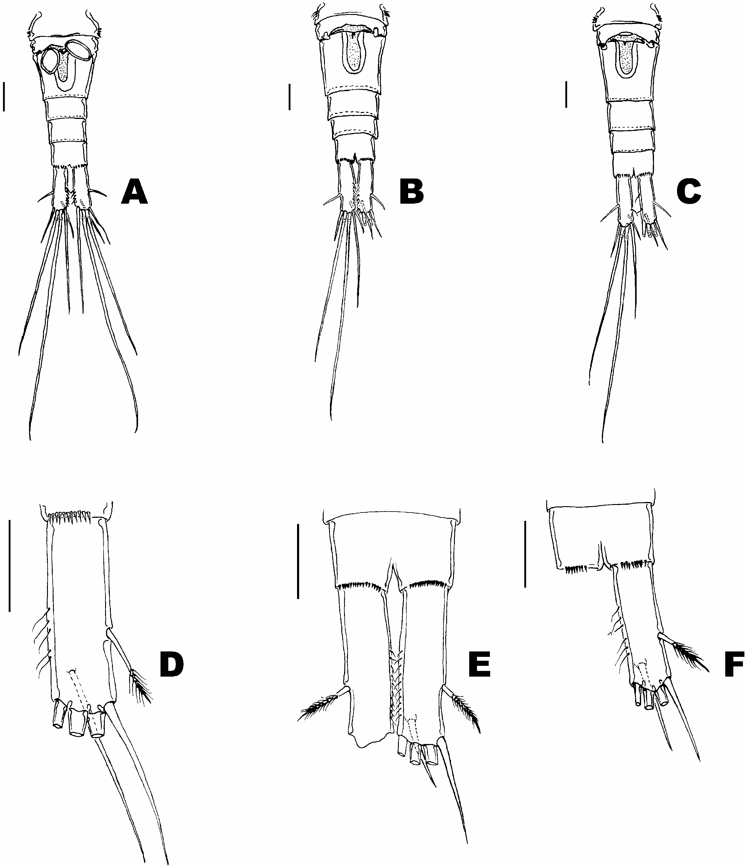
? M. venezolanus: Reid and Reid 1994, p 83 –87, Figures 3 View Figure 3 , 4 View Figure 4 .
The bad condition of the type specimens of M. brasilianus prevented a detailed analysis; however, we based our criteria on the observations of 10 adult females from Itacoatiara , Amazonas, Brazil (03 ° 10.807 9 S, 58 ° 14.631 9 W, west from Manaus, established by Kiefer 1936 as the type locality) .
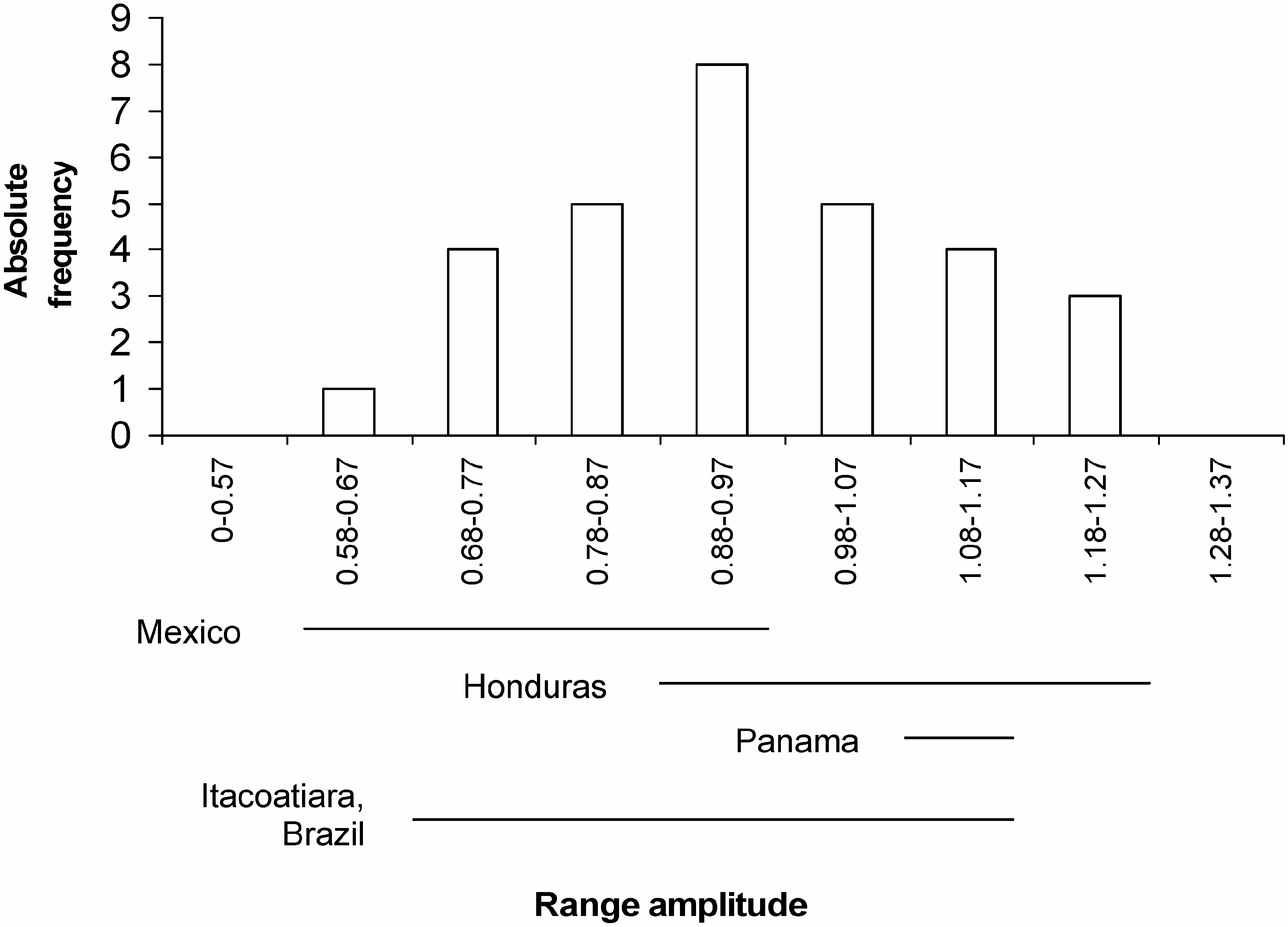
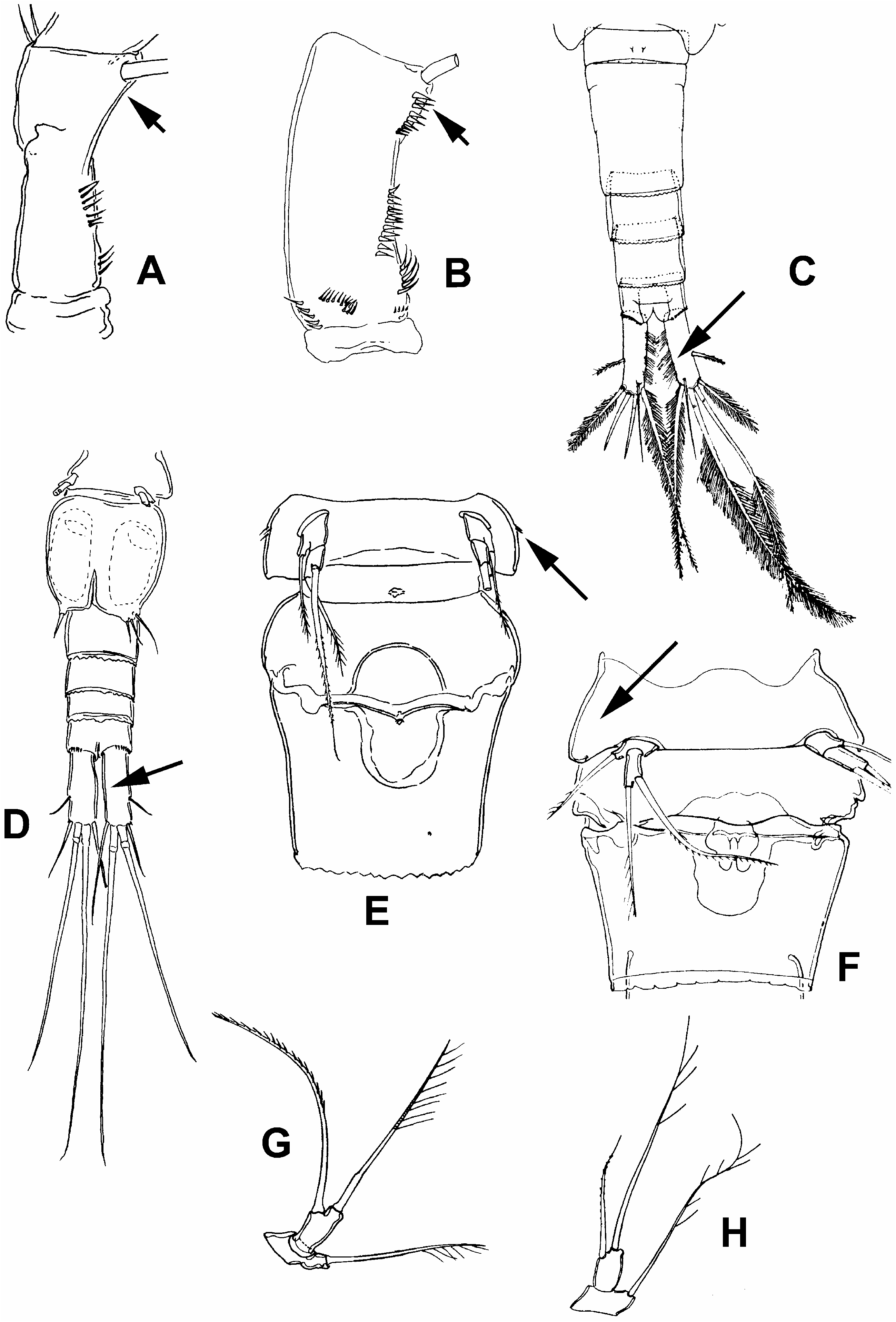
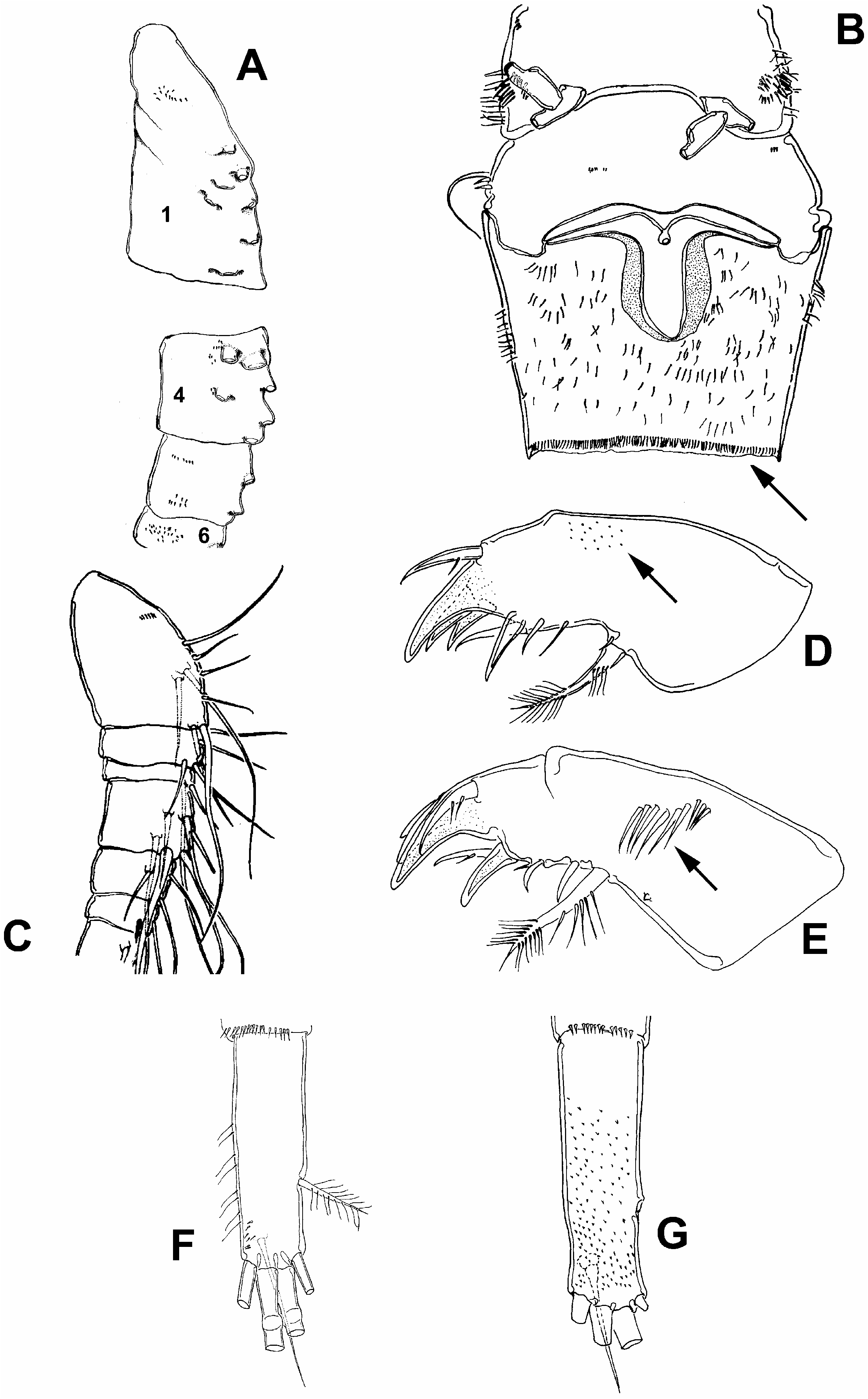
There is only one female labelled as M. varius in the world, from Taxisco, Guatemala (see Table I). Dussart (1987) recognized the resemblance between this specimen and M. brasilianus . Hołyńska et al. (2003) stated that its taxonomical status remains questionable; however, they included this species as part of the M. meridianus – brasilianus complex. Our analysis indicates that the morphological features used by Dussart (1987) to separate these species are not consistent: the presumed absence of an acute angle on the outer margin of the first endopodal segment of the first swimming leg has proved to be erroneous ( Figure 1A–C View Figure 1 ). The same is true with respect to the assumed absence of ornamentation on the coxa and inner margin of the fourth leg basis ( Figure 1D–F View Figure 1 ). The holotype of M. varius bears a spermatophore fixed to its genital aperture ( Figure 1G View Figure 1 ), but no differences were observed on the structure of the seminal receptacle between M. varius and M. brasilianus ( Figure 1G, H View Figure 1 ). Another character used to separate M. varius was the length ratio of two caudal setae: dorsal seta/lateral seta [relatively longer in comparison with M. brasilianus ( Dussart 1987) ]. We found that the proportional length observed in the holotype specimen of M. varius shows a lower figure than that determined from the original description of M. brasilianus (1.2 versus 1.3). In addition, the comparison of the seta length proportions ( Figure 2B, E View Figure 2 ) in different populations of M. brasilianus observed here (Table I), through an exploration of frequencies, shows a wide variability, even in the same population ( Figure 3 View Figure 3 ); therefore, we consider that this proportion is a weak feature for separating species.
Dussart (1987) described another species, M. venezolanus ; this was also recognized as being similar to M. brasilianus . The description was based on specimens collected in Lake Valencia, Venezuela. The differences by which M. venezolanus was separated from M. brasilianus were: caudal rami without spines on distal–lateral corner; abdominal somites with serrated hyaline fringe; dorsal seta on caudal rami shorter than outer seta; fifth thoracic somite with dispersed hair-like setae; seminal receptacle with two ‘‘bumps’’, followed by two curved horns. All these features are present in M. brasilianus too ( Figures 1B, C, E, F, H, I View Figure 1 , 2B, C, E, F View Figure 2 ), including the lack of spines on the distal–lateral corner of the caudal rami: no adult female of M. brasilianus examined here, even the specimens identified by Kiefer, showed those spines. The type specimen is useless for taxonomical analysis, and now we speculate that the spines shown in the illustrations, but not mentioned in the original description of M. brasilianus , indicate that this specimen is an immature CV female (Gutiérrez-Aguirre and Suárez-Morales 2003). Another specimen collected in Managuiri, Amazonas, Brazil, and identified by Kiefer (see Table I), does not have these spines. Therefore, the spine presence/absence pattern that would separate M. brasilianus and M. venezolanus is not a usable character. Therefore, we conclude that both M. varius and M. venezolanus are junior synonyms of M. brasilianus and should be considered as that in future taxonomic accounts of neotropical Mesocyclops . However, the identity of the specimens recorded by Reid and Reed (1994) as M. venezolanus from the Yukon Territory remains unverified, taking into account the differences of the hairornament observed by Hołyńska et al. (2003) in comparison with M. brasilianus .
Number of neotropical species of Mesocyclops
Before this study, up to 20 species, two varieties, and one subspecies of Mesocylops had been recorded in the neotropics. With the addition of the recently described M. evadomingoi ( Gutiérrez-Aguirre and Suárez-Morales 2001a) and the new record of the Asian M. pehpeiensis in Mexico ( Suárez-Morales et al. 2005), the junior synonymy of M. varius and M. venezolanus with M. brasilianus , the record of M. kieferi in Brazil, assignable to M. ogunnus (see Gutiérrez-Aguirre et al. 2003a), and the reassignation of M. longisetus var. araucanus to species rank by Pilati and Menu-Marque (2002), the revised, current account of neotropical records of Mesocyclops is: 20 species, one variety, and probably one species known as M. annulatus diversus (see section on distributional remarks later).
Key to the neotropical species of Mesocyclops
1. Inner basis of first trunk limb without seta („„, ♀♀) ( Figure 4A View Figure 4 )...... 2
– Inner basis of first trunk limb with seta, or spine-like seta („„, ♀♀) ( Figure 4B View Figure 4 ). 6
2. Fifth pediger naked ventrally ( Figure 4C View Figure 4 ); intercoxal sclerite of fourth trunk limb with high, acute projections („„, ♀♀) ( Figure 4D View Figure 4 ).......... 3
– Fifth pediger pilose ventrally ( Figure 4E View Figure 4 ); intercoxal sclerite of fourth trunk limb with low, non-acute projection („„, ♀♀) ( Figure 4F View Figure 4 )......... 4
3. Ducts connected with genital aperture straight (♀♀) ( Figure 4C View Figure 4 ), inner margin of fourth trunk limb basis pilose („„, ♀♀) ( Figure 4D View Figure 4 )....... M. pescei
– Ducts connected with genital aperture directed in rectum angle (♀♀) ( Figure 4G View Figure 4 ), inner margin of fourth trunk limb basis naked („„, ♀♀) ( Figure 4H View Figure 4 ).. M. pehpeiensis
4. Maxillular palp with spines on inner margin („„, ♀♀) ( Figure 5A View Figure 5 ).. M. ogunnus
– Maxillular palp without spines on inner margin („„, ♀♀) ( Figure 5B View Figure 5 ).... 5
5. Inner margin of caudal rami naked („„, ♀♀) ( Figure 5C View Figure 5 ). Antennal basis with a row of large spines (♀♀) ( Figure 5D View Figure 5 ) or a row of tiny spines („„) ( Figure 5E View Figure 5 ) near insertion of setae opposite to exopod........ M. thermocyclopoides
– Inner margin of caudal rami pilose („„, ♀♀) ( Figure 5F View Figure 5 ). Antennal basis with a dense group of tiny spines („„, ♀♀) ( Figure 5G View Figure 5 ) near insertion of setae opposite to exopod.................. M. aspericornis
6. Intercoxal sclerite of fourth trunk limb with pointed projections („„, ♀♀) ( Figure 5H View Figure 5 )..................... 7
– Intercoxal sclerite of fourth trunk limb smooth („„, ♀♀) ( Figure 5I View Figure 5 )... 17
7. Antennal basis without spines next to exopod („„, ♀♀) ( Figure 6A View Figure 6 ).... 8
– Antennal basis with spines next to exopod („„, ♀♀) ( Figure 6B View Figure 6 ).... 11
8. Inner margin of caudal rami pilose („„, ♀♀) ( Figure 6C View Figure 6 )..... M. edax
– Inner margin of caudal rami naked („„, ♀♀) ( Figure 6D View Figure 6 )....... 9
9. Fifth pediger with spines on ventro-lateral margins (♀♀) ( Figure 6E View Figure 6 ). M. reidae
– Fifth pediger naked ventro-laterally (♀♀) ( Figure 6F View Figure 6 )........ 10
10. Medial spine longer than apical seta on distal segment of fifth leg („„, ♀♀) ( Figure 6G View Figure 6 )................... M. chaci
– Medial spine shorter than apical seta on distal segment of fifth leg („„, ♀♀) ( Figure 6H View Figure 6 )................... M. yutsil
11. Intercoxal sclerite of fourth trunk limb with low, non-acute projections („„, ♀♀) ( Figure 7A View Figure 7 ).................... 12
– Intercoxal sclerite of fourth trunk limb with high, acute projections („„, ♀♀) ( Figure 7B View Figure 7 ).................... 15
12. Anterior margin of seminal receptacle convex, with lateral arms thin (♀♀) ( Figure 7C View Figure 7 )................. M. annulatus
– Anterior margin of seminal receptacle concave, wide lateral arms (♀♀) ( Figure 7D View Figure 7 ).................... 13
13. Lateral arms of seminal receptacle not strongly curved (♀♀) ( Figure 7D View Figure 7 ); inner surface of 16th antennular segment with more than two rows of spines („„) ( Figure 7E View Figure 7 ). Length/width ratio of caudal rami is 3.06 (2.8–3.27) (♀♀) and 2.93 (2.84–3.06) („„); Length/width ratio of third endopodal segment of fourth trunk limb is 2.52 (2.0–2.8) (♀♀) and 2.94 (2.75–3.05) („„); base of lateralmost terminal caudal seta without spines (♀♀) (see Pilati and Menu-Marque 2002) ( Figure 7I View Figure 7 )................ M. longisetus s. str.
– Lateral arms of seminal receptacle strongly curved (♀♀) ( Figure 7F View Figure 7 ); inner surface of 16th ( Figure 7G View Figure 7 ) or 15th antennular segment (see Pilati and Menu-Marque 2002) with two rows of spines („„)............. 14
14. Length/width ratio of third endopodal segment of fourth trunk limb is 2.6–3.2 (♀♀) ( Figure 7H View Figure 7 ); length/width ratio of caudal rami is 2.6–3.2 (♀♀) ( Figure 7I View Figure 7 ).............. M. longisetus var. curvatus
– Length/width ratio of third endopodal segment of fourth trunk limb is 3.48 (3.3– 3.8) (♀♀) ( Figure 7J View Figure 7 ) and 3.21 (3.14–3.46) („„); length/width ratio of caudal rami is 3.79 (3.5–4.2) ( Figure 7K View Figure 7 ) (♀♀) and 3.27 (3.13–3.30) („„); base of lateralmost terminal caudal seta with spines (♀♀) (see Pilati and Menu-Marque 2002)................. M. araucanus
15. Anterior margin of seminal receptacle strongly convex ( Figure 8A View Figure 8 ), anal somite ornamented dorsally (♀♀) ( Figure 8B View Figure 8 ).......... M. ellipticus
– Anterior margin of seminal receptacle weakly convex ( Figure 8C View Figure 8 ), anal somite naked dorsally (♀♀) ( Figure 8D View Figure 8 ).............. 16
16. Row of spines on posterior margin of anal somite not dorso-ventrally continuous (♀♀) ( Figure 8E View Figure 8 )............... M. intermedius
– Row of spines on posterior margin of anal somite dorso-ventrally continuous (♀♀) ( Figure 8F View Figure 8 )................. M. paranaensis
17. Anterior margin of seminal receptacle concave (♀♀) ( Figure 8G View Figure 8 ).... 18
– Anterior margin of seminal receptacle convex (♀♀) ( Figure 8H View Figure 8 )..... 19
18. Spines on antennular segments 4–6 ( Figure 9A View Figure 9 ); abdominal somites with a row of setae along posterior margins (♀♀) ( Figure 9B View Figure 9 )....... M. meridionalis
– Antennular segments 4–6 without spines ( Figure 9C View Figure 9 ); abdominal somites without a row of setae along posterior margins (♀♀) ( Figure 8G View Figure 8 ).... M. brasilianus
19. Praecoxal surface of maxillulae with short scales on posterior surface („„, ♀♀) ( Figure 9D View Figure 9 )................. M. evadomingoi
– Praecoxal surface of maxillulae with long setae in posterior surface („„, ♀♀) ( Figure 9E View Figure 9 ).................... 20
20. Caudal rami with inner margin pilose, spines on ventro-posterior surface (♀♀) ( Figure 9F View Figure 9 )............... M. pseudomeridianus
– Caudal rami with inner margin naked, scales on entire ventral surface (♀♀) ( Figure 9G View Figure 9 )................. M. meridianus


No known copyright restrictions apply. See Agosti, D., Egloff, W., 2009. Taxonomic information exchange and copyright: the Plazi approach. BMC Research Notes 2009, 2:53 for further explanation.
|
Kingdom |
|
|
Phylum |
|
|
Class |
|
|
Order |
|
|
Family |
|
|
Genus |
Mesocyclops brasilianus Kiefer, 1933
| Gutiérrez-Aguirre, M. A., Suárez-Morales, E., Martínez, A. Cervantes-, Elías-Gutiérrez, M. & Previattelli, D. 2006 |
M. venezolanus
| : Reid and Reid 1994: 83 |
M. varius
| Dussart 1987 |
M. venezolanus
| Dussart 1987 |
M. brasilianus
| Kiefer 1933 |