
Mastogloia belaensis M.Voigt
|
publication ID |
https://doi.org/ 10.5852/ejt.2017.374 |
|
DOI |
https://doi.org/10.5281/zenodo.3852194 |
|
persistent identifier |
https://treatment.plazi.org/id/039FFA2B-FFEE-FFD6-4E19-FC3BE81E3302 |
|
treatment provided by |
Carolina |
|
scientific name |
Mastogloia belaensis M.Voigt |
| status |
|
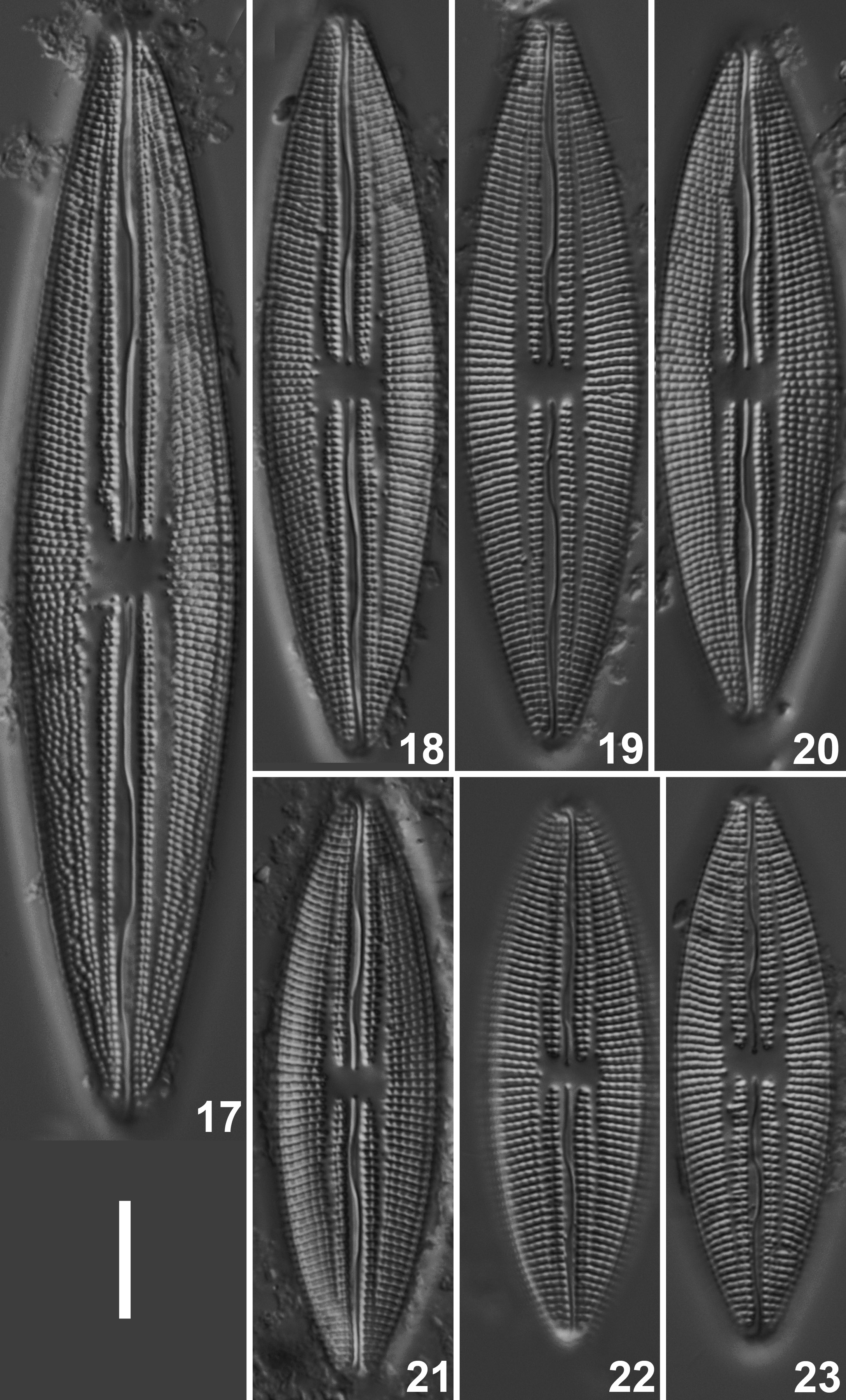
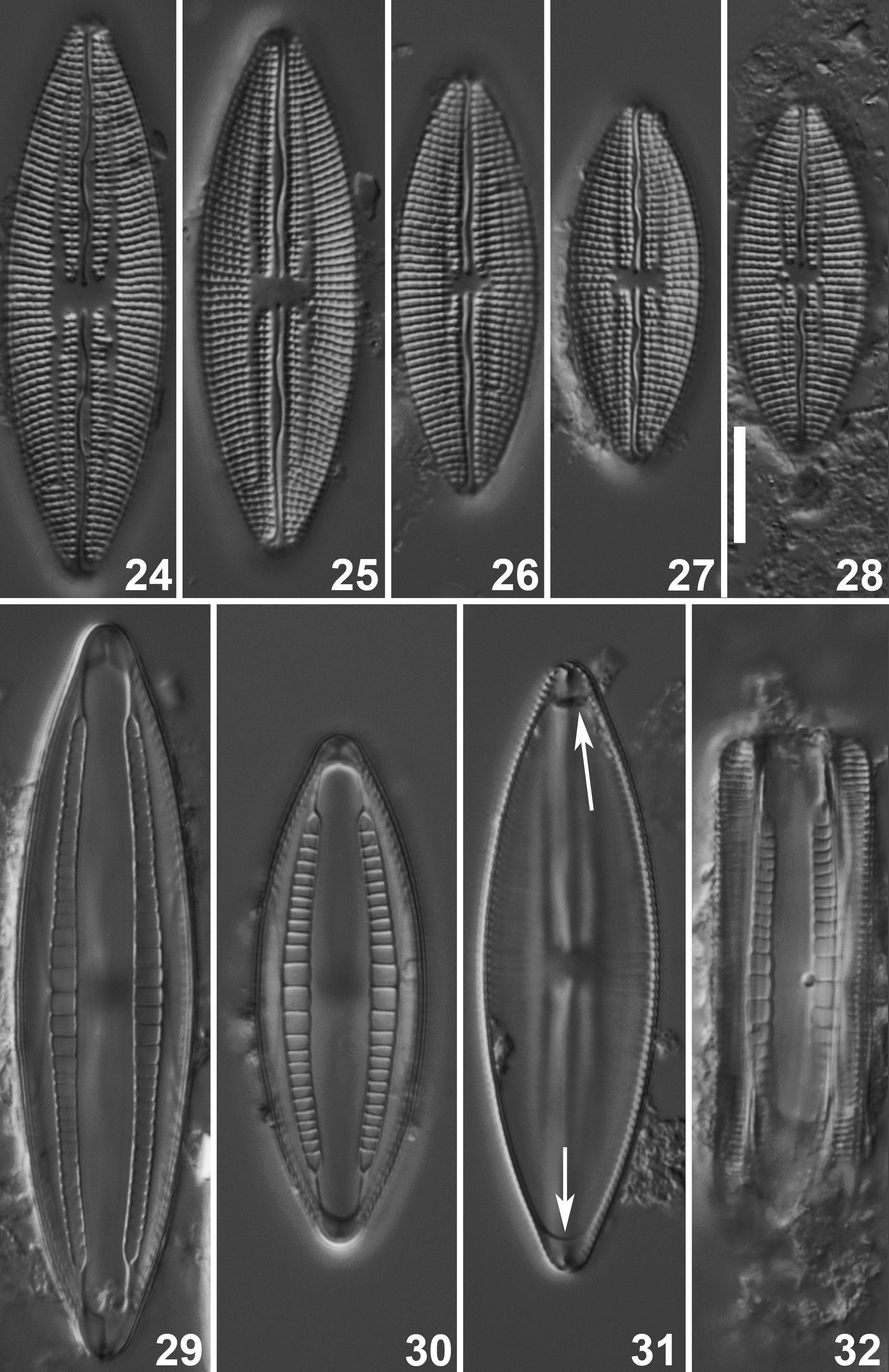
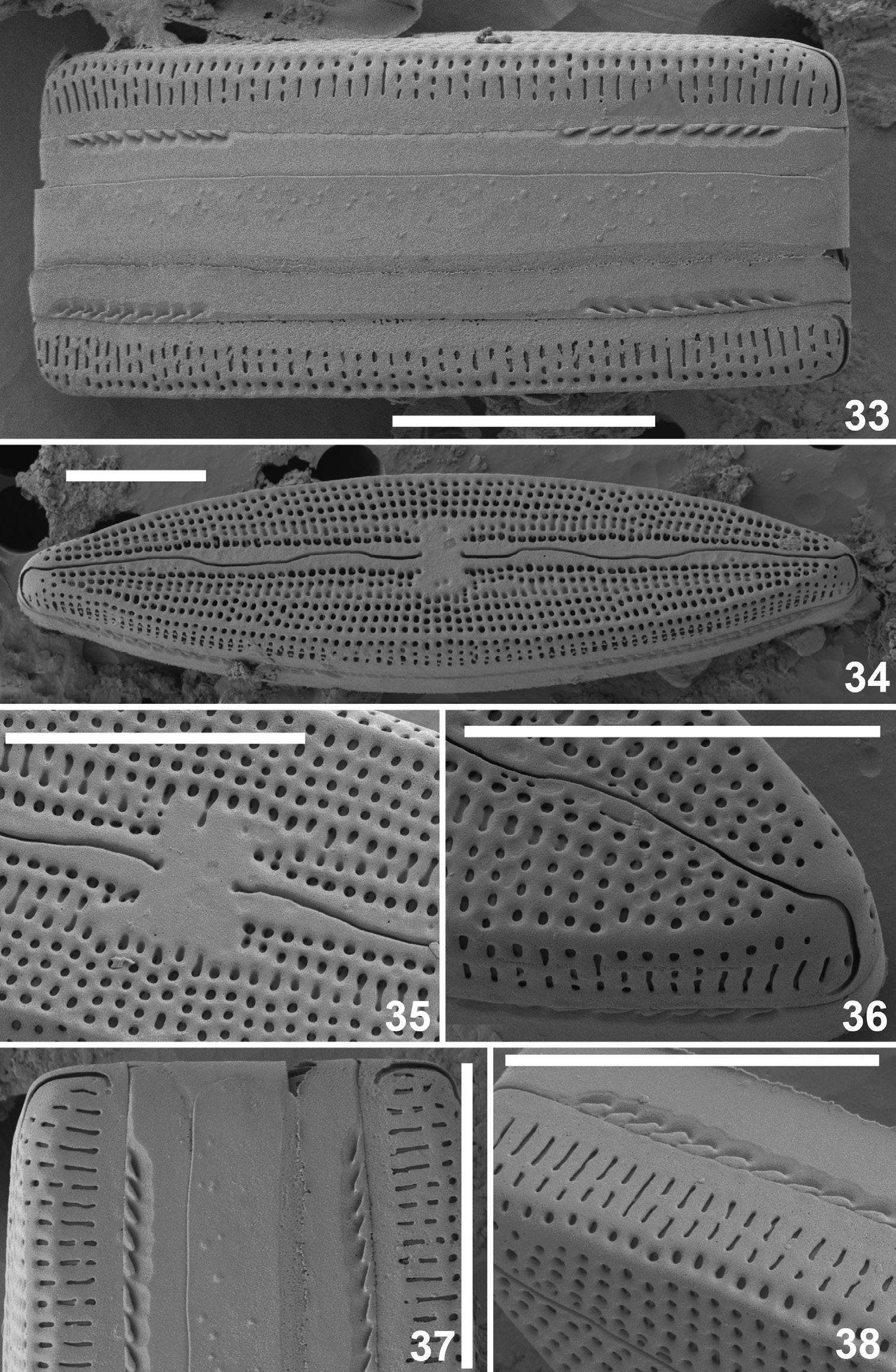
Figs 17–45 View Figs 17–23 View Figs 24–32 View Figs 33–38 View Figs 39–45
Journal of the Royal Microscopical Society ser. 3 75: 189, pl. 1 fig. 1, 5, 6, 7 ( Voigt 1956).
Description ( Senegal population)
Light microscopy ( Figs 17–32 View Figs 17–23 View Figs 24–32 )
Valves lanceolate to elliptic-lanceolate with convex margins. Apices non-protracted, acutely rounded to slight protracted, subrostrate. Valve dimensions (n = 50): length 31–99 µm, width 11.5–20.0 µm. Axial area narrow, lanceolate, narrowing towards the apices. Lyre-shaped hyaline zone present close to the axial area, separating one row of pseudoloculi from the striae. Central area rather small, rectangular. Raphe lateral with clearly undulating branches. Proximal raphe endings indistinct, straight. Distal endings hooked towards the same direction. Striae radiate throughout, becoming less radiate and even parallel to slightly divergent ( Fig. 25 View Figs 24–32 ) towards the apices, 13–15 in 10 µm. Pseudoloculi quite large, well visible in LM, 15–20 in 10 µm. Partectal ring clearly displaced towards the middle of the valve, composed of partecta of different size (6–8 in 10 µm): the middle 4–8 partecta (length 1.9–2.9 µm, width 1.8–3.9 µm) considerable larger than the outer partecta (length 0.9–1.4 µm, width 1.2–1.8 µm).
Scanning electron microscopy ( Figs 33–45 View Figs 33–38 View Figs 39–45 )
External raphe branches clearly undulating ( Fig. 34 View Figs 33–38 ). Proximal raphe endings simple, very weakly expanding, slightly deflected ( Figs 34–35 View Figs 33–38 ). Distal raphe fissures centrally crossing the terminal nodule, elongated, hooked towards the same direction, continuing onto the valve mantle, terminating almost near the mantle edge ( Fig. 36 View Figs 33–38 ). Very low, slightly thickened marginal crest visible on the valve face/ mantle junction separating the striae on the valve face from the mantle areolae by a hyaline line ( Fig. 34 View Figs 33–38 ). Mantle striae entirely uniseriate, composed of several, usually transapically elongated to slit-like pseudoloculi ( Figs 33–34, 36, 38 View Figs 33–38 ). First pseudoloculi near the junction rounded ( Figs 36, 38 View Figs 33–38 ). Valve face almost flat, subdivided into two zones: outer zone composed of uniseriate striae, with up to four rounded pseudoloculi, central zone formed by one row of rounded pseudoloculi close to the axial area and one row of transapically elongated rectangular pseudoloculi ( Figs 34–35 View Figs 33–38 ). Near the central area, rectangular pseudoloculi replaced by 1–2 small, rounded ones whereas near the apices, central transapically elongated pseudoloculi absent, outer zone with rounded pseudoloculi reaching the axial area ( Fig. 36 View Figs 33–38 ). Shallow depressions sometimes visible in the axial area ( Figs 34–36 View Figs 33–38 ). Hyaline areas present on each valve apex ( Fig. 36 View Figs 33–38 ). Internally, raphe sternum lyriform, slightly raised ( Figs 39, 42 View Figs 39–45 ). Raised thickened costa-like interstriae interrupted by the raphe-sternum extending from the axial area towards the valve margins, separating the areolae ( Figs 42–43 View Figs 39–45 ). Inner areolae arranged in groups of 4–8 per pseudoloculus ( Fig. 45 View Figs 39–45 ). Raphe branches straight with indistinct, coaxial proximal endings, terminating on a raised central nodule ( Figs 39, 42 View Figs 39–45 ). Valve apices with clear pseudosepta covering the distal raphe endings ( Fig. 43 View Figs 39–45 ).
Girdle composed of the valvocopulae and one open, non-perforated copula ( Figs 33, 37 View Figs 33–38 ). Small siliceous papillae present on the copula ( Fig. 37 View Figs 33–38 ). Valvocopulae with typical partectal ring, opening near the apices through a series of elongated partectal pores ( Figs 33, 37–38 View Figs 33–38 ). Partectal ring open, one end closed but with a cleft forming a narrow lacuna ( Fig. 40 View Figs 39–45 ), other split with a cleft ( Fig. 41 View Figs 39–45 ), covering entirely the pseudosepta. Partecta extending to almost ¼ to 1/6 from the valve apex, clearly separated from the valve margin by a siliceous flange ( Fig. 39 View Figs 39–45 ). Partecta subequal in size with the large ones grouped in the middle, the smaller ones closer to the apices ( Fig. 39 View Figs 39–45 ). Partecta ornamented with several series of small, rounded areolae, loosely aggregated in very unclear plaques and arranged inwardly ( Fig. 44 View Figs 39–45 ).

No known copyright restrictions apply. See Agosti, D., Egloff, W., 2009. Taxonomic information exchange and copyright: the Plazi approach. BMC Research Notes 2009, 2:53 for further explanation.