
Marionina aestuum, STEPHENSON, 1932
|
publication ID |
https://doi.org/ 10.1093/zoolinnean/zlab073 |
|
publication LSID |
lsid:zoobank.org:pub:3FB3FBB8-4112-463A-ADEF-35CD427C8AF4 |
|
DOI |
https://doi.org/10.5281/zenodo.6461129 |
|
persistent identifier |
https://treatment.plazi.org/id/039DC377-FFA3-FFC1-4DC8-F9C8FEA159A1 |
|
treatment provided by |
Plazi |
|
scientific name |
Marionina aestuum |
| status |
|
MARIONINA AESTUUM STEPHENSON, 1932 View in CoL
( FIGS 9B, C View Figure 9 , 10 View Figure 10 , 11 View Figure 11 )
Marionina aestuum Stephenson, 1932: 246–251 View in CoL .
Lumbricillus aestuum View in CoL – Nielsen & Christensen, 1959: 96.
Type material: BMNH 1931:6:23:42–43 (in alcohol, not studied), BMNH 1933.2.23.893–896, four mature sectioned specimens (studied). Syntypes. Loc. Shore of Bay of Isles, South Georgia. Leg. ‘Discovery’ 1925–1927 ( Stephenson, 1932) ( Boros & Sherlock, 2010).
Type locality: Shore of Bay of Isles, South Georgia Island .
New material examined: SMNH 198154 ( CE 12477) one mature specimen collected in 2010 from South Georgia. For information on collection localities and GenBank accession numbers for COI barcodes, see Table 1 View Table 1 and the Supporting Information ( Table S1 View Table 1 ).
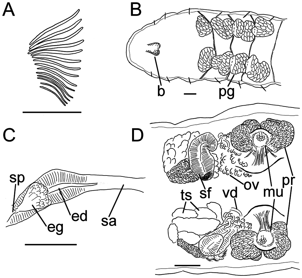
Description of new, mounted, material with comparative notes to Stephenson’s sectioned material: Dark grey worm (at least when preserved), with subepithelial black pigmentation, densest dorsally, decreasing in intensity ventrally. Length of first 20 segments 2.1 mm (fixed, amputated specimen); first 15 segments 1.9 mm long; width at clitellum 0.65 mm. Chaetae sigmoid ( Fig. 10A View Figure 10 ). Upper bundles dorsolateral (closer to lateral line than the ventral bundles), with five to eight (possibly more) chaetae anterior to clitellum; numbers not discernable in postclitellar segments. Ventral bundles with 11–15 chaetae anterior to clitellum, nine to 11 chaetae posteriorly, at least to XX. The longest measured chaetae 95 µm long, ~5 µm wide. Epidermis densely covered with rows of pale gland cells; sectioned material also showed dense, deeply staining granular gland cells. Clitellum with reticulate pattern of gland cells, extending over XII–XIII, absent ventrally. Head pore not observed.
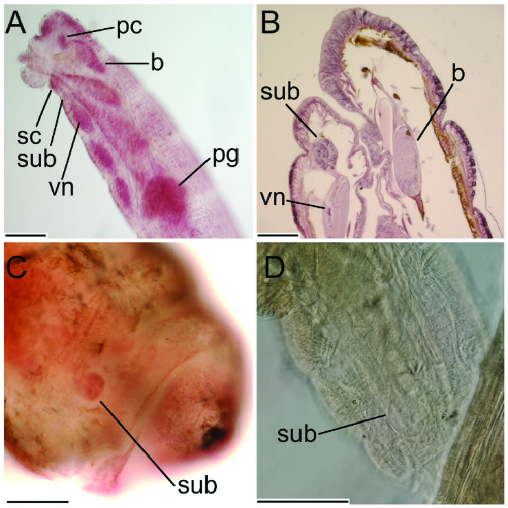
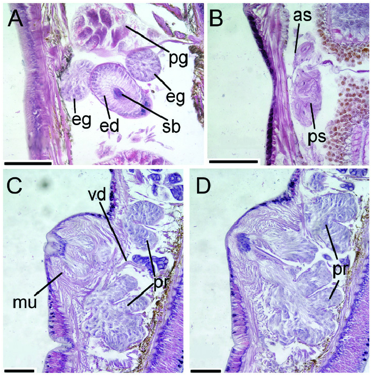
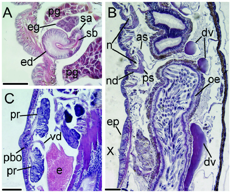
Coelomocytes numerous, ~15 µm long; round, oval or spindle shaped; granulated, with distinct nucleus. Paired pharyngeal glands ( Fig. 10B View Figure 10 ) present in IV, V and VI, with third pair extending back into VII; each pair dorsally separate. Origin of dorsal vessel not discernable, with peristomial bifurcation. Nephridia ~95 µm long (longer in sectioned material: Fig. 11D View Figure 11 ), observed in 7/8–9/10. Anteseptale narrower than, and about half as long as, postseptale, consisting of distinct funnel and part of the nephridial body, seen in sectioned material ( Fig. 11B View Figure 11 ). Postseptale oval, tapering into posteroventral efferent duct. Brain with posterior incision, with clusters of perikarya on prostomial nerves and at ventral ends of circumoesophageal connectives. Ventral nerve cord frontally bearing a large subbuccal bulb ( Fig. 9B, C View Figure 9 ); see Marionina discussion above.
Male genitalia paired ( Fig. 10D View Figure 10 ). Testes originating in anterior of XI, with maturing male cells enveloped in testis sacs, cleft lengthwise into irregular lobes, distally breaking up into free-floating cysts. Sperm funnels in XI, 240 µm long, 110 µm wide, that is, about two times longer than wide; funnels tapering towards vasa deferentia. Most of vasa irregularly coiled in XII, 15 µm wide. Penial bodies indistinct; each male pore surrounded by a small ring of gland cells embedded in a muscular framework, bearing two prostate glands, one anterior and one posterior, 125 and 160 µm long, respectively; each prostate with thread-like ventral connections to the male pore. The sectioned material ( Fig. 11C, D View Figure 11 ) shows these structures in more detail, with a few gland cells surrounding the pore through which the vasa discharge; the gland cells connected via two stalks to two prostate glands that widen gradually and end in several dorsal lobes, and the muscular framework that surrounds all but the dorsal part of the prostates. The muscles cause the body wall to protrude around the male pore. Ovaries in XII. No mature eggs observed.
Spermathecae ( Figs 10C View Figure 10 , 11A View Figure 11 ) in V, spindle or tube shaped; in our new material 315 µm long, 65 µm wide at the ectal duct and 30 µm wide at widest part of ampulla, and in the sectioned material 85 µm wide at the ectal duct and 60 µm wide at widest part of ampulla. Ectal duct difficult to distinguish from ampulla, but according to Stephenson (1932) the ectal duct is the wider part with tall epithelial cells, whereas the ampulla has a thinner epithelium, and this is supported in his sectioned material. In our mounted specimen ( Fig. 10C View Figure 10 ), the (thick-walled) duct is about as long as the (thin, apparently still developing) ampulla; in the sectioned material, the ampulla is 1.5–2.0 times longer than the duct. Ampulla connected to oesophagus. No sperm observed in the new material. Two separate, pedunculate glands flanking (one dorsal and the other ventral) each spermathecal pore (both glands shown in Fig. 11A View Figure 11 ); these glands ≤ 55 µm in diameter in our worm and ≤ 100 µm in the sectioned material. No midventral subneural glands were observed in the new material.
Geographical distribution and habitat: Morphologically identified only from South Georgia. The etymology of the epithet aestuum (Latin, ‘of the tides’) refers to the position of the type locality ‘between tide marks’, which coincides with the habitat of our specimen. The BMNH collection has specimens collected by E. M. van Zinderen Bakker at five localities on Marion Island in 1976, determined as L. aestuum by E. G. Easton ( Boros & Sherlock, 2010), but we have not been able to validate this find.
Remarks: Our single specimen is similar to Stephenson’s (1932) in most characters, but we did not observed the two small subneural glands midventral in segments XIII and XIV, sperm filling the spermathecae or developing male cells bulging forward into X reported by Stephenson, which indicates that our specimen was not fully mature. Sections in Stephenson’s slides clearly show sperm filling the spermathecal lumen, mostly in the ectal, thick-walled portion, that is, the portion referred to as the ectal duct by Stephenson (1932).
Marionina aestuum View in CoL can be distinguished from other subantarctic pigmented Marionina View in CoL in the following characters: M. werthi View in CoL has only a single prostate gland and spermathecae without glands; M. aestuum View in CoL has two prostate glands and spermathecae with glands. Marionina benhami View in CoL has minute penial bodies, similar to those of M. aestuum View in CoL , but instead of two large prostates, M. benhami View in CoL has a number of associated glandular masses (prostates), some in front and some behind the male pores. Marionina aestuum View in CoL is more similar to M. grisea View in CoL (sectioned material of which we studied too; Fig. 12 View Figure 12 ; for a full description, see Stephenson, 1932), but the former: (1) has more chaetae per bundle ( M. grisea View in CoL has up to eight in ventral bundles, five in laterals); (2) spermathecae of about even width ( M. grisea View in CoL has a shorter spermathecal duct that is twice as thick as the ampullae; in the fully matured sectioned material of M. aestuum View in CoL it is 1.5 times thicker); and (3) lacks the thickened ventral epithelial plate in X ( Fig. 12B View Figure 12 ; described for mature specimens by Stephenson, 1932). Nevertheless, our species is still morphologically closest to the new species M. fusca View in CoL , which will be discussed below.
| SMNH |
Department of Paleozoology, Swedish Museum of Natural History |
| V |
Royal British Columbia Museum - Herbarium |
| VI |
Mykotektet, National Veterinary Institute |
No known copyright restrictions apply. See Agosti, D., Egloff, W., 2009. Taxonomic information exchange and copyright: the Plazi approach. BMC Research Notes 2009, 2:53 for further explanation.
|
Kingdom |
|
|
Phylum |
|
|
Class |
|
|
Order |
|
|
Family |
|
|
Genus |
Marionina aestuum
| Klinth, Mårten J., Rota, Emilia, Martinsson, Svante, Prantoni, Alessandro L. & Erséus, Christer 2022 |
Lumbricillus aestuum
| Nielsen CO & Christensen B 1959: 96 |
Marionina aestuum
| Stephenson J 1932: 251 |