
COCCULINIDAE, Dall, 1882
|
publication ID |
https://doi.org/ 10.1046/j.1096-3642.2003.00058.x |
|
persistent identifier |
https://treatment.plazi.org/id/03B6B923-EE35-FFE6-8E01-62ED3E786FFE |
|
treatment provided by |
Carolina |
|
scientific name |
COCCULINIDAE |
| status |
|
FAMILY COCCULINIDAE View in CoL
MACLEANIELLA MOSKALEVI LEAL & HARASEWYCH
1999
Material Examined
Puerto Rico Trench (BMSM 1000). Two preserved paratypes were sectioned for anatomical reconstructions (see details in Leal & Harasewych, 1999; Strong & Harasewych, 1999). No specimens were available for scanning electron microscopic investigations of midgut morphology; midgut morphology is inferred from sections. Ciliary currents within the midgut are unknown.
External anatomy and mantle cavity
Weakly developed anterior pedal gland opening anteriorly and laterally to foot sole. Operculum absent. Mantle penetrated by haemal sinuses. Hypobranchial gland weakly developed, comprising several large cells in pallial roof. Pseudoplicatid gill forming small ciliated papilla present in front of anus in right mantle roof.
Reproductive system
Adults simultaneous hermaphrodites. Gonopericardial canal absent. Gonad forming voluminous sac, containing both eggs and sperm. Sac-like portion of gonad connecting to visceral glandular gonoduct by non-ciliated duct. Glandular gonoduct opening to enclosed duct of seminal receptacle under base of mantle cavity. Duct of seminal receptacle opening to seminal receptacle posteriorly and mantle cavity anteriorly. Receptacle containing mass of unorientated sperm. Seminal groove absent. Copulatory organ formed by enlarged right cephalic tentacle.
Alimentary System
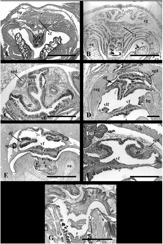
Foregut. Radula rhipidoglossate. Single pair of odontophoral cartilages present. Subradular region cuticularized; subradular organ absent. Sublingual cavity bearing two lateral, glandular pouches. Salivary glands comprising small pouches. Small, unpaired jaw above ventral mouth. Jaw homogeneous, not composed of rods. Ventral ciliary tract beginning within posteriormost buccal cavity as weakly glandular, ciliated thickening. Ventral tract elaborated into large T-shaped, mid-ventral fold for short distance upon emerging from buccal cavity. T-shaped, mid-ventral fold rapidly becoming obscured by underlying mass of glandular tissue ( Fig. 8A View Figure 8 , vf); large mass lens-shaped, not bilobed. Similar to ventro-lateral folds ( Fig. 9A View Figure 9 , vlf), mid-ventral fold and glandular mass not continuing posteriorly into mid-oesophagus. Paired oesophageal pouches present. Two glandular, ventro-lateral folds present at inner margin of pouch ducts, persisting only short distance within anterior oesophagus. Oesophageal pouches histologically differentiable into two regions. Anteriorly, pouches simple and weakly glandular. Tissue of the pouches first becoming more significantly glandular along ventral surface, forming convoluted epithelium. Convoluted glandular tissue expanding posteriorly, completely lining pouches as mid-ventral glandular mass diminishing. Glandular tissue of mid-oesophageal pouches and mid-ventral fold histologically distinct; the former comprising low, vacuolated cells, the latter dense, elongate prismatic cells.
Midgut. Posterior oesophagus entering midgut postero-dorsally. Extensive cuticle lining gastric chamber, elaborated into prominent gastric shield mid-dorsally. Grooved tract across midgut roof continuous with oesophagus; sorting area absent. Paired digestive gland ducts opening to posterior oesophagus. Caecum absent.
Hindgut. Intestine exiting midgut posteriorly. Style sac and intestinal groove absent. Hind gut completing three, large loops.
Reno-pericardial system
Single, left, sac-like kidney; excretory lamellae absent. Nephridial gland absent.
Nervous system and sensory structures
Broad circum-oesophageal nerve ring lacking secondary connections between ganglia; nerve ring weakly dystenoid. Buccal connectives passing anteriorly to buccal ganglia lying below oesophageal pouches. Single commissure connecting pedal ganglia. Supraoesophageal ganglion innervating osphradial ganglion within mantle roof. Possibly sensory, ciliated, prismatic cells overlying osphradial nerve. Single visceral ganglion present. Tentacular nerve single. Eyes modified into basitentacular gland on small stalks at outer bases of cephalic tentacles. Statocysts containing single statolith in one animal, several tiny statocones (~3) in another.
Remarks
Internal anatomy was described by Strong & Harasewych (1999); the shell, radula, and external aspects of the copulatory organ were described by Leal & Harasewych (1999). This redescription uses terminology to promote homology comparisons in a higher order framework and elaborates on the salivary glands, ventral folding of the foregut, the ventral glandular mass, and the histological differentiation of the oesophageal pouches. Given the histological differentiation between the anterior and posterior portion of the oesophageal pouches, these structures are interpreted as continuous buccal pouches and non-sepatate oesophageal glands.
Discussion
The family Cocculinidae is remarkably morphologically diverse, considering that so few comprehensive anatomical accounts exist. All cocculinids have a highly vascularized pallial roof, most likely playing a significant role in gas exchange. The mantle cavity bears the apertures of the genital system and alimentary system on the right and a hypobranchial gland in the centre of the pallial roof that may be weakly developed or well-developed and contained within a pocket, ventrally enclosed by the kidney. Just in front of the anus is a pseudoplicatid gill that ranges from large and foliated to small and papillate. Other species lack a gill, bearing only a ciliary tract or a series of leaflets ( Haszprunar, 1987, 1988c, 1998a; unpublished data; Sasaki, 1998; Strong & Harasewych, 1999).
All cocculinids are simultaneous hermaphrodites. A pallial glandular gonoduct is lacking, instead the gonoduct that conveys gametes from the gonad to the base of the mantle cavity is glandular. A seminal groove may be present or absent, and leads from the genital aperture to a copulatory organ formed by or associated with the right cephalic tentacle, the foot, or the oral lappet. There may be one or two ‘seminal receptacles’ that store unorientated sperm ( Haszprunar, 1987, 1988c, 1998a; Sasaki, 1998; Strong & Harasewych, 1999).
Radular cartilages comprise a single pair. Jaws typically consist of a single, small, unpaired plate on the anterior buccal cavity roof, or may be robust and weakly paired. The jaws are homogeneous and lack rods ( Haszprunar, 1987, 1988c, 1998a). Although early studies reported cocculinoidean salivary glands as absent ( Haszprunar, 1987, 1988c; Strong & Harasewych, 1999), it is now recognized that the salivary glands form simple, glandular pouches in most taxa ( Haszprunar, 1998a). However, some possess prominent, tubular salivary glands (Haszprunar, unpublished data). In those species with pouch-like salivary glands, the sublingual cavity is highly glandular ( Haszprunar, 1987, 1988c; Strong & Harasewych, 1999). Outpocketings of the cocculinid oesophagus have been referred to as ‘oesophageal pouches’ and/or ‘oesophageal glands’ ( Haszprunar, 1987, 1988c, 1998a; Salvini-Plawen, 1988; Sasaki, 1998; Strong & Harasewych, 1999). Due to the internal differentiation of the pouches (anteriorly simple, posteriorly convoluted), they are here interpreted as representing continuous buccal pouches and oesophageal glands.
The Cocculinoidean circulatory system is characterized by a rectum that does not penetrate the ventricle. A nephridial gland is lacking ( Haszprunar, 1987, 1988c; Strong & Harasewych, 1999).
The nervous system of cocculinoideans may be hypoathroid with un-fused supra-esophageal and visceral ganglia or weakly dystenoid with fused or unfused visceral and supra-esophageal ganglia. No secondary connections are formed between the pleural and oesophageal ganglia. Sensory structures include osphradia and statocysts. The osphradium is present or absent; in some the osphradium lacks a discrete sensory epithelium, consisting only of ciliated cells overlying the osphradial nerve. Cocculinoideans possess statocysts bearing a single statolith. However, Macleaniella is polymorphic for this feature. Eyes rarely contain pigment and are typically modified into a mucus-secreting basitentacular gland ( Haszprunar, 1987, 1988c, 1998a; Strong & Harasewych, 1999).

No known copyright restrictions apply. See Agosti, D., Egloff, W., 2009. Taxonomic information exchange and copyright: the Plazi approach. BMC Research Notes 2009, 2:53 for further explanation.
|
Kingdom |
|
|
Phylum |
|
|
Class |
|
|
Order |
|
|
Family |