
Luciogobius punctilineatus, Koreeda & Motomura, 2022
|
publication ID |
https://doi.org/ 10.11646/zootaxa.5138.2.2 |
|
publication LSID |
lsid:zoobank.org:pub:B2A26575-FE6C-41FB-9510-8BC19DBAB792 |
|
DOI |
https://doi.org/10.5281/zenodo.6560545 |
|
persistent identifier |
https://treatment.plazi.org/id/9A25FB47-2B86-450C-8D1B-183BE27D5AE7 |
|
taxon LSID |
lsid:zoobank.org:act:9A25FB47-2B86-450C-8D1B-183BE27D5AE7 |
|
treatment provided by |
Plazi |
|
scientific name |
Luciogobius punctilineatus |
| status |
sp. nov. |
Luciogobius punctilineatus n. sp.
[New English name: Dot-lined Earthworm Goby; new standard Japanese name: Shiranui-mimizuhaze]
Figures 1–4 View FIGURE 1 View FIGURE 2 View FIGURE 3 View FIGURE 4 , 5A View FIGURE 5 , 6 View FIGURE 6 ; Tables 1–3 View TABLE 1 View TABLE 2 View TABLE 3
Luciogobius sp. : Koreeda & Motomura, 2021: 47, fig. 8A–E (Shimokoshiki-shima island, Koshiki Islands, Kagoshima Pref., Japan).
Luciogobius sp. 11 (not of Shibukawa et al., 2019): Ito & Okumura, 2021: 4, fig. 4 (Kaifu, Tokushima Pref., Japan).
Holotype. KAUM –I. 156161. 29.8 mm SL, mouth of Okawa River , Okawa , Akune , Kagoshima, southwestern Kyushu , Japan, coll. by R. Koreeda & R. Furuhashi, 1 Apr. 2021.
Paratypes. 20 specimens (20.9–33.1 mm SL). Kyushu : KAUM –I. 156162. 33.1 mm SL, KAUM –I. 156163. 27.7 mm SL, KAUM –I. 156164. 28.2 mm SL, KAUM –I. 156165. 29.9 mm SL, KAUM –I. 156166. 28.4 mm SL, KAUM –I. 156167. 25.1 mm SL, collected with holotype; KAUM –I. 158051, 27.6 mm SL, KAUM –I. 158052, 28.0 mm SL, KAUM –I. 158053, 25.8 mm SL, KAUM –I. 158054, 26.4 mm SL, KAUM –I. 158055, 24.5 mm SL, KAUM –I. 158056, 24.9 mm SL, KAUM –I. 158057, 23.5 mm SL, KAUM –I. 158058, 25.4 mm SL, KAUM –I. 159066, 28.6 mm SL, KAUM –I. 159067, 26.7 mm SL, KAUM –I. 159068, 25.5 mm SL, mouth of Shirinashi River , Okawa , Akune, Kagoshima, Japan, coll. by R. Koreeda, 26 June 2021 . Koshiki Islands : KAUM –I. 147759, 24.7 mm SL, south of fishing port at east side of Nagahama Bay, Nagahama, Shimokoshiki, Satsuma-sendai, Shimokoshiki-shima island, Koshiki Islands, Kagoshima, Japan, coll. by R. Nakagawa, 17 Oct. 2020 . Osumi Islands : KAUM –I. 146246, 24.9 mm SL, north of Shojiura Fishing Port, Genna, Nishinoomote, Kumage, Tanega-shima island, Osumi Islands, Kagoshima, Japan, coll. by R. Furuhashi, 17 Sept. 2020 ; KAUM –I. 163150, 20.9 mm SL, mouth of Otoko River , Koseda , Yakushima , Kumage-gun, Yaku-shima island, Osumi Islands, Kagoshima, Japan, coll. by D. Kato, 4 Dec. 2021 .
Non-type specimens. 15 specimens (12.1–33.4 mm SL). Shikoku : KAUM –I. 159034, 24.7 mm SL, KAUM –I. 159035, 22.9 mm SL, KAUM –I. 159036, 20.9 mm SL, mouth of Shinjo River , Susaki , Kochi, Japan, coll. by H. Saito, 22 July , 2021. Kyushu : KAUM –I. 156187, 26.3 mm SL, north of mouth of Shimadomari River , Sataizashiki , Minami-osumi, Kimotsuki, Kagoshima, Japan, coll. by R. Koreeda, 10 Apr. 2021 ; KAUM –I. 157124, 33.4 mm SL, KAUM –I. 157125, 28.7 mm SL, KAUM –I. 157126, 26.3 mm SL, same locality as KAUM–I. 156187, 26 Apr. 2021 . Amami Islands : KAUM –I. 145562, 12.1 mm SL, KAUM –I. 145563, 15.0 mm SL, KAUM –I. 145564, 25.7 mm SL, KAUM –I. 145565, 26.8 mm SL, KAUM –I. 145566, 24.0 mm SL, KAUM –I. 145567, 28.1 mm SL, KAUM –I. 145568, 24.8 mm SL, KAUM –I. 145569, 18.6 mm SL, south of mouth of Heda River, Uken, Oshima-gun, Amamioshima island, Kagoshima, Japan, coll. by R. Koreeda & N. Shimizu, 23 July 2020 .
Diagnosis. A species of Luciogobius with the following combination of characters: total second dorsal-fin rays 10–12 (modally 11); total anal-fin rays 12–14 (modally 13); pectoral-fin rays 8–12 (modally 10); vertebrae 16–18 + 22–24 = 39–42 (modally 17 + 23 = 40); pectoral-fin posterior margin slightly concave; pelvic fins united, forming a disc; snout relatively short, length 3.1–4.3% of SL; AAA distance twice body depth at anus, 11.4–16.9% of SL; snout length less than 34.7% of AAA distance; pre-anus length less than 85.5% of pre-anal-fin length; single poorly defined black longitudinal line along mid-lateral body region from behind pectoral fin to caudal-fin base, indistinct anteriorly (line embedded, visible through semi-transparent muscle tissue in fresh or live specimens); black spots forming a single longitudinal row on mid-lateral body surface from behind pectoral fin to caudal-fin base (more distinct in preserved specimens).
Description. Counts and measurements given in Tables 1 View TABLE 1 and 2 View TABLE 2 . In this description, data for holotype presented first, followed by data for paratypes in parentheses (if different). Body elongate, slightly compressed. Head depressed. Anterior nostril with short tube reaching to upper lip; posterior nostril horizontally ovate ( Fig. 2 View FIGURE 2 ). Snout slightly swollen dorsally, especially above nostril. Eye small, rounded, covered with thin skin, located anterodorsally on head; dorsal and posterior surface of skin covering eyes rounded, not projecting posteriorly. Dorsal profile of head between eye and nape swollen (centrally concave) in dorsal view. Interorbital space wider than snout length, slightly swollen anteriorly (or with a transverse ridge anteriorly, rarely completely flat). Single low longitudinal ridge extending from below anterior nostril to behind eye ( Fig. 2 View FIGURE 2 ). Mouth oblique; posterior end of maxilla extending beyond vertical through anterior margin of eye. Pair of symphysial flaps fused anteriorly, rounded. Jaws with bands of 4–6 rows of small conical teeth; outer teeth larger; upper jaw band wider. Tongue free, anteriorly bilobed. Gill opening narrow, extending below upper end of pectoral-fin base to level with posterior end of jaw.Anus located slightly behind midpoint of body. Urogenital papilla just behind anus, small and round (in both sexes; median slit in one mature female paratype slightly larger than in others). Origin of second dorsal fin posterior to anal-fin origin. Distance between anus and anal-fin origin twice body depth at anus. First and second dorsal-fin rays of second dorsal fin spinous (or only first ray spinous), remaining rays soft, segmented; sixth ray longest (fifth to seventh). First and second anal-fin rays spinous (or only first ray spinous), remaining rays soft, segmented. Pectoral fin small; membranes between adjacent rays moderately incised posteriorly for one-sixth to one-fourth (rarely one-third) of each ray pair; membrane between 2 uppermost and 2 lowermost rays not incised (sometimes incised); all rays lacking serrations; free rays absent. Sucker-shaped pelvic fins small; paired fins fused, frenum with smooth margin. Caudal fin rounded (sometimes slightly elongate). Zygapophyses on anterior base of haemal spines well developed.
Cephalic sensory system: Canals and pores absent. Cephalic sensory papillae (shown in Fig. 2 View FIGURE 2 ) in four rows; row a located behind eye; row b extending from below anterior nostril to behind eye; row c located at highest point of longitudinal skin ridge on cheek, extending to behind eye; and row d extending from snout tip to above midpoint of maxilla, along upper margin of maxilla, subsequently curving dorsoposteriorly to middle of head. Row cp, longitudinal papillae row on lower margin of cheek swelling between rows c and d. Row f located behind symphysial flaps. Rows e and i extending along weak flap on lower margin of lower jaw to lower part of preopercle. Row ot discontinuous. Rows os and oi located behind row ot.
Coloration (live and fresh specimens.) Based on Figures 1 View FIGURE 1 , 3 View FIGURE 3 and Koreeda & Motomura (2021: fig. 8A–C, E): Body yellowish-green to brown dorsally, whitish ventrally (yellow to orange ventrally in gravid females). A single poorly defined black longitudinal line mid-laterally on body from behind pectoral fin to caudal-fin base, embedded internally and visible through semi-transparent muscle issue. Small black spots forming a single longitudinal row mid-laterally on body surface from behind pectoral fin to caudal-fin base, more distinct posteriorly, discontinuous anteriorly (sometimes indistinct in live specimens). Black to brown spots scattered on dorsal body surface and caudal peduncle. Dorsal-, anal-, pectoral-, and pelvic-fin rays and membranes semitransparent white to pale yellow. Caudal fin semitransparent white, with yellow-margined black rounded blotch basally; posterior margin of black blotch reaching posteriorly to between one-fourth and half of caudal-fin length. In juveniles (12.1–15.0 mm SL), body pale yellow to beige with few pigments; black stripe and dotted line indistinct; caudal-fin blotch reaching posteriorly to less than one-fifth of fin length; yellow margin absent.
Coloration of preserved specimens. Based on Figure 4 View FIGURE 4 and Koreeda & Motomura (2021: fig. 8D): Body whitish, with a single distinct longitudinal line on mid-lateral surface, and small brown blotches scattered dorsally and on caudal peduncle. Caudal fin with black to brown blotch basally. Single embedded longitudinal line not visible.
Etymology. The specific name punctilineatus is derived from Latin meaning “dotted line,” and refers to the single longitudinal dotted line on the lateral surface of the body.
Distribution and ecological notes. Currently known from southern Japan, specimens having been collected from Tokushima ( Ito & Okumura, 2021) and Kochi prefectures (both southern Shikoku), Kagoshima mainland (southern Kyushu), Shimokoshiki-shima island (Koshiki Islands, off Kagoshima mainland, East China Sea), Tanegashima and Yakushima islands (Osumi Islands, south of Kyushu), and Amami-oshima island (Amami Islands, Ryukyu Islands).
Most specimens were collected from gravel in the vicinity of freshwater springs near coastal river mouths ( Fig. 7 View FIGURE 7 ), but not from the rivers or river mouths themselves. On Amami-oshima island, all specimens were collected from the gravel layer (with a freshwater spring 10 cm below the surface) on a sandy beach 100 m west of the Heda River mouth. Luciogobius punctilineatus was abundant in the above-mentioned habitats, often occurring together with Luciogobius sp. 4 sensu Maeda et al., 2008, although suitable habitats for L. punctilineatus were more limited than those of the latter.
Juveniles of L. punctilineatus (12.1–15.0 mm SL; Fig. 1C View FIGURE 1 ) were collected in July on Amami-oshima island, and fully matured females ( Fig. 3A View FIGURE 3 ) collected from April to June on Kagoshima mainland. The reproductive season for this species is likely to be spring to early summer.
Comparisons. Luciogobius punctilineatus belongs to the Luciogobius elongatus complex, defined by Shibukawa et al. (2019) as having no free pectoral-fin rays, total vertebrae 39–42, the AAA distance greater than body depth at the anus, first to third anal-fin rays unbranched and spine-like, and anteriormost pterygiophore of the anal fin inserted behind the fourth haemal spine. Luciogobius punctilineatus is easily distinguished from all other members of the complex (except Luciogobius sp. 11 sensu Shibukawa et al., 2019), by having 10–12 total dorsal-fin rays, 12–14 total anal-fin rays, 8–12 pectoral-fin rays with moderately incised membranes, pelvic fins united, forming a ventral disc, 39–42 total vertebrae, and a single poorly defined longitudinal black line (embedded in muscle tissue) and dotted line (on body surface) mid-laterally along the body.
Luciogobius sp. 11 ( Fig. 8 View FIGURE 8 ) is similar to L. punctilineatus in sharing the above-mentioned characters ( Tables 2 View TABLE 2 , 3 View TABLE 3 ), the two species co-occurring at least in Kochi Prefecture ( Shibukawa et al., 2019; Koreeda & Motomura, 2021; this study). However, the latter has a greater AAA distance [11.4–16.9% (mean 13.3%) of SL vs. 9.1–10.4% (9.6%) in Luciogobius sp. 11; Fig. 9B View FIGURE 9 ; Table 3 View TABLE 3 ], and shorter snout [19.2–34.7% (26.4%) of AAA vs. 36.7–44.8% (40.9%); Fig. 9E View FIGURE 9 ; Table 3 View TABLE 3 ] and pre-anus lengths [75.0–82.0% (79.3%) of preanal-fin length (except in juveniles less than 15.0 mm SL) vs. 84.4–88.8% (86.1%); Fig. 9F View FIGURE 9 ; Table 3 View TABLE 3 ]. The membranes between adjacent pectoral-fin rays (except 2 uppermost and 2 lowermost rays) are incised posteriorly for one-sixth to one-fourth of each ray pair in L. punctilineatus , to one-third to half in Luciogobius sp. 11 ( Shibukawa et al., 2019; Koreeda & Motomura, 2021; Figs. 1 View FIGURE 1 , 2 View FIGURE 2 , 5 View FIGURE 5 , 8 View FIGURE 8 ), and the first anal-fin pterygiophore is usually inserted behind the fifth haemal spine in L. punctilineatus and behind the fourth spine in Luciogobius sp. 11 ( Table 2 View TABLE 2 ). The new species further differs from Luciogobius sp. 11 in having relatively lower numbers of anal-fin rays [12–14 (modally 13) vs. 14–16 (15) in Luciogobius sp. 11; Shibukawa et al., 2019; Tables 1 View TABLE 1 , 2 View TABLE 2 ] and vertebrae [16–18 (18) + 22–24 (23) = 39–42 (40) vs. 17–18 (18) + 22–25 (23) = 40–43 (41); Shibukawa et al., 2019; Koreeda & Motomura, 2021; Tables 1 View TABLE 1 ], and a relatively higher number of pectoral-fin rays [8–12 (10) vs. 8–9 (9); Shibukawa et al., 2019; Tables 1 View TABLE 1 ]. The anus and anal-fin origin of L. punctilineatus are located more anteriorly and posteriorly, respectively, compared with Luciogobius sp. 11 ( Fig. 9C, D View FIGURE 9 ). In preserved specimens, a single longitudinal row formed by black spots on the mid-lateral surface of the body is more distinct in L. punctilineatus than in Luciogobius sp. 11 (see Koreeda & Motomura, 2021: fig. 8; Figs. 4 View FIGURE 4 , 8 View FIGURE 8 ), although this difference is unclear in live individuals. Luciogobius sp. 11 also exhibits sexual dichromatism (yellow males and brown females; Shibukawa et al., 2019), which is unknown in L. punctilineatus . Accordingly, individuals of L. punctilineatus are similar to males of Luciogobius sp. 11 in having a yellow body ( Shibukawa et al., 2019: figs. 26D–E, 27D; Koreeda & Motomura, 2021: fig. 8; Fig. 3A View FIGURE 3 ).
Ito and Okumura (2021) reported a single specimen (20.1 mm SL) of Luciogobius sp. 11 sensu Shibukawa et al., 2019 from Tokushima Prefecture, Shikoku, Japan. However, based on their description and photograph, the specimen is most likely to have been L. punctilineatus , having a relatively greater AAA distance (twice body depth at anus) and relatively shorter snout length (25.4% as HL).
Luciogobius sp. 3 sensu Maeda et al., 2008 is also similar to L. punctilineatus in most meristics, pectoral-fin membrane structure, and coloration ( Maeda et al., 2008; Maeda, 2014). However, the latter is easily distinguished from Luciogobius sp. 3 by vertebral number (16–18 + 22–24 = 39–42 vs. 21–23 + 19–21 = 41–43; Maeda et al., 2008; Maeda, 2014) and greater AAA distance (about twice that of Luciogobius sp. 3 ; Maeda et al., 2008).
The p -distances among the four species of Luciogobius and one species of Inu , estimated from the partial sequences of the 12S rRNA gene, showed that differences between L. punctilineatus n. sp. and Inu koma , L. martellii , L. elongatus , and Luciogobius sp. 11 were 17.34–18.28%, 5.50–6.32%, 3.91–5.53%, and 1.53–3.90%, respectively ( Table 4 View TABLE 4 ). Luciogobius punctilineatus and Luciogobius sp. 11 were recovered in different groups, although it was supported by a low bootstrap value ( Fig. 6 View FIGURE 6 ). Although the molecular differences among L. punctilineatu s, Luciogobius sp. 11, and L. elongatus were less clear, the species are differentiated from each other by AAA distance, pectoral- and pelvic-fin shapes, and coloration.
Geographic variation. Geographic variations in some meristics and coloration were apparent in L. punctilineatus , the dorsal-, anal-, and pectoral-fin rays in specimens from Amami-oshima island tending to be fewer than in specimens from other localities ( Table 2 View TABLE 2 ). Similarly, vertebral numbers in specimens from Shikoku (Tokushima and Kochi prefectures) tended to be lower than in specimens from other localities [16–17 (mainly 17) + 22–23 (22) = 39 in specimens from Shikoku ( Ito and Okumura, 2021; this study) vs. 17–18 + 22–24 (23) = 40–42 (40–41) in the latter]. Specimens from Kochi Prefecture and the Osumi Peninsula ( Figs. 1D, E View FIGURE 1 , 3C View FIGURE 3 , 4D, E View FIGURE 4 ) had a relatively larger black caudal-fin blotch with a narrower yellowish margin, compared with specimens from other localities ( Figs. 1A–C View FIGURE 1 , 3A, B View FIGURE 3 , 4A–C View FIGURE 4 ). Although these minor differences are regarded here as representing geographic variations within L. punctilineatus , specimens from Shikoku, Osumi Peninsula, and Amami-oshima island were excluded from the type series of the new species.
Comparative material examined. Luciogobius sp. 11 sensu Shibukawa et al., 2019: KAUM –I. 142153, 29.3 mm SL, Iifuchi , Yaizu, Shizuoka, Japan, coll. by S. Yamashita, 9 Mar. 2020 ; KAUM–I. 145507, 26.4 mm SL, KAUM–I. 145508, 28.6 mm SL, KAUM –I. 145509, 28.3 mm SL, same locality of KAUM –I. 142153, S. Yamashita , 10 Mar. 2020 ; KAUM –I. 159027, 25.9 mm SL, mouth of Niyodo River , Haruno, Kochi, Japan, coll. by H. Saito, 13 July 2021 .


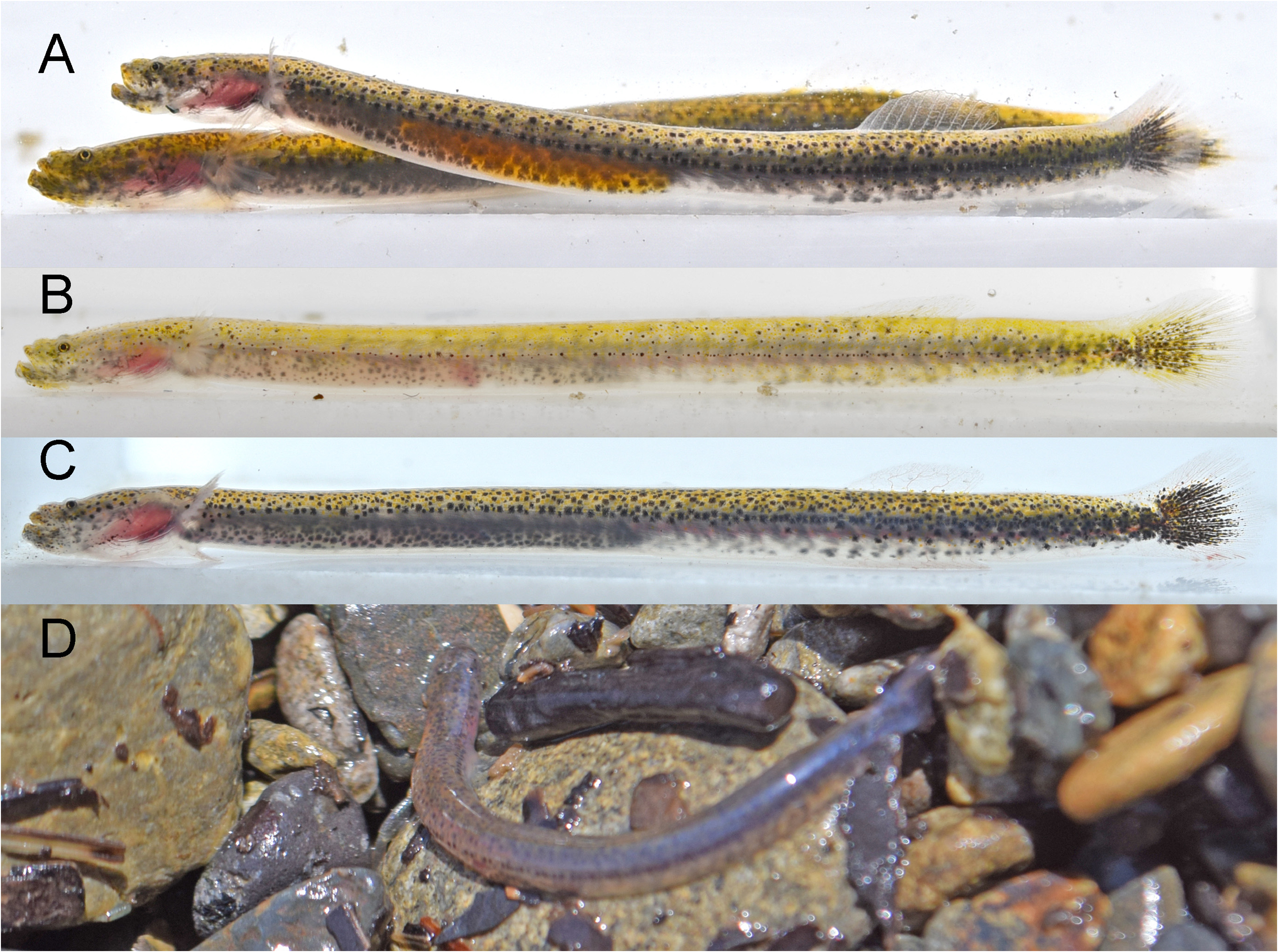
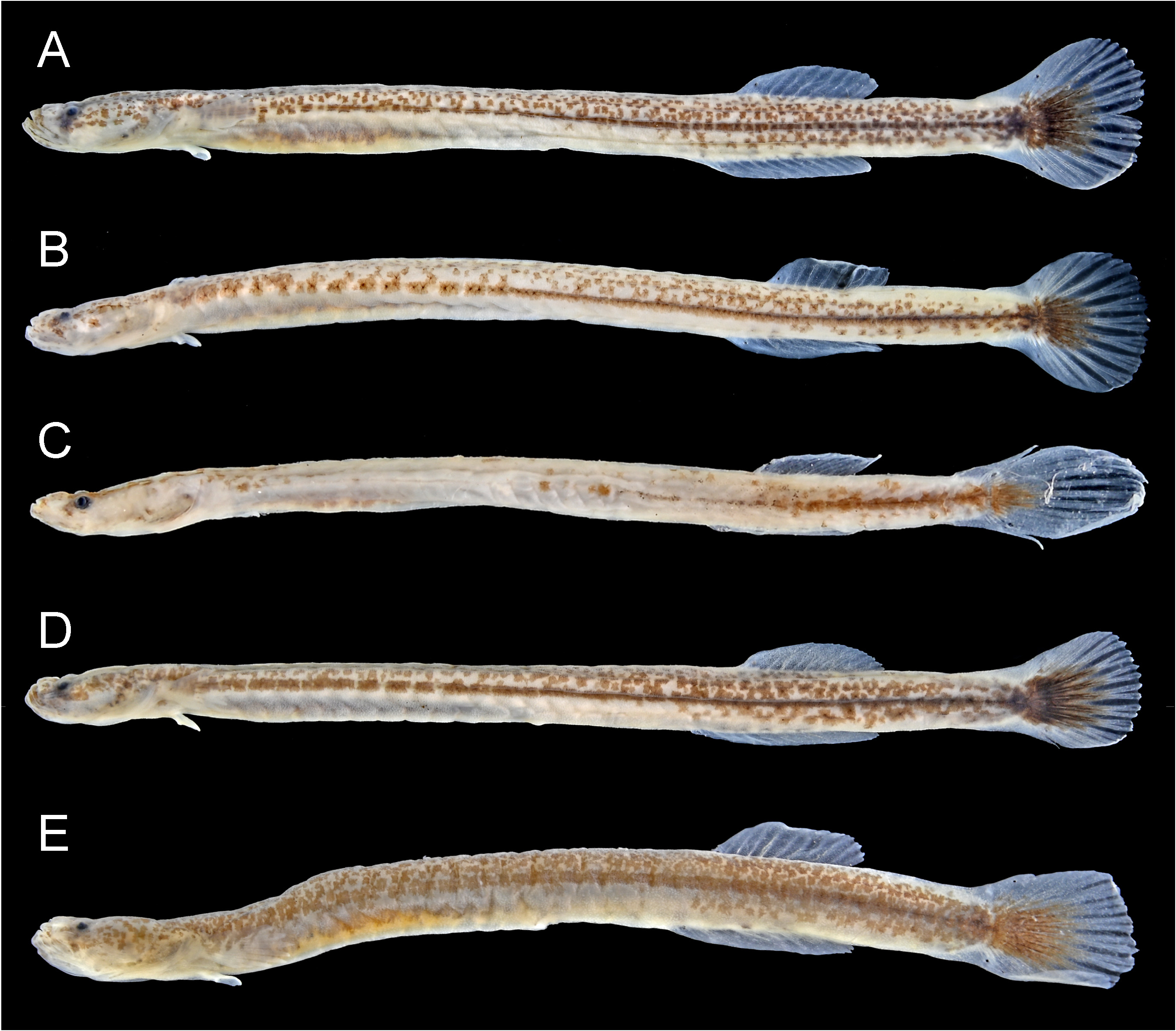
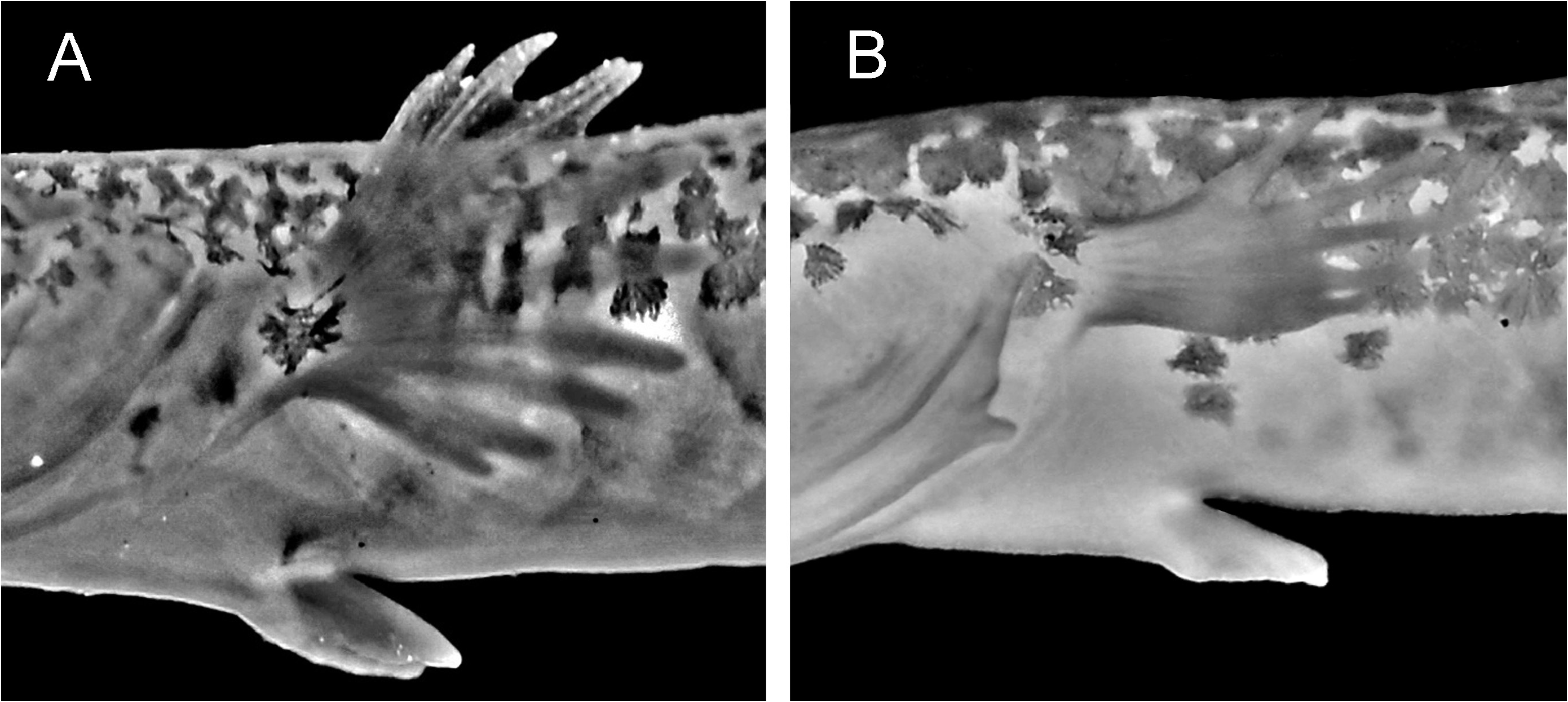
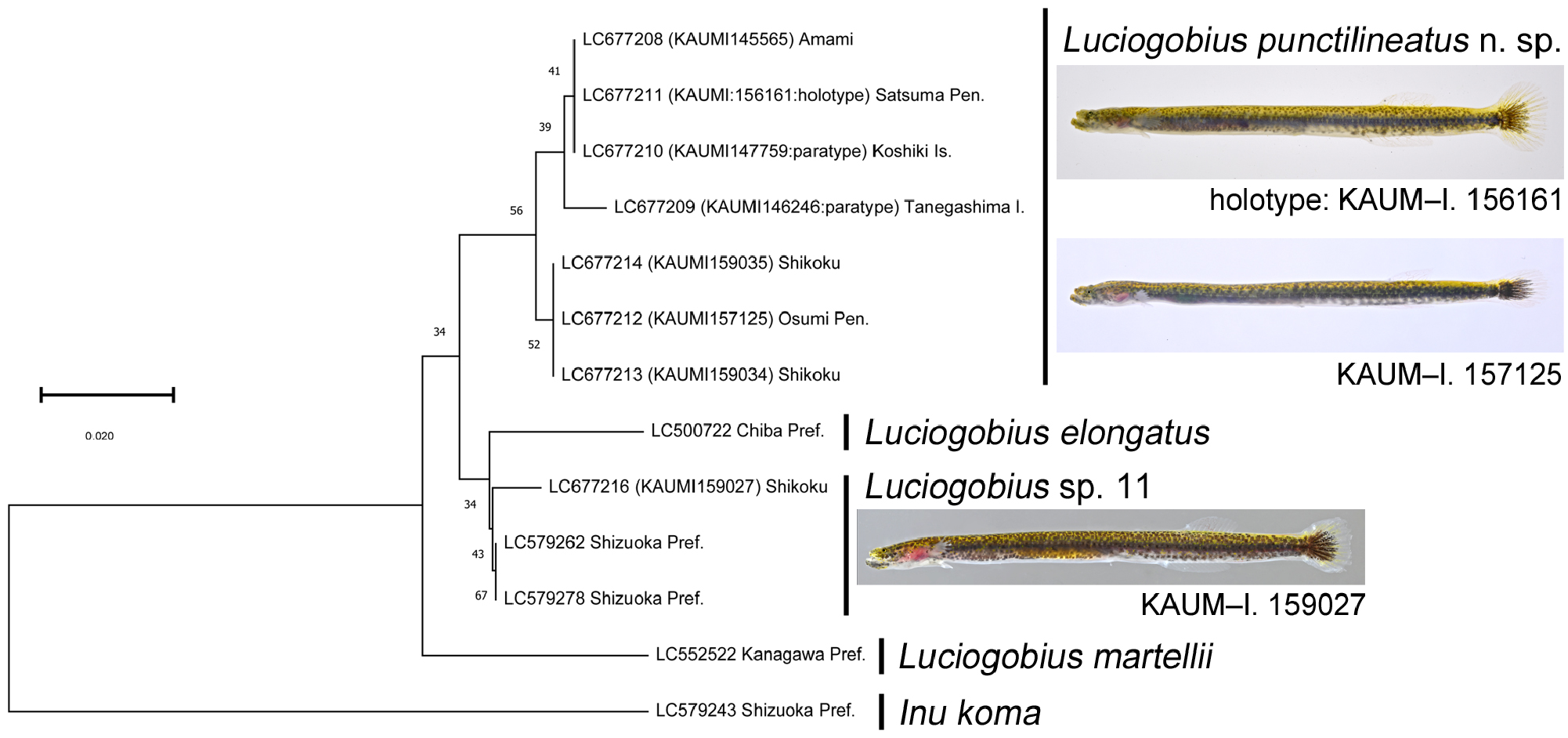

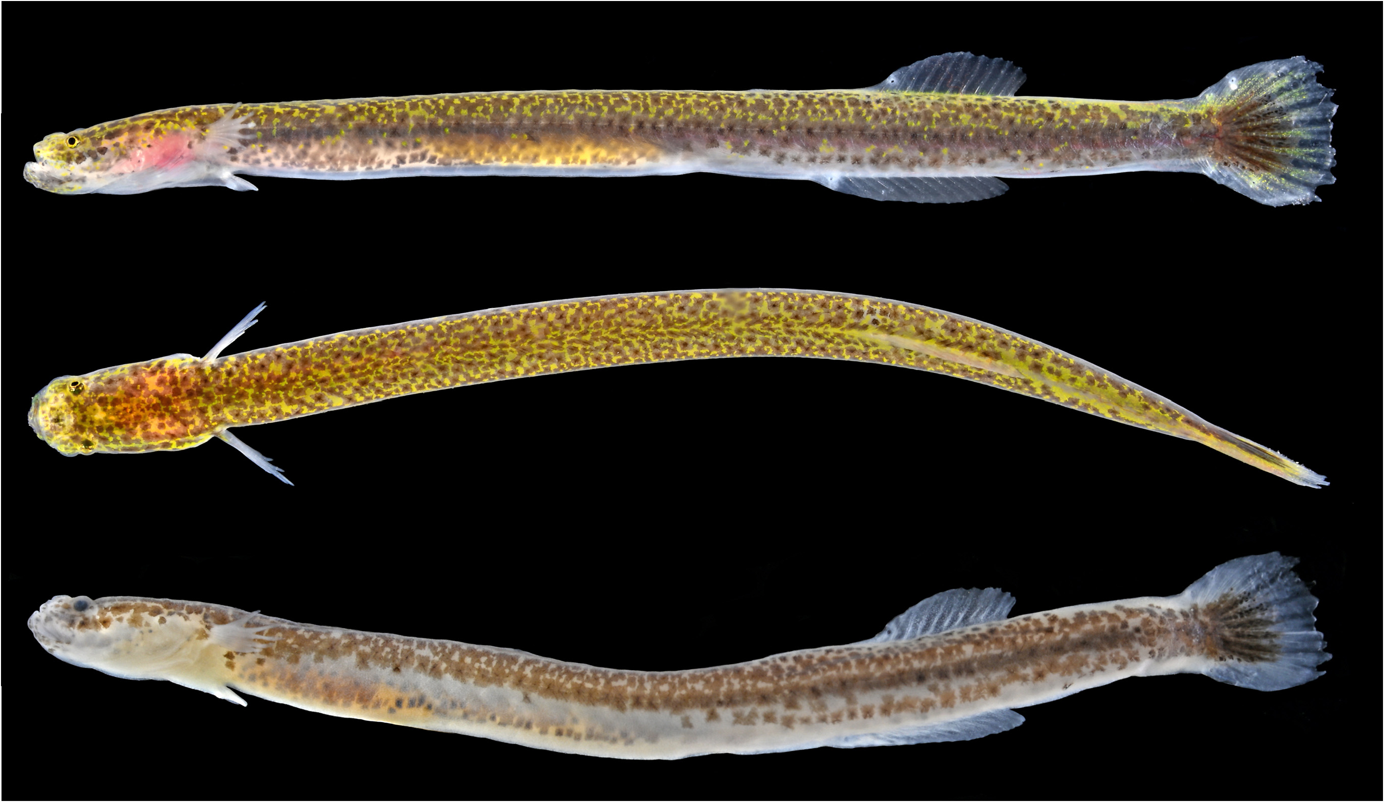
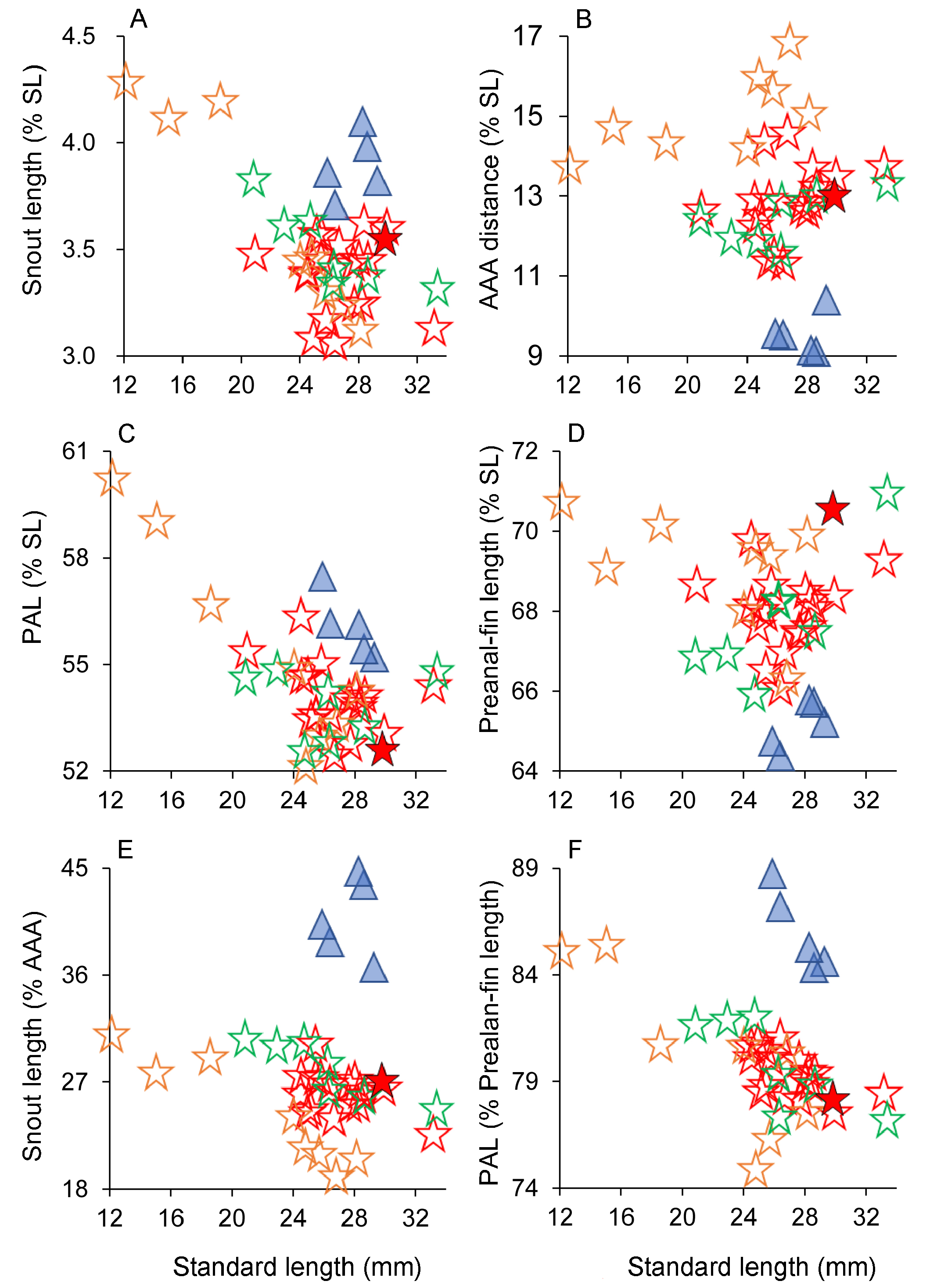
TABLE 1. Counts and proportional measurements of Luciogobius punctilineatus n. sp. (modes and means ± standard deviation shown in parentheses).
| Holotype | Paratypes1 | Non-types | ||
|---|---|---|---|---|
| Amami-oshima island | Kochi Pref. and Osumi Pen. | |||
| n = 18 | n = 8 | n = 7 | ||
| Standard length (SL; mm) | 29.8 | 20.9–33.1 | 12.1–28.1 | 20.9–33.4 |
| Counts | ||||
| Total dorsal-fin elements | 11 | 10–12 | 10–11 (10) | 10–12 (11) |
| Total anal-fin elements | 13 | 12–14 | 12–14 (12) | 12–14 (13) |
| Pectoral-fin rays | 11 | 9–12 | 8–12 (9) | 9–11 (10) |
| Caudal-fin segmented rays | 9 + 8 | 9–10 + 7–8 | 9–10 + 7–8 | 9–10 + 8 |
| Pelvic-fin rays | I, 5 | I, 5 | I, 5 | I, 5 |
| Vertebrae | 17 + 23 = 40 | 17 + 23 = 40 (8), 17 + 24 = 41 (1), 18 + 22 = 40 (1), 18 + 23 = 41 (6), 18 + 24 = 42 (1) | 18 + 24 = 42 (6) | 16 + 23 = 39 (1), 16 + 22 = 39 (2), 17 + 23 = 40 (1), 18 + 23 = 41 (2), 18 + 24 = 42 (1) |
| Dorsal-fin pterygiophore formula (P-V) | 24 | 23–24-25 (23-24) | 24-25–25-26 | 22-23–25 |
| First anal-fin pterygiophore insertion behind | 5th haemal spine | 4–6th (5th) haemal spine | 5–6th (5th) haemal spine | 4–6th (5th) haemal spine |
| Measurements (% SL) | ||||
| Head length | 15.1 | 13.1–14.8 (13.9±0.4) | 13.2–16.4 (14.6±1.1) | 12.7–16.6 (14.8±1.7) |
| Head depth | 5.6 | 4.8–6.6 (5.6±0.5) | 5.0–6.0 (5.4±0.4) | 5.3–7.6 (6.1±0.8) |
| Head width | 8.1 | 6.4–8.2 (7.4±0.5) | 5.3–7.1 (6.3±0.6) | 7.0–9.3 (8.1±0.9) |
| Snout length | 3.6 | 3.1–3.6 (3.4±0.2) | 3.1–4.3 (3.7±0.5) | 3.3–3.8 (3.5±0.2) |
| Upper-jaw length | 4.8 | 3.8–5.2 (4.6±0.3) | 4.1–5.2 (4.7±0.4) | 4.2–5.3 (4.8±0.4) |
| Eye diameter | 1.0 | 0.9–1.2 (1.1±0.1) | 0.7–1.6 (1.0±0.3) | 0.9–1.3 (1.1±0.2) |
| Interorbital width | 2.7 | 1.8–2.5 (2.1±0.2) | 1.7–2.3 (2.0±0.2) | 1.8–3.1 (2.5±0.5) |
| Body depth at pelvic-fin origin | 5.9 | 4.7–6.5 (5.5±0.5) | 4.0–5.7 (5.1±0.6) | 5.1–6.9 (6.1±0.6) |
| Body depth at anus | 6.4 | 5.1–7.3 (6.1±0.7) | 4.9–6.5 (5.4±0.5) | 6.1–7.2 (6.6±0.5) |
| Body depth at anal-fin origin | 6.8 | 5.6–7.5 (6.7±0.6) | 5.1–6.7 (5.8±0.6) | 6.3–8.1 (7.2±0.7) |
| Body width | 6.0 | 5.1–6.6 (5.7±0.4) | 3.9–5.7 (4.9±0.6) | 5.4–6.7 (6.1±0.4) |
| AAA distance | 13.1 | 11.4–14.6 (12.9±0.9) | 13.8–16.9 (15.1±1.0) | 11.7–13.4 (12.5±0.6) |
| Least caudal-peduncle depth | 6.1 | 4.9–6.8 (5.9±0.5) | 4.4–5.7 (5.3±0.5) | 5.7–7.3 (6.5±0.7) |
| Maximum caudal-peduncle depth | 6.4 | 5.5–6.7 (6.1±0.4) | 4.8–5.9 (5.6±0.3) | 5.5–8.1 (6.8±1.1) |
| Caudal-peduncle length | 18.2 | 15.8–19.1 (17.6±1.0) | 15.7–18.5 (17.0±1.0) | 17.0–18.8 (17.5±0.6) |
| Pre-anus length | 52.7 | 52.5–56.4 (54.1±0.9) | 52.2–60.3 (55.5±2.9) | 52.6–54.9 (53.9±1.0) |
| Pre-second dorsal-fin length | 70.6 | 69.2–73.2 (71.2±1.3) | 71.3–75.7 (73.9±1.4) | 70.2–73.4 (71.8±1.3) |
| Pre-anal-fin length | 67.3 | 66.1–69.8 (68.0±0.9) | 66.4–70.8 (69.2±1.4) | 66.0–71.0 (67.6±2.1) |
| Pre-pelvic-fin length | 15.5 | 14.2–16.8 (15.4±0.7) | 13.8–18.2 (15.5±1.6) | 14.9–18.1 (15.9±1.3) |
| Second dorsal-fin base length | 12.4 | 10.2–13.2 (12.0±0.8) | 9.2–11.7 (10.2±1.0) | 10.5–14.1 (12.5±1.3) |
| Anal-fin base length | 15.4 | 14.0–17.1 (15.5±0.9) | 11.9–15.0 (13.7±1.1) | 14.8–17.1 (16.1±1.0) |
| Second dorsal-fin length | 4.1 | 3.2–5.8 (4.2±0.7) | 3.1–4.7 (4.0±0.7) | 3.8–5.6 (4.6±0.5) |
| Anal-fin length | 3.5 | 3.0–4.7 (3.7±0.5) | 2.8–3.9 (3.4±0.5) | 2.7–5.5 (4.1±1.0) |
......continued on the next page
TABLE 2. Frequency distribution of dorsal-, anal-, and pectoral-fin ray counts, and insertion of first anal-fin pterygiophore relative to haemal spines in Luciogobius punctilineatus n. sp. and Luciogobius sp. 11. Gray shaded values include data from Shibukawa et al. (2019). *Including holotype of L. punctilineatus.
| Dorsal-fin rays | Anal-fin rays | |||||||||
|---|---|---|---|---|---|---|---|---|---|---|
| 10 | 11 | 12 | 13 | 11 | 12 | 13 | 14 | 15 | 16 | |
| L. punctilineatus Kochi to Tanegashima | 4 | 16* | 5 | 2 | 14* | 9 | ||||
| L. punctilineatus Amami-oshima | 6 | 2 | 4 | 3 | 1 | |||||
| Luciogobius sp. 11 Shizuoka and Kochi | 4 | 1 | 2 | 3 | ||||||
| continued. | ||||||||||
| Pectral-fin rays | First anal-fin pterygiophore inserted behind haemal spine | |||||||||
| 8 | 9 | 10 | 11 | 12 | 4 | 5 | 6 | 7 | 8 | |
| L. punctilineatus Kochi to Tanegashima | 6 | 9 | 9* | 1 | 4 | 15* | 2 | |||
| L. punctilineatus Amami-oshima | 1 | 3 | 2 | 1 | 3 | 2 | ||||
| Luciogobius sp. 11 Shizuoka and Kochi | 1 | 4 | 4 | |||||||
TABLE 3. Comparisons of selected morphometrics between Luciogobius punctilineatus n. sp. and Luciogobius sp. 11.
| Luciogobius punctilineatus | Luciogobius sp. 11 | |
|---|---|---|
| Snout length (%SL) | 3.1–4.3% (3.5%) | 3.7–4.2% (3.9%) |
| AAA distance (%SL) | 11.4–16.9% (13.3%)e | 9.1–10.4% (9.6%) |
| Pre-anus length (%SL) | 52.2–60.3% (54.5%) | 55.2–57.5% (56.1%) |
| Pre-anal-fin length (%SL) | 66.0–71.0% (68.2%) | 64.4–66.3% (65.1%) |
| Snout length (%AAA distance) | 19.2–34.7% (26.4%) | 36.7–44.8% (40.9%) |
| Pre-anus length (%pre-anal-fin length) | 75.0–82.0% (79.3%)* | 84.4–88.8% (86.1%) |
*excluding data for specimens less than 15.0 mm SL.
| KAUM |
Kagoshima University Museum |
| R |
Departamento de Geologia, Universidad de Chile |
No known copyright restrictions apply. See Agosti, D., Egloff, W., 2009. Taxonomic information exchange and copyright: the Plazi approach. BMC Research Notes 2009, 2:53 for further explanation.
|
Kingdom |
|
|
Phylum |
|
|
Class |
|
|
Order |
|
|
Family |
|
|
Genus |
Luciogobius punctilineatus
| Koreeda, Reo & Motomura, Hiroyuki 2022 |
Luciogobius sp.
| Koreeda, R. & Motomura, H. 2021: 47 |
Luciogobius sp.
| Ito, T. & Okumura, D. 2021: 4 |