
Listriodon lockharti Pomel, 1848
|
publication ID |
https://doi.org/ 10.5281/zenodo.5377612 |
|
persistent identifier |
https://treatment.plazi.org/id/038987C1-F35A-FFCE-9AC1-FB04FEDBC842 |
|
treatment provided by |
Marcus |
|
scientific name |
Listriodon lockharti Pomel, 1848 |
| status |
|
Listriodon lockharti Pomel, 1848
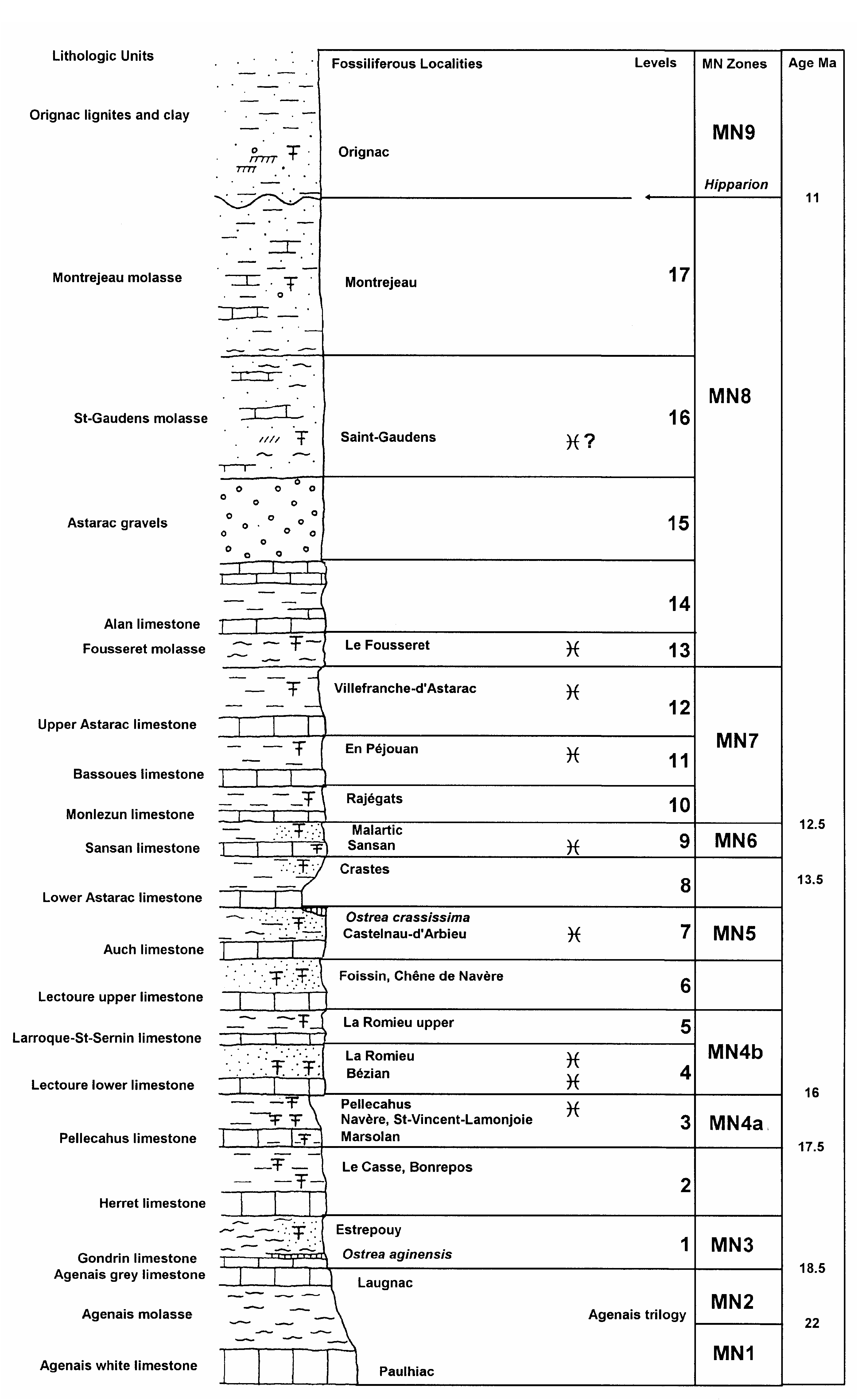
Listriodonts with bunodont to bunolophodont dentitions are common in European basal middle Miocene deposits such as Pellecahus (MN4a) and La Romieu (MN4b) in the Aquitaine Basin ( Fig. 11 View FIG ). The main distinguishing feature of the species is the degree of bunodonty in the cheek teeth, but the mesiodistal expansion of the upper central incisor and the number of subdivisions that it has are additional factors that separate this species from more lophodont forms such as Listriodon splendens . Some undescribed material from Pellecahus in the Muséum national d’Histoire naturelle, Paris, is more complete than previously described specimens ( Van der Made 1996) and adds considerably to our understanding of the morphology of the lower jaw.
MATERIAL EXAMINED
Lower dentition
LRM 557, i1; LRM 842, i1; LRM 796, i1; LRM 843, i1; LRM 556, i1; LRM 558, i1; LRM 841, left i2; LRM 844, left i2 (unerupted); LRM 555, left i3; LRM 536, mandible with left p4-m3 (m3 broken); LRM 538, right mandible with p2-m3 (m2-m3 broken); LRM 544, p2-p4; LRM 968, right p2; LRM 849, right p3; LRM 550, right p4; LRM 551, left p4; LRM 850, left p4; LRM 965, right m1-m3 (m1 broken); LRM 542, left m1-m2; LRM 1054, right m1; LRM 548, worn m1; LRM 546, right m1 (same individual as LRM 542); LRM 547, left m2; LRM 543, right
Length i1 i2 i3 p2 p3 p 4 m 1 m 2 m 3 i1 i2 i3 p2 p3 p 4 m 1 m 2 m 3 Breadth
FIG. 8. — Size variation (in mm) of the lower dentition of Listriodon lockharti Pomel, 1848 . Symbols:, maximum and minimum measures; l, range of variation;´, Retama fossils; ➔, worn specimens.
m3; LRM 545, left m3; LRM 852, left m3; LRM 568, left di2; LRM 569, left di2; LRM 571, left dm4. All the specimens listed here are housed in the Muséum national d’Histoire naturelle, Paris.
Upper dentition
LRM 845, right I2; LRM 966, left I3; LRM 563, left I3; LRM 851, right upper male canine; LRM 561, left P2; LRM 562, left P3; LRM 549, broken left upper molar; LRM 573, left M1; LRM 554, left M2 (broken); LRM 967, right M3; LRM 572, right dM3.
Postcranial skeleton
LRM 585, distal right humerus (fragmentary); LRM 586, proximal right ulna; LRM 587, proximal left ulna; LRM 853, proximal right radius; LRM 588, proximal right radius (same individual as LRM 586); LRM 537, proximal left third metacarpal; LRM 591, right fourth metacarpal; LRM 854, right fourth metacarpal; LRM 592, proximal left fifth metacarpal; LRM 593, distal left tibia; LRM 594, distal left tibia; LRM 597, right calcaneum (broken); LRM 595, left talus; LRM 596, right talus; LRM 599, left cuboid; LRM 600, right navicular; LRM 603, proximal left third metatarsal; LRM 602, proximal right third metatarsal; LRM 751, right third tarsal bone; LRM 601, right third tarsal bone; LRM 755, right third tarsal bone; LRM 604, distal end first phalanx; LRM 605, second phalanx; LRM 606, second phalanx; LRM 607, second phalanx; LRM 608, third phalanx.
DESCRIPTION
Upper dentition ( Table 2)
In labial view the crown of I2 is low and triangular. It possesses a beaded cingulum lingually and
Upper dentition Length Breadth I3 13.8 7.3 I3 15.2 7.8 I3 14.0 7.2 P2 15.6 12.3 P3 17.4 16.3 M1 21.5 20.0 M3 26.0 23.6 M3 22.7 17.3 Lower dentition Length Breadth i1 10.4 10.3 i1 10.5 10.2 i1 10.2 9.8 i1 10.2 9.7 i1 10.2 10.0 i1 10.6 10.3 i2 12.2 10.7 i3 13.2 8.7 p2 16.9 9.0 p2 16.5 9.1 p2 16.3 8.8 p3 18.4 10.5 p3 18.7 10.5 p3 18.3 11.5 p3 18.9 11.2 p4 17.8 12.6 p4 19.0 13.1 p4 17.0 13.4 p4 17.3 13.1 p4 16.8 12.7 p4 18.2 14.0 m1 20.4 14.2 m 1 20.4 15.7 m 1 19.8 15.3 m 2 23.4 19.0 m2 23.6 18.6 m 2 23.2 18.3 m 2 22.6 19.1 m 2 22.9 18.3 m 3 30.5 19.0 m3 31.0 18.6 m 3 32.5 19.2 m 3 29.4 18.2
the root is short and curves distolingually. Both upper third incisors in the sample from Pellecahus possess triangular crowns in lateral view. In occlusal view the teeth are trenchant. Lingually there is a sharp basal cingulum. Both specimens have long conical roots that curve sharply lingually near their apices.
The upper male canine is almost circular in section, and is strongly curved from root to tip. There is a prominent, almost flat wear facet anteriorly caused by abrasion against the lower canine. A remnant of wrinkled enamel is present ventrally but it does not extend onto the root.
The P2 has an occlusal outline which is triangular with rounded corners. The tip of the main cusp is in the centre of the crown and has ridges that reach the anterolingual and posterolabial corners of the tooth. A cingulum completely surrounds the tooth apart from a small gap in the middle of its buccal surface, and in the distolingual corner there is a small accessory cusplet between the cingulum and the main cusp.
The P3 is basically an enlarged version of the P2. It has three roots.
Upper molars from Pellecahus differ from those of fully lophodont listriodonts by having clear anterior and median accessory cusplets, even if these are small and located between the anterior ends of the main cusps in each loph rather than anterior to the lophs as in other suids. In fully lophodont listriodonts a ridge connects the tips of the main cusps in each loph. In the Pellecahus material, in contrast, ridges descend anteriorly towards the anterior and median accessory cusplets, and it is only in medium wear that a lophlike morphology is expressed. “Furchen” (Hünerman 1968) are present but weakly developed in the upper molars, but in any case are more distinct than they are in fully lophodont listriodonts. In the M3, the talon is located in a lingual position, almost in line with the protocone and hypocone. The Pellecahus upper molars are usually adorned with more or less complete buccal cingula.
Mandible
LRM 536 is the most complete of the mandibles from Pellecahus and it could well belong to the same individual as LRM 538. The anterior part of the symphysis is broken as far back as the right canine alveolus on the right and the middle part of the postcanine diastema on the left side. The symphysis reaches back to the rear of the p2. Judging from the remnant of the canine alveolus preserved the mandible probably represents a male individual. Immediately behind the canine alveolus there is a prominent circular, but not very deep, alveolus for the p1. Between the p1 and the p2 there is a diastema of about 58 mm. An accurate measurement is not possible due to the crushed condition of the specimen, but in any case the diastema is long. The body of the mandible is deep (58 mm below p3 and 65 mm below m3) and it is bucco-lingually relatively narrow, but this may be due to the crushing that the jaw has undergone. There is a mental foramen below the anterior end of p4 positioned about two thirds the height of the body. The overall morphology of the mandible is typical of Listriodon species and departs radically from the Libycochoerus plan ( Pickford 1986) and we see no substantive reason to consider that it should be assigned to a separate genus Bunolistriodon . In this respect we are in agreement with Leinders (1975) who studied listriodont mandibles from Torralba 2 and Munebrega 1 in Spain.
Lower dentition ( Table 2)
The lower incisors from Pellecahus are generally more robust than those of lophodont listriodonts. The i1s are low crowned with a curved cutting edge, the mesial and distal ends of the cusp curving lingually in unworn specimens. There is a prominent centrally positioned lingual pillar which is large at its base and dies out apically, so that with wear the cutting edge of the crown becomes more and more square in outline. The roots are straight, robust and elongated. The i2 is slightly higher crowned and less symmetrical than the i1, the distal edge protruding slightly to form a shallow scoop-like basin between the central pillar and the distal edge of the crown. Its occlusal edge is almost straight, even in unworn specimens. The root is also slightly bowed, its apex curving mesially. The i3 is even more asymmetrical than the i2 and lower crowned and more spatulate in labial aspect. Its cutting edge is virtually straight when unworn. The mesial height of the crown is appreciably less than the distal height, the central pillar is low but strong, and the root is short and straight.
The p2 is located at the distal end of a diastema. It is a two-rooted tooth with a single main cusp accompanied distally by an enlarged distal accessory cusp which is located slightly to the buccal side of midline. There is a tiny anterior accessory cusplet and a low, sharp, cingulum distally. The distal cusplet is about one third the height of the main cusp.
The p3 is basically a larger version of the p2, except that the distal accessory cusplet is about half the height of the crown and the main cusp has a hint of the development of an “innenhugel” (Hünerman 1968). The p4 is shorter than the p3 and is more rectangular in occlusal outline. The anterior accessory cusplet is more strongly developed than it is in the anterior premolars, and there is a strong crest running from it to the apex of the main cusp. There is a well developed “innenhugel” which is closely fused to the main cusp. In unworn specimens the tips of the main cusp and the “innenhugel” are separated from each other, but even with slight wear they fuse together to form a loph-like structure. The distal accessory cusplet is two thirds the height of the crown, almost forming a separate cusp, which however, is joined to the main cusp by a crest. The distal cingulum fades into the lingual and labial surfaces of the crown.
The lower molars are bunodont with the usual suid layout of four main cusps arranged in two lophs, with anterior, median and posterior accessory cusplets in the midline of the crown. The median accessory cusp is particularly prominent and blocks the median transverse valley to well over half the height of the crown. Several of the teeth retain cingular remnants on their buccal surfaces, but in none of them is the cingulum complete. The “furchen” are present but weakly expressed, and the main cusps in each pair are positioned in close proximity to each other, so that even in the early wear stages, a loph-like structure is developed. The talonid of the third lower molar consists of a dune-shaped cusp with the horns of the dune pointing anteriorly, the buccal horn joining the posterior accessory cusplet which is well developed.
Pickford M. & Morales J.
No known copyright restrictions apply. See Agosti, D., Egloff, W., 2009. Taxonomic information exchange and copyright: the Plazi approach. BMC Research Notes 2009, 2:53 for further explanation.
|
Kingdom |
|
|
Phylum |
|
|
Class |
|
|
Order |
|
|
Family |
|
|
Genus |