
Lissoclinum guinense, Monniot & Monniot, 2001
|
publication ID |
https://doi.org/ 10.5281/zenodo.5391440 |
|
DOI |
https://doi.org/10.5281/zenodo.5468057 |
|
persistent identifier |
https://treatment.plazi.org/id/F57D87A3-FF8D-3168-E805-FC63FB5E15A0 |
|
treatment provided by |
Marcus |
|
scientific name |
Lissoclinum guinense |
| status |
sp. nov. |
Lissoclinum guinense View in CoL n. sp.
( Figs 66 View FIG ; 67A View FIG ; 122D View FIG )
TYPE MATERIAL. — Papua New Guinea. Cape Rodney Pass, 10°15.66’S, 148°22.27’E, 31 m, 11. VI.1998 ( MNHN A2 LIS 152).
ETYMOLOGY. — From New Guinea.
DESCRIPTION
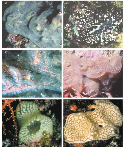
Inflated underwater ( Fig. 122D View FIG ), the colonies become flat encrusting sheets when fixed. They are colourless with a cloudy aspect due to irregularly distributed small spicules. The wide common cloacal apertures ( Fig. 122D View FIG ) correspond to an extensive common cloacal cavity.
The zooids are small and colourless but the eggs and larvae have a brown pigment in formalin preservative. The oral siphon is short and narrow with six pointed lobes ( Fig. 66A View FIG ). The cloacal aperture is narrowed by a strong thoracic musculature. There is no cloacal languet. Lateral thoracic organs were not clearly observed. There is no retractor muscle.
The abdomen is folded under the thorax and the waist is short ( Fig. 66A View FIG ). The gut has the usual compartments. The single testis follicle lies beneath the gut loop. The sperm duct is straight. The ovary is appended in a pouch attached anteriorly to the stomach and contains one large egg ( Fig. 66A View FIG ). The larvae ( Fig. 66B View FIG ) are incubated in the basal layer of the colony. The eggs and the posterior half of larvae contain brown droplets, probably pigment cells.
The larvae are small, 0.55 mm for the trunk. They have three anterior adhesive papillae and four pairs of intercalated ampullae. The larval gut is posterior to a large vitellus vesicle. There are no buds.
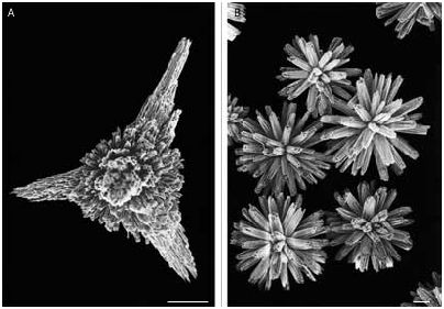
The spicules ( Fig. 67A View FIG ) have generally four stellate branches made of needles. They measure 60 to 70 µm. They are very similar to those of Lissoclinum verrilli (Van Name, 1902) .
REMARKS
By numerous characters this species is closely allied to the Polynesian Lissoclinum tuheiavae Monniot C. & Monniot F., 1987 . The latter has larger zooids and much larger spicules, and its larvae have no pigment. But both species share the absence of a cloacal languet, the absence of a retractor muscle, the shape of the ovary, and a non-gemmiparous larva with four pairs of anterior ampullae.
Lissoclinum verrilli has the same shape of spicules, but its larvae are gemmiparous.
Lissoclinum triangulum Sluiter, 1909 View in CoL has different spicules and a larva with six pairs of larval ampullae.
| VI |
Mykotektet, National Veterinary Institute |
| MNHN |
Museum National d'Histoire Naturelle |
No known copyright restrictions apply. See Agosti, D., Egloff, W., 2009. Taxonomic information exchange and copyright: the Plazi approach. BMC Research Notes 2009, 2:53 for further explanation.
|
Kingdom |
|
|
Phylum |
|
|
Class |
|
|
Order |
|
|
Family |
|
|
Genus |
Lissoclinum guinense
| Monniot, Françoise & Monniot, Claude 2001 |
Lissoclinum triangulum
| Sluiter 1909 |