
Lernanthropus breviculus Kabata, 1979
|
publication ID |
https://doi.org/ 10.11646/zootaxa.4736.1.1 |
|
publication LSID |
lsid:zoobank.org:pub:970D7D36-6D8C-4463-B9EA-D3B8E191BE72 |
|
DOI |
https://doi.org/10.5281/zenodo.3671132 |
|
persistent identifier |
https://treatment.plazi.org/id/554BDB52-737C-FFEE-5FC9-FD572A41FE1C |
|
treatment provided by |
Plazi |
|
scientific name |
Lernanthropus breviculus Kabata, 1979 |
| status |
|
Lernanthropus breviculus Kabata, 1979
( Figs. 12–14 View FIGURE 12 View FIGURE 13 View FIGURE 14 )
Material examined: Holotype 1♀ on Cheilinus chlorourus (Bloch, 1791) , Heron Island , Queensland, 24 August 1963; collected by P.C. Young; NHMUK Reg. No. 1977.121.
Comparative material examined: 1♀ from C. chlorourus, Baie de Koutio , New Caledonia; collected by J.–L. Justine, NHMUK Reg. No. 2010.657 .
7♀♀, 2♂♂ from Choerodon graphicus De Vis, 1885 , New Caledonia, collected by J.–L. Justine, NHMUK Reg. No. 2012.249–257 . 1♀ from C. graphicus , New Caledonia, collected by J.–L. Justine, NHMUK Reg. No. 2012.248 .
1♀ from Cheilinus trilobatus Lacepède, 1801 , New Caledonia, collected by J.–L. Justine, NHMUK Reg. No. 2012.261 .
3♀♀ from Oxycheilinus unifasciatus (Streets, 1877) , New Caledonia, collected by J.–L. Justine, NHMUK 2012.258 View Materials – 260 View Materials .
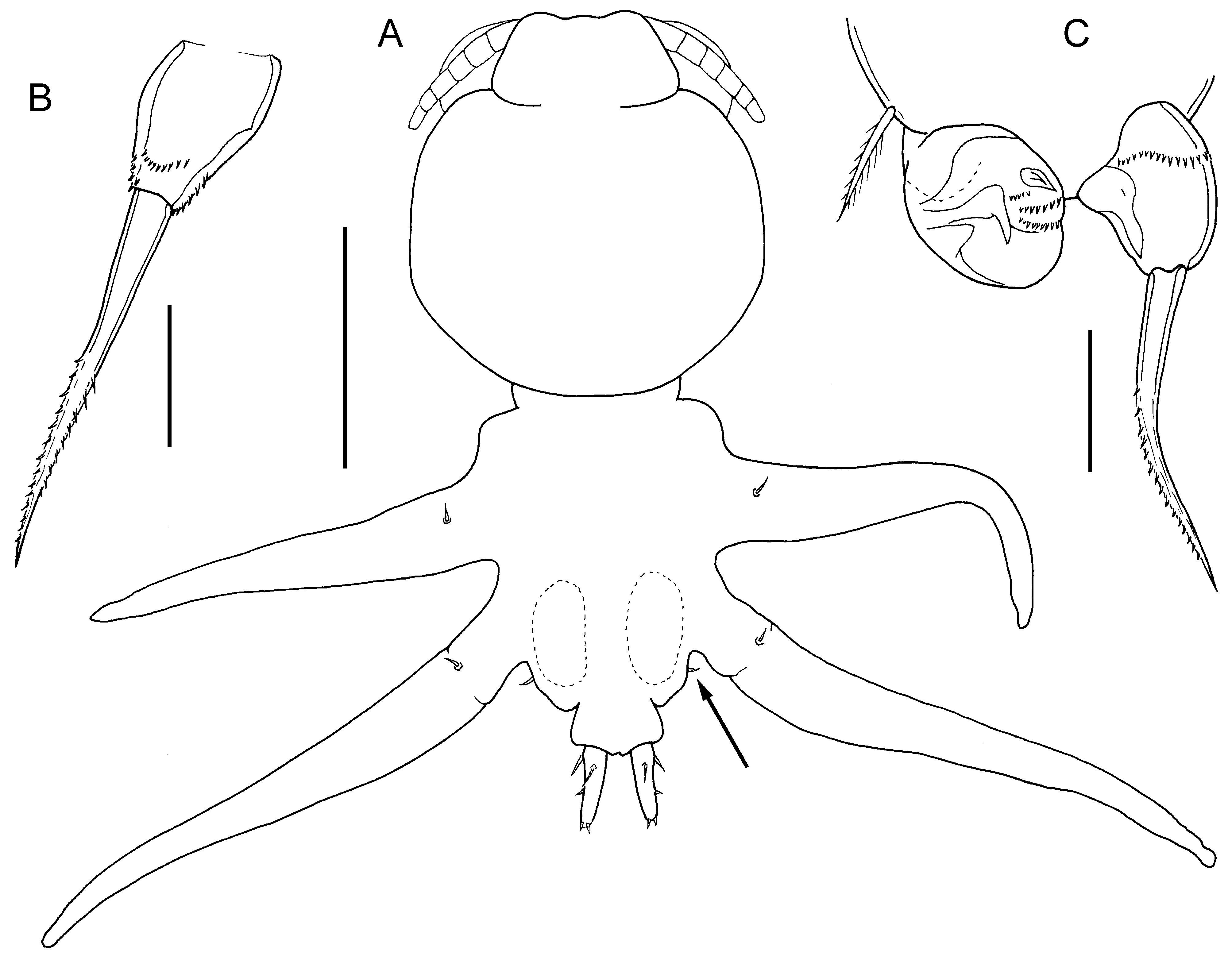
Supplementary description of female: Cephalothorax about as long as wide, with slightly angular convex lateral margins ( Fig. 12A View FIGURE 12 ). Anterior part of trunk (second and third pedigerous somites) broader than cephalothorax and broader than posterior part (fourth pedigerous somite), covered by narrow dorsal trunk plate. Dorsal trunk plate longer than wide, with slightly angular lateral margins and free posterior margin. Urosome comprising fifth pedigerous somite, genital complex and 1-segmented abdomen, all fused ( Fig. 13A View FIGURE 13 ); genital complex about twice as wide as long, with large paired genital apertures located dorsally and paired copulatory pores at posterolateral corners; surface ornamented with pair of sensillae. Paired caudal rami elongate, about 4.1 times longer than wide; tapering towards blunt apex; bearing 2 plumose setae dorsally, one short naked seta on mid-lateral margin, and 2 short naked setae apically.
Antennule ( Fig. 13B View FIGURE 13 ) indistinctly 6-segmented; armed with 1, 3, 2, 1, 1, 10 + 2ae. Antenna comprising long corpus with shallow papilla on medial surface, plus strongly recurved distal subchela ( Fig. 13C View FIGURE 13 ). Parabasal flagellum absent. Maxilla with terminal claw ornamented with spinules ( Fig. 13D View FIGURE 13 ). Maxilliped ( Fig. 13E View FIGURE 13 ) 2-segmented; comprising massive corpus with papilliform process and proximal swelling on myxal surface, and strong distal subchela. Leg 1 biramous ( Fig. 13F View FIGURE 13 ), joined by intercoxal sclerite; protopod with outer seta on papilla plus inner margin spine: exopod 1-segmented with 5 spines on distal margin; endopod 1-segmented, unarmed but with internal glandular structure at apex. Leg 2 forming large ventrally directed lobe ( Fig. 13G View FIGURE 13 ) derived from protopod armed with outer seta, and carrying small, 1-segmented rami distally; exopod armed with 4 vestigial spines; endopod lobate, unarmed. Leg 3 forming fleshy lamella, orientated horizontally and directed-posteriorly; members of leg pair fully fused along midline ( Fig. 12B View FIGURE 12 ). Leg 4 bilobate; inner and outer lobes subequal, protruding well beyond posterior margin of dorsal trunk plate ( Fig. 12B View FIGURE 12 ). Leg 5 absent. Body length of holotype ♀ 1.90 mm ( Kabata, 1979a).
Description of male: Body smaller than female ( Fig. 14A View FIGURE 14 ), total length about 1.70 mm (based on 2 specimens). Cephalothorax large, comprising about 46% of total body length, with convex lateral margins. Frontal area of cephalothorax carrying antennule and antennae, defined by indentation. Trunk comprising all fused post-cephalothoracic somites ( Fig. 14A View FIGURE 14 ), including urosome. Anal somite defined, bearing paired caudal rami; each ramus elongate, about 3.0 times longer than wide, armed with 2 plumose setae proximally on dorsal surface, 1 short lateral seta located about at mid-length, plus 2 apical setae.
Antennule 6-segmented as in female; setal formula: 1, 3, 2, 0, 1, 3 + ae, 7 + ae. Parabasal flagellum absent. Antenna comprising long, slender corpus and distal subchela terminating in strongly recurved claw: corpus armed with broad process proximally on medial surface plus inner distal process; subchela armed with strong accessory claw proximally and another accessory claw near middle. Mandible stylet-like, with 8 marginal teeth near apex. Maxillule bilobate, larger lobe armed with 3 unequal elements distally; smaller lobe with strong apical element. Maxilla with 2 rows of denticles on distal claw. Maxilliped comprising robust corpus bearing small pointed myxal process and ornamented with patches of blunt spinules proximally on medial surface, and distal subchela armed with inner seta about at mid-length plus blunt process at base of terminal claw.
Leg 1 robust, members of leg pair joined by intercoxal sclerite as in female: basis armed with outer seta on papilla and stout inner spine; exopod 1-segmented, broadening distally, armed with 5 distal spines, as in female; endopod 1-segmented ( Fig. 14B View FIGURE 14 ), tapering distally, armed with spinulose apical seta about 2.5 times longer than segment; segment ornamented with spinules distally. Leg 2 ( Fig. 14C View FIGURE 14 ) lacking intercoxal sclerite; basis with outer seta; both rami 1-segmented; exopod lobate, modified with spinous structures and rows of spinules on distal surface; endopod just longer than wide and armed with long unilaterally spinulose seta apically; seta about 2.0 times longer than segment; surface of segment ornamented with spinule row proximally. Leg 3 ( Fig. 14A View FIGURE 14 ) uniramous, forming long cylindrical process directed posterolaterally from ventrolateral origin on trunk, armed with basal seta dorsally at base of limb; surface of leg 3 densely ornamented with rounded tubercles. Leg 4 ( Fig. 14A View FIGURE 14 ) uniramous, forming long cylindrical process, outer basal seta present dorsally at base of limb. Leg 5 represented by minute papilla with apical seta (arrowed in Fig. 14A View FIGURE 14 ).
Distribution: Kabata (1979a) established L. breviculus based on a single female collected from the gills of the labrid Cheilinus chlorourus (as C. chlorurus ) caught off Heron Island. It has been collected subsequently by J.-L. Justine from the same host, C. chlorourus , caught in the Baie de Koutio, New Caledonia, and from three other labrid species, Choerodon graphicus (NHMUK 2012.248 and 2012.249–257), Oxycheilinus unifasciatus (NHMUK 2012.258–260) and Cheilinus trilobatus (NHMUK 2012.261).
Remarks: Kabata (1979a) had only a single specimen, the holotype, which he did not dissect, so this species has never been fully described. On the basis of material in the collections of the Natural History Museum, we here provide a supplementary description of the female including details of appendage structure, plus the first description of the male. The material described here was collected by J.-L. Justine from Choerodon graphicus caught off New Caledonia.
Considering only the habitus of the female, L. breviculus appears to be closely related to L. callionymicola El-Rashidy & Boxshall, 2012 described from Callionymus filamentosus Valenciennes, 1837 caught in the Mediterranean Sea ( El-Rashidy & Boxshall, 2012), but the dorsal trunk plate of L. callionymicola is very short and the tips of the caudal rami are visible in dorsal view, whereas in L. breviculus the dorsal trunk plate is relatively longer and the caudal rami are completely concealed ( Fig. 11 View FIGURE 11 A–B).


| NHMUK |
Natural History Museum, London |
No known copyright restrictions apply. See Agosti, D., Egloff, W., 2009. Taxonomic information exchange and copyright: the Plazi approach. BMC Research Notes 2009, 2:53 for further explanation.