
Integripelta shirayamai, Gordon, Dennis P., Mawatari, Shunsuke F. & Kajihara, Hiroshi, 2002
|
publication ID |
https://doi.org/ 10.1046/j.1096-3642.2002.00020.x |
|
DOI |
https://doi.org/10.5281/zenodo.5106369 |
|
persistent identifier |
https://treatment.plazi.org/id/039B3920-2867-7745-FF73-F9D8FE7CD2BC |
|
treatment provided by |
Carolina |
|
scientific name |
Integripelta shirayamai |
| status |
sp. nov. |
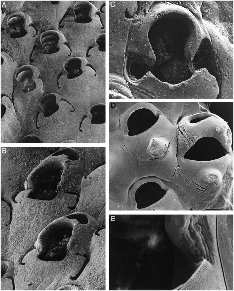
INTEGRIPELTA SHIRAYAMAI View in CoL SP. NOV.
( FIG. 3A–C View Figure 3 )
Lepralia bilabiata: Okada 1929: 24 , fig. 10, pl. 2, fig. 3; Sakakura 1935: 25, fig. 7.
Eurystomella bilabiata: Mawatari 1952: 280 .
Material examined
Holotype: SMBL Type no. 401, 33°02.326¢N, 132°05.608¢E, 80–86 m, off the island of Saiki-wan, Oita Prefecture, eastern Kyushu [Seto Marine Laboratory Stn 5, 26 September 1998].
Paratypes: SMBL Type no. 402, and NIWA P-1219, same locality as holotype .
Description
Colony encrusting, unilaminar, multiserial. Self-overgrowth not seen. Colony colour unknown. Autozooids contiguous, quincuncially arranged, 0.47–0.75 mm long (0.58 ± 0.07 mm), 0.28–0.55 mm wide (0.41 ± 0.06 mm). Gymnocystal frontal shield flat, centrally smooth and imperforate. Orifice longer than wide, somewhat dumbbell-shaped, the anter high-arched and rounded with the proximal corners somewhat condyle-like; poster not wider than anter, the proximal rim gently and evenly concave. Conspicuous crescentic slits curve proximolaterally from corners of poster; below the outer edge of each slit is a narrow shelf. Traces of these slits, paired or distally continuous, occur in incompletely formed autozooids, i.e. kenozooids. No peristome, umbones, spines, or avicularia. Orifice of maternal zooids larger overall than in autozooids, the distal kenozooid with a large, transversely elongate foramen sloping distad. Interzooidal communication via tiny uniporous mural septula. Ancestrula not known.
Etymology
After Professor Yoshihisa Shirayama, director of the Seto Marine Laboratory, Shirahama, Kii Peninsula, in recognition of his contributions to biodiversity appreciation in Japan.
Remarks
This is a very striking species, easily recognizable by the crescentic lateral slits, which are very conspicuous in dead zooids. Okada (1929) attributed specimens in his collection from Mutsu Bay to Eurystomella bilabiata but illustrated the slits and, in one zooid, a spine-like umbo, described in the text as keel-like or carinate and restricted to older zooids. None of the specimens we have examined show this latter feature. Eurystomella shirayamai is also distinguished on the basis of the ‘ooecial kenozooid’, the foramen of which is larger and more bean-shaped than in E. bilabiata .
Cook & Chimonides (1981) noted the crescentic lateral slits illustrated by Okada (1929) and Sakakura (1935) and also discovered them in a specimen in The Natural History Museum, London ( BMNH 1885.8.29.1) from “Sio-u-whu Bay”. [This name, not found in modern atlases, refers to a locality south of Vladivostok, Russia, in the Japan Sea. The coordinates on the label give the following data: “Sio-u-whn(u) Bay, Gulf of Tartary, 42°N, 133°S ”.] Cook & Chimonides (1981) wrote: “Some of the Japanese populations have been reported to show characters [sic] states which vary somewhat from those of the eastern Pacific specimens. [The slits] are covered by brown cuticle which appears to be continuous distally with that of the operculum”. This observation accords with our interpretation that the slits are lateral extensions of the indentations seen at the proximolateral corners of most eurystomellids (see Cook & Chimonides, 1981). Unfortunately, the specimens available to us were all dead and lacked opercula and membranes. The narrow shelf below the outer edge of each slit is clearly homologous with the excavations that occur in the gymnocysts of species like I. novella sp. nov. and I. sextaria sp. nov. (cf. Figs 2A, B, F View Figure 2 and 3C View Figure 3 ). One other possibility is that the slits represent frontal foramina that have migrated laterally; this is suggested by their presence in kenozooids lacking orifices, but, in one instance, the kenozooidal slits are distally continuous and the distal part of the inverse U-shaped slit is suggestive of aborted orificial development. Further, the distalmost parts of the frontal gymnocyst merely abut, and do not fuse, with the proximal corners of the ‘ooecial kenozooid’, such that organic continuity between the operculum and the slits is possible just at or under the loci of abutment.
Distribution
Integripelta shirayamai View in CoL is endemic to east Asian waters. It has been reported (as Eurystomella bilabiata ) from numerous localities in Mutsu Bay, northern Honshu ( Okada, 1929), from south-eastern Honshu ( Mawatari, 1952), and south of Vladivostok in the Japan Sea. Pleistocene material was illustrated by Sakakura (1935), who found specimens to be very rare to common in the Dizôdô beds of the Bôsô Peninsula, eastern Honshu. Specimens examined for this paper were collected off Saiki-wan, eastern Kyushu (courtesy of the Seto Marine Laboratory).

No known copyright restrictions apply. See Agosti, D., Egloff, W., 2009. Taxonomic information exchange and copyright: the Plazi approach. BMC Research Notes 2009, 2:53 for further explanation.
|
Kingdom |
|
|
Phylum |
|
|
Class |
|
|
Order |
|
|
Family |
|
|
Genus |
Integripelta shirayamai
| Gordon, Dennis P., Mawatari, Shunsuke F. & Kajihara, Hiroshi 2002 |
Eurystomella bilabiata: Mawatari 1952: 280
| Mawatari S 1952: 280 |
Lepralia bilabiata:
| Sakakura K 1935: 25 |
| Okada Y 1929: 24 |