
Indoartemon medius Siriboon & Panha, 2014
|
publication ID |
https://doi.org/ 10.5281/zenodo.4504075 |
|
publication LSID |
lsid:zoobank.org:pub:2300F64E-BD30-4E06-A24F-A226185A4BD3 |
|
persistent identifier |
https://treatment.plazi.org/id/ABA299D2-E829-4245-B70C-4763BA98A058 |
|
taxon LSID |
lsid:zoobank.org:act:ABA299D2-E829-4245-B70C-4763BA98A058 |
|
treatment provided by |
Carolina |
|
scientific name |
Indoartemon medius Siriboon & Panha |
| status |
sp. nov. |
Indoartemon medius Siriboon & Panha View in CoL , new species
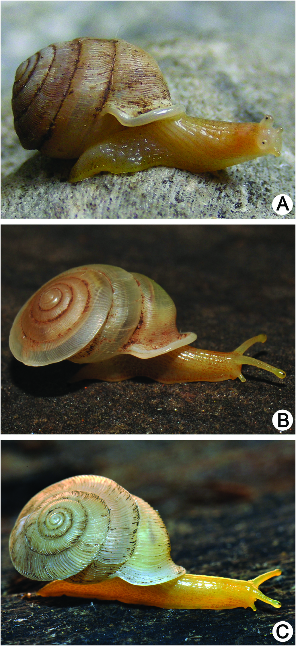
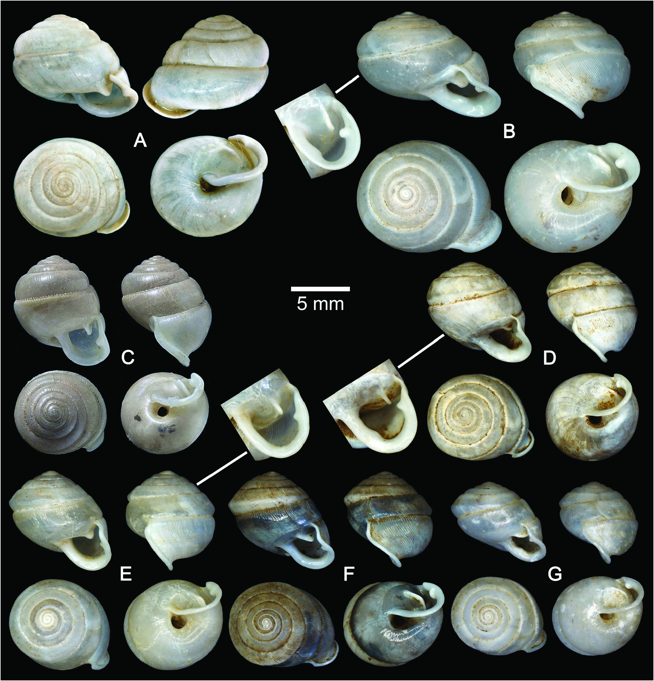
( Figs 1 View Fig , 2A View Fig , 3 View Fig E–G, 4A–C, 5A–I, Table 1)
Material examined. Holotype CUMZ 5016 View Materials ( Fig. 3E View Fig ). Measurement: height 8.8 mm, width 9.2 mm, 7 whorls . Paratypes: CUMZ 5017 View Materials (56 shells; Fig. 3F View Fig ) , CUMZ 6206 View Materials (28 specimens in ethanol) , NHMUK 20130076 View Materials (2 shells) , SMF (2 shells), and ZRC (2 shells).
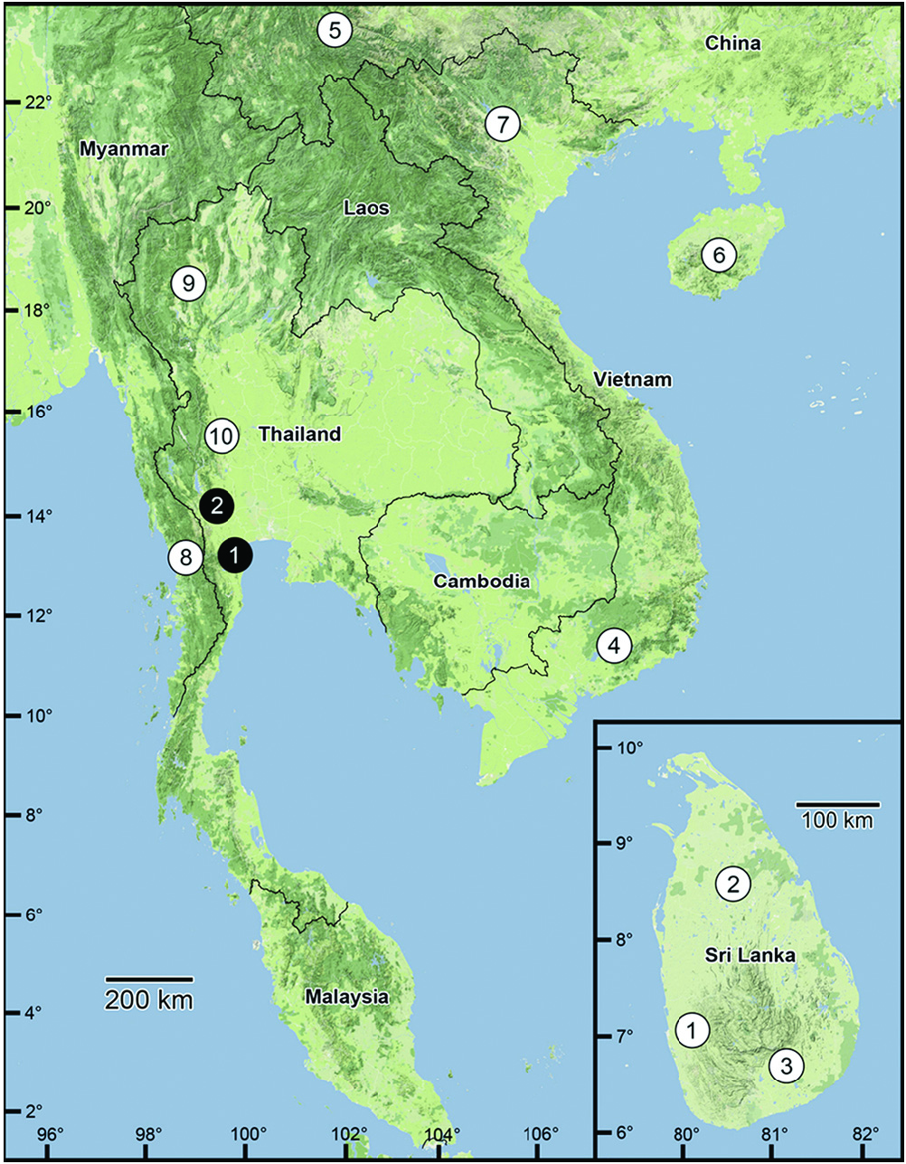
Type locality. Wat Chuak Charoentham, Ban Rai , Uthai Thani, an isolated limestone hill about 200 m above mean sea level (15°16'26.3"N, 99°41'43.4"E) GoogleMaps .
Other material examined. Tam Lom-Tam Wang, Si Samrong, Sukhothai. Shells slightly smaller than specimens collected from Uthai Thani ( CUMZ 5018 View Materials , Fig. 3G View Fig ) .
Etymology. The specific epithet “ medius ” is derived from the Latin “ medius ” meaning “middle”. It refers to the distribution range of this new species in central Thailand.
indicated in parentheses.
Diagnosis. Indoartemon medius , new species, can be distinguished from I. cingalensis and I. fuchsianus ( Gredler, 1881) in its higher spire, the last whorl being less deviated from the vertical axis, the left periphery of the penultimate whorl being keeled, and the absence of a columellar lamella. Indoartemon laevis differs from I. medius , new species, in its lower spire, the left periphery of the penultimate whorl being rounded and not extending beyond the diameter of the last whorl, and the presence of a basal lamella. Indoartemon prestoni possesses a distinct suture, the left periphery of the penultimate whorl extends beyond the diameter of the last whorl, and the umbilicus is wider. Indoartemon medius , new species, can be distinguished from the type species by its larger shell, the left periphery of the penultimate whorl being shouldered, last whorl being more deviated from the vertical axis, its wider umbilicus, and semi-ovate aperture.
Description. Shell oblique-heliciform, white, and translucent; whorls 7, spire conical with indistinct suture. Shell surface glossy, with fine transverse ridges that diminish below the periphery. Embryonic shell with about 2½ whorls and smooth surface; following whorls regularly expanded. Shell periphery keeled; last whorl axially deflected. Umbilicus narrow and deep. Aperture subcircular; peristome discontinuous, thickened and slightly expanded. Apertural dentition with one parietal lamella and one palatal lamella ( Fig. 3E View Fig ).
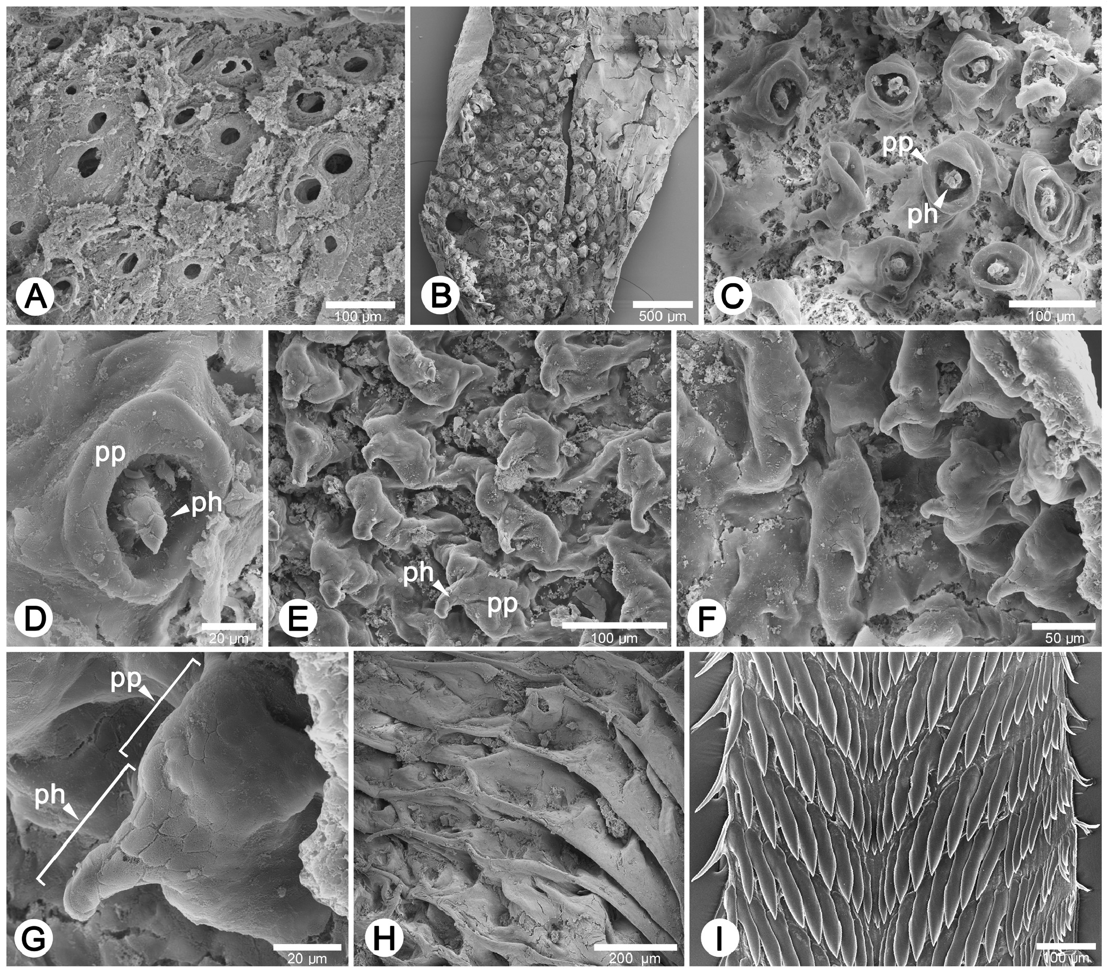
Radula. Teeth arranged in anteriorly V-shaped rows, each row containing 33–37 teeth with the formula (16-18)-1- (16-18); central tooth small, short, triangular with pointed cusp. Lateral and marginal teeth undifferentiated, unicuspid and lanceolate; lateral teeth gradually reduced in length and size with outer teeth much smaller and shorter than inner teeth ( Fig. 5I View Fig ).
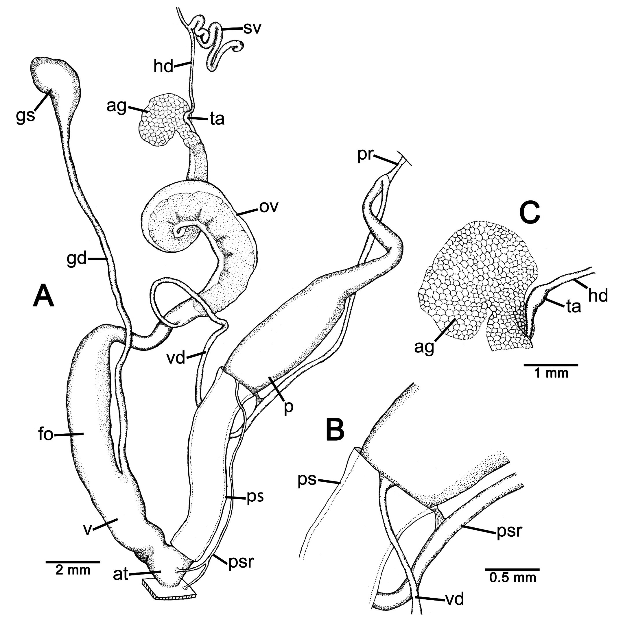
Genital organs. Atrium (at) short. Proximal penis (p) long slender; becoming slightly broader before tapering distally. Penial sheath (ps) thin, extending about half of total penis length, penial sheath retractor muscle (psr) very thin, originating at atrium, inserting distally on penial sheath ( Fig. 4A View Fig ). Vas deferens (vd) slender, attached to distal end of penial sheath by a narrow band of connective tissue ( Fig. 4B View Fig ). Penial retractor muscle (pr) thin and very long, inserting distally on penis at the penis and vas deferens junction.
Internal wall of atrium possesses numerous atrial pores ( Fig. 5A View Fig ). Proximal penial wall with scattered and pale brown penial hooks about 3 hooks/200 μm 2; hooks withdrawn into penial papillae separated by low reticulated folds. Proximal penial hooks are minute (<0.01 mm in length), tips obtuse and curved towards genital orifice ( Fig. 5C, D View Fig ). Distal penial wall with pale brown penial hooks about 5 hooks/200 μm 2; hooks located on penial papillae (pl). Penial papillae may extend across penial hook and adjacent areas with low reticulated folds ( Fig. 5E, F View Fig ). Distal minute penial hooks with obtuse tips (<0.01 mm in length) expanded at base and curved towards genital orifice ( Fig. 5G View Fig ).
Vagina (v) short, stout, about quarter of total penis length. Gametolytic duct (gd) long, extending as far as albumin gland; gametolytic sac ovate (gs). Free oviduct (fo) stout and long. Oviduct (ov) enlarged and folded; prostate gland inconspicuous and bound to oviduct ( Fig. 4A View Fig ). Talon (ta) small, very short, and club shaped ( Fig. 4C View Fig ). Hermaphroditic duct (hd) bearing long seminal vesicle (sv) about one and half times longer than the length from talon to branching point of seminal vesicle. Vaginal wall with oblique parallel vaginal folds; vaginal hooks absent ( Fig. 5H View Fig ).
Animal. Live specimens exhibit yellowish to orange reticulated skin, and orange tentacular retractor muscles are visible through the semi-transparent body ( Fig. 2A View Fig ).
No known copyright restrictions apply. See Agosti, D., Egloff, W., 2009. Taxonomic information exchange and copyright: the Plazi approach. BMC Research Notes 2009, 2:53 for further explanation.