
Gadigaleyrodes, Dooley Iii, John W. & Gillespie, Peter, 2013
|
publication ID |
https://doi.org/ 10.11646/zootaxa.3608.2.4 |
|
publication LSID |
lsid:zoobank.org:pub:9206E4A9-6FFE-41FD-A99F-430183B85775 |
|
DOI |
https://doi.org/10.5281/zenodo.6163928 |
|
persistent identifier |
https://treatment.plazi.org/id/03C787CD-7960-FFCB-FF57-5847E92BF780 |
|
treatment provided by |
Plazi |
|
scientific name |
Gadigaleyrodes |
| status |
gen. nov. |
Gadigaleyrodes gen. n.
Type species: Gadigaleyrodes froggatti sp. n.
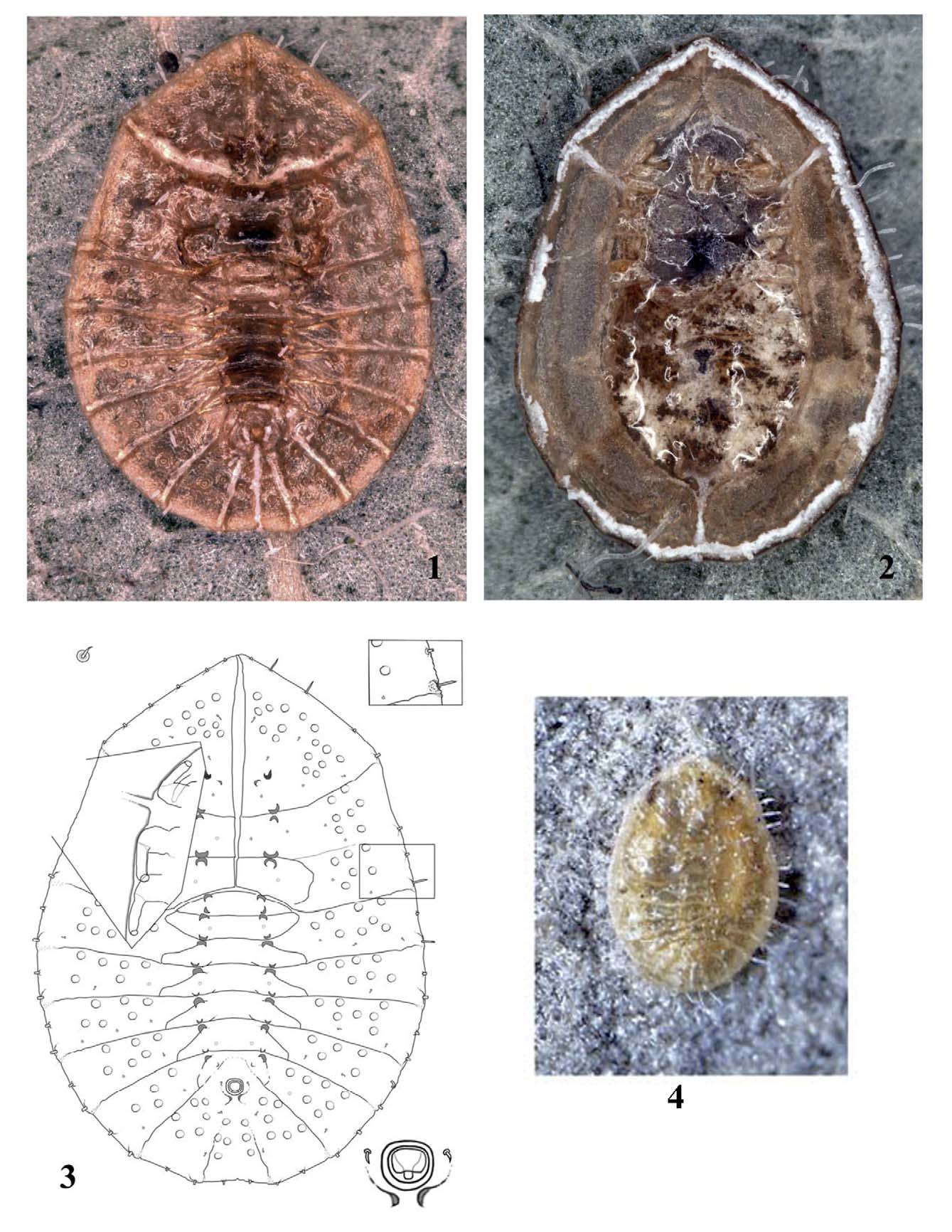
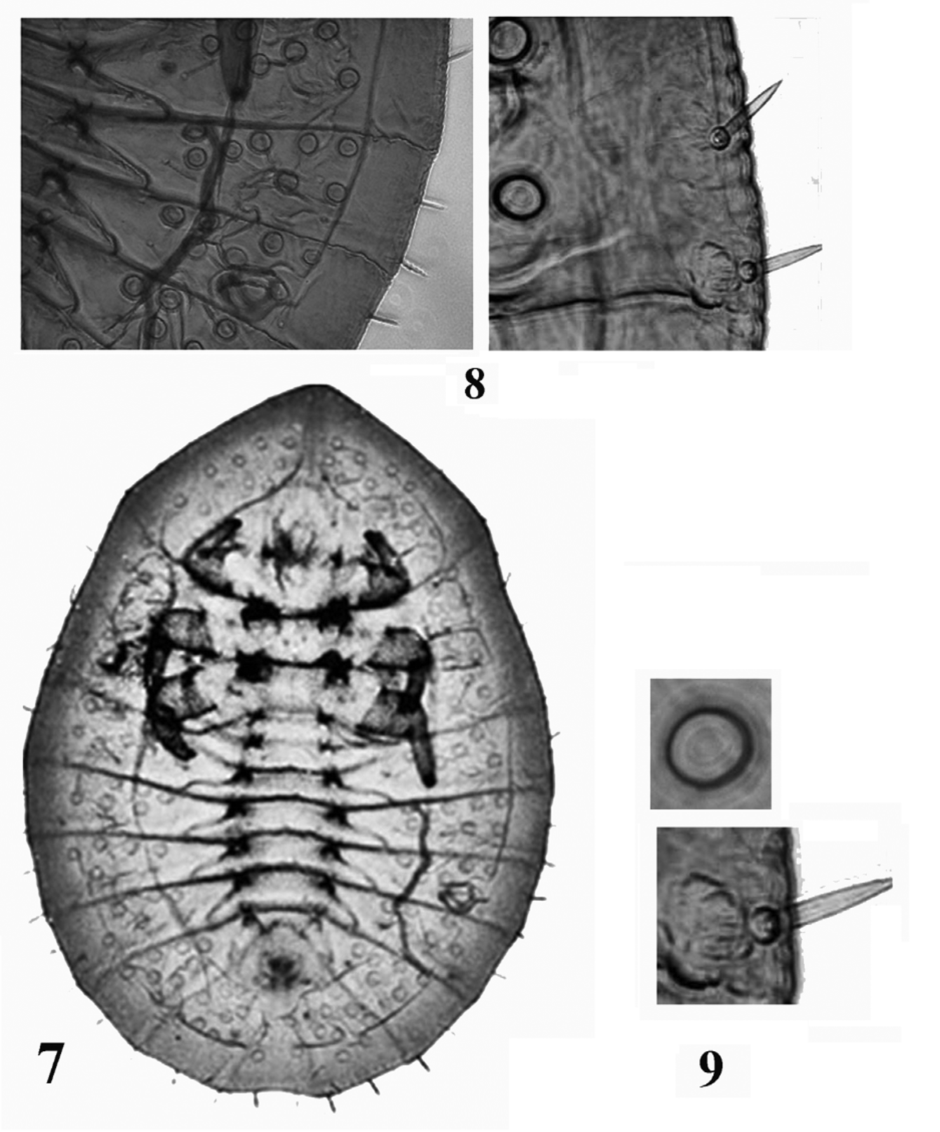
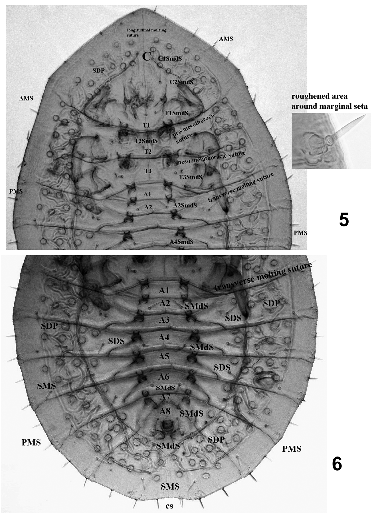
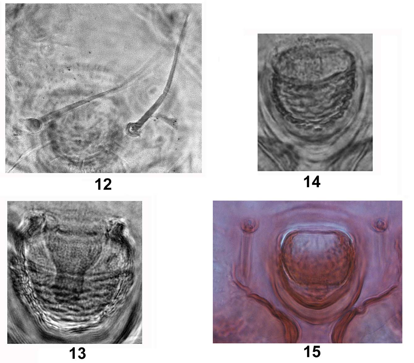
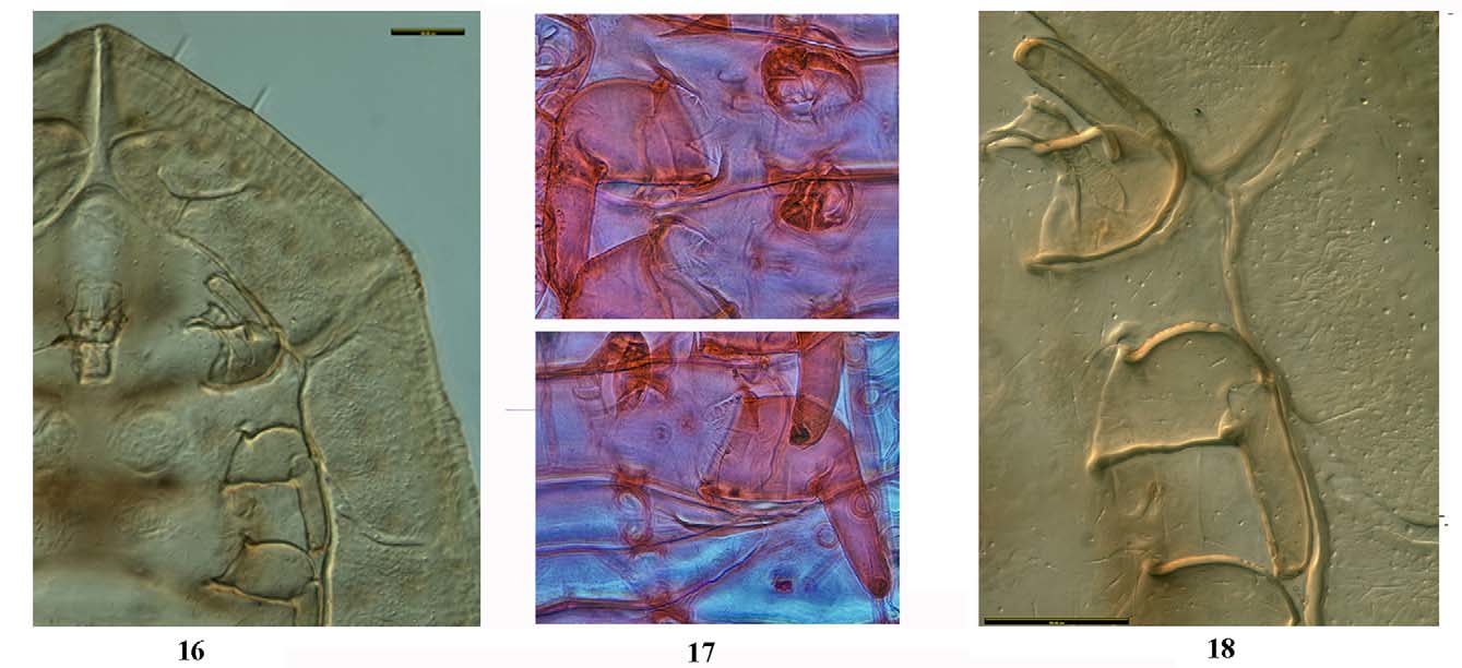
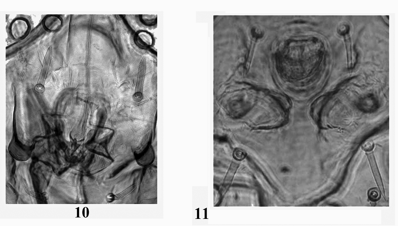
Puparium. Oval shaped measuring 1279 – 1610 [1371] long by 939 – 1219 [1010] wide with the holotype 1588 long by 1194 wide. Cephalic margin straight narrowing to an obtuse angle medially; caudal margin becoming slightly concave between the pair of marginal caudal setae ( Figs 1, 3 View FIGURES 1 – 4 , 7 View FIGURES 7 – 9 )). Margin smooth with slightly roughened and expanded marginal areas at posterior margin of each segment immediately mesad of intersegmental sutures, particularly noticeable in abdominal segments ( Figs 3 View FIGURES 1 – 4 , 9 View FIGURES 7 – 9 ). Dorsum. Longitudinal and transverse sutures on dorsum terminate at margin; pro-mesothoracic suture terminates at margin anteriorly to a position opposite rostrum; mesometathoracic suture terminates at subdorsum. Abdominal rhachis present with lateral arms extending submedially, coalescing with pronounced intersegmental sutures on III–VIII, each rhachis reaching margin ( Figs 1, 3–6 View FIGURES 1 – 4 View FIGURES 5 – 6 ). Submedian tubercles on dorsum present from cephalothorax extending in a series to abdominal segment VIII ( Figs 5, 6 View FIGURES 5 – 6 ). Median width of abdominal segment VII much narrower than preceding segments. Vasiform orifice cordate ( Figs 14, 15 View FIGURES 12 – 15 ), operculum ( Fig. 15 View FIGURES 12 – 15 ) covering most of orifice and situated 5–6 times the width of the orifice from posterior margin. Lingula ( Fig. 13 View FIGURES 12 – 15 ) included within vasiform orifice with head rounded and partially exposed beyond operculum. Chaetotaxy. Cephalothoracic and abdominal setae present on submedian, subdorsal, submarginal and pupal case margin. Two pairs of dorsal submedian cephalic setae and one pair each on T1, T2, and T3 present ( Fig. 5 View FIGURES 5 – 6 ). Meso-metathoracic legs with one ventral seta each on basal segment of leg ( Fig. 17 View FIGURES 16 – 18 ). Abdominal submedian setae on abdominal A1, A3, A5, and A7 absent. Pairs of dorsal submedian abdominal setae present on A2, A4, A6, and A8 (P(n) SmdS where n = 2, 4, 6, 8). Two pairs of P(8)SmdS present with one pair directly anterior and one directly posterior to the vasiform orifice. Also one ventral pair, PVSMdS, of submedian setae ( Fig. 14 View FIGURES 12 – 15 ) present lateral to midpoint of vasiform orifice. Margin with more than 20 pairs of evenly distributed setae; submargin with one dorsal abdominal seta paired between each lateral arm of rhachis from A1 to A8; subdorsum with two abdominal setae between each lateral arm of rhachis from A1 to A7 (one inside and one outside of the ventral fold). ( Figs 3 View FIGURES 1 – 4 , 6 View FIGURES 5 – 6 ). Ve n te r. Legs two segmented, suture between leg segments visible only under high magnification; leg segments angular with their outer lateral angle oriented downward and slightly toward the median, each terminating in adhesive pad ( Figs 3 View FIGURES 1 – 4 , 16, 18 View FIGURES 16 – 18 ); antennae short, overlapping prothoracic leg segment but not extending beyond leg margin ( Figs 16, 18 View FIGURES 16 – 18 ). Distinctive ventral margin-concentric fold present slightly distal to legs and at some distance from margin. In vivo, the external margin and the concentric and radiating folds are waxy ( Fig. 2 View FIGURES 1 – 4 ).
Etymology. The genus is named in honor of the Gadigal tribe, the indigenous inhabitants of the Sydney basin, the area from which the original specimens were collected.
Comments. This genus is erected to contain a single unusual new species. It is remarkable for the well-defined segmental sutures including the rhachis, subdorsal papillae, submedian tubercles, two segmented legs, ventral fold, and the setation on the dorsum, venter, margin of the pupal case, subdorsum, and submedian. The unusual ventral fold appears to enclose and protect the legs. The variation in length, diameter, and structure of the dorsal and ventral setae and papillae into different sizes may be the result of effects of host substrates. This species appears to be quite uncommon, and was not found in a repeated search at both collection sites.
Gadigaleyrodes froggatti sp. n.
Puparium habitus. Thin veneer of clear wax present on dorsum but unapparent in life. Marginal wax not obvious, but whitish wax present ventrally ( Fig. 2 View FIGURES 1 – 4 ); puparium found on upper side of leaf; puparium oval shaped, pale brown with two pigmented areas medially, one from mesothoracic to metathoracic segments, and one on abdominal segments V and VI ( Fig. 1 View FIGURES 1 – 4 ). Slide mounted puparium. Puparium 1279 – 1610 [1371] long by 939 – 1219 [1010] wide with the holotype 1588 long by 1194 wide. Cephalic margin narrows, caudal margin slightly concave ( Figs 1 – 4 View FIGURES 1 – 4 ) between paired caudal setae (CS). Margin apparently smooth except for roughened or corrugated area mesad of junction of lateral margin and segmental suture, at base of associated marginal seta ( Figs 3 View FIGURES 1 – 4 , 9 View FIGURES 7 – 9 ). Under high magnification, margin shallowly and inconsistently crenulate. Tracheal and caudal clefts undifferentiated ventrally and dorsally. Dorsum. ( Figs 1 View FIGURES 1 – 4 , 5 View FIGURES 5 – 6 ), longitudinal molting suture terminates at the anterior margin of the pupal case. The transverse molting suture terminates at corrugated area mesad of or adjacent to last thoracic marginal seta. Promesothoracic suture terminates at corrugated area mesad of marginal seta, arching anterior to opposite the rostrum ( Fig. 5 View FIGURES 5 – 6 ); meso-metathoracic suture terminates just beyond second pair of legs; transverse suture terminates at corrugated area mesad of associated marginal seta, or margin slightly arching upwards ( Figs 3 View FIGURES 1 – 4 , 5 View FIGURES 5 – 6 , 9 View FIGURES 7 – 9 ). Rhachis present with lateral arms coalescing with intersegmental sutures and these reaching margin. Subdorsum punctuated by wide band of rimmed, subcircular flat papillae ( Figs 3 View FIGURES 1 – 4 , 5,6 View FIGURES 5 – 6 , 8 View FIGURES 7 – 9 ) from cephalon to below vasiform orifice. Cephalothoracic papillae numbering from 19–31 [26] on each side with the holotype numbering 24 and 28; abdominal papillae numbering from 26 – 44 [33] on each side of the abdomen with the holotype numbering 41 and 44 papillae. Diameter of papillae, taken from 12 samples each of the subdorsal, cephalothoracic and abdominal papillae, from holotype and 3 paratypes: cephalothoracic papillae range from 26–36 [29] wide and the abdominal papillae from 26 – 40 [32] wide; holotype cephalothoracic papillae range from 26–34 wide and the abdominal papillae measuring 26–40 wide. Submedian tubercles present on cephalothorax and the 8 abdominal segments ( Figs 5 – 6 View FIGURES 5 – 6 ). Cephalon with pair of submedian tubercles posterior to C2SmdS setae; pair of tubercles along promesothoracic suture of T1; T2 through A7 segments with an anterior and posterior pair of submedian tubercles; A8 with pair of tubercles posterior and lateral to the vasiform orifice. Vasiform orifice. cordate 38–50 [43] long by 32–47 with holotype 47 long by 32 wide; with transverse scallops along the lateral margin to base of orifice ( Figs 3 View FIGURES 1 – 4 , 7, 9 View FIGURES 7 – 9 , 10 View FIGURES 10 – 11 ); operculum cordate 27–37 long [31] by 31–41 wide [36] with the holotype 32 long by 37 wide, covering most of orifice. Lingula included with vasiform orifice; Lingula head small, rounded, partially exposed beyond operculum. Chaetotaxy ( Fig. 3 View FIGURES 1 – 4 , 5, 6 View FIGURES 5 – 6 ). Lateral margin of pupal case with evenly distributed, stout lanceolate setae ( Figs 5, 6 View FIGURES 5 – 6 , 9 View FIGURES 7 – 9 ) ranging in numbers 9–13 [10] setae (AMS) on each side from C1 to T3 with the holotype having 21 on both sides; Two pairs of cephalic submedian setae present: C1SmdS 27 – 40 [34] long broken off of holotype and C2SmdS (located anterior to the mouth parts) 25–54 [43] long with the holotype 54 and 50 long. Three pairs of thoracic submedian setae present: T1SmdS 30–46 [36] long with holotype pair 40 and 46 long; T2SmdS is 36–51 [41] long with holotype 51; and T3SmdS 35–46 [41] with the holotype 35 long. Cephalothoracic subdorsal and submarginal setae number 3–10 [6] on each side with holotype numbering 4 setae on one side and 6 on the other side. Posterior (abdominal) marginal setae (PMS) 7–12 [10] setae on each side from A1 to A8 excluding caudal setae (CS) with holotype having 12 pairs of setae. Abdominal submedian setae on abdominal A1, A3, A5, and A7 absent. Pairs of submedian abdominal setae present A2, A4, A6, and A8 (P(n)SmdS where n = 2,4,6,8): P2SmdS 30–51 [42] with holotype 51 long, P4SmdS 36–48 [42] with holotype 47 and 48, P6SmdS 32–45 [39] with holotype 45, P8SmdS(a) 23–39 [34] anterolateral to the vasiform orifice with holotype each 39 long, and P8SmdS(b) 31–47 [38] posterolateral to the vasiform orifice with holotype each 47 long. CS (caudal setae) 27 to 43 [36] with holotype 41 and 42 long ( Fig 6 View FIGURES 5 – 6 ). A pair of ventral submedian setae on A8 (P8VSMdS) lateral to midpoint of vasiform orifice 50 – 126 long ( Fig. 12 View FIGURES 12 – 15 ) [84] with the holotype 75 and 126 long. Venter ( Figs 3 View FIGURES 1 – 4 , 8 View FIGURES 7 – 9 , 12–14 View FIGURES 12 – 15 ). Cuticle smooth except apical submargin which has a narrow band with a finely stippled pattern. A broad sub medial margin-concentric ventral fold is present, more prominent laterally and interrupted below vasiform orifice, at mesothoracic and longitudinal molting suture; ventral fold patterned internally. Legs bi-segmented with apical adhesive pad ( Figs 16 – 18 View FIGURES 16 – 18 ); basal segment of meso- and metathoracic legs each armed with 1 bristle-like seta with length 1/3 the width of basal segment ( Fig. 17 View FIGURES 16 – 18 ). Antenna parallel to and subequal to length of second segment of prothoracic leg overlapping first segment.
Etymology. This species is named in honor of the original collector, William Wallace Froggatt.
Hosts. Syncarpia glomulifera (Myrtaceae) ; also an unknown rainforest climber.
Material examined: Australia, New South Wales: Holotype puparium slide, “Aleurodes sp.”/ “ Syncarpia laurifol ”/ “Bot Gard. Sydney”/ “21.12.[18]99” (apwca091012638002—CASC).
Paratypes: 20 puparia on 14 slides same data as holotype, 4 puparia on 3 slides (ASCU), 2 puparia on 1 slide (BMNH), 7 puparia on 5 slides (CASC), 1 puparium slide (CDFA), 4 puparia on 2 slides (PPQC), 1 puparium slide (USNM); 1 puparium slide, Unknown host (climber), Jerusalem Falls, Barrington Tops National Park, NSW, 32.245618o S 151.724361o E, elev 347m, 23 Jan 2008, P.S. Gillespie (ASCT00167723—ASCU)
Comments. Despite the slightly damaged label, the handwriting is clearly that of Froggatt (P. Gillespie pers. obs.) and the “Bot Gard. Sydney” he refers to is clearly (the now) Royal Botanic Gardens, in Sydney central district on the shore between the current Sydney harbor bridge and Opera house. Many other specimens similarly labeled are in the collections of ASCU and Froggatt’s interest in these gardens (Froggatt, 1932) showed he liked to collect there. The name of the host, “ Syncarpia laurifol ”, is presumably a misspelling of Syncarpia laurifolia , a junior synonym of Syncarpia glomulifera .
No known copyright restrictions apply. See Agosti, D., Egloff, W., 2009. Taxonomic information exchange and copyright: the Plazi approach. BMC Research Notes 2009, 2:53 for further explanation.