
Fusicolla gigas C. Liu, Z.Q. Zeng & W.Y. Zhuang, 2022
|
publication ID |
https://doi.org/ 10.11646/phytotaxa.536.2.5 |
|
DOI |
https://doi.org/10.5281/zenodo.6323875 |
|
persistent identifier |
https://treatment.plazi.org/id/03A887EA-FFBF-0A75-FF41-F9E957E4F84A |
|
treatment provided by |
Plazi |
|
scientific name |
Fusicolla gigas C. Liu, Z.Q. Zeng & W.Y. Zhuang |
| status |
sp. nov. |
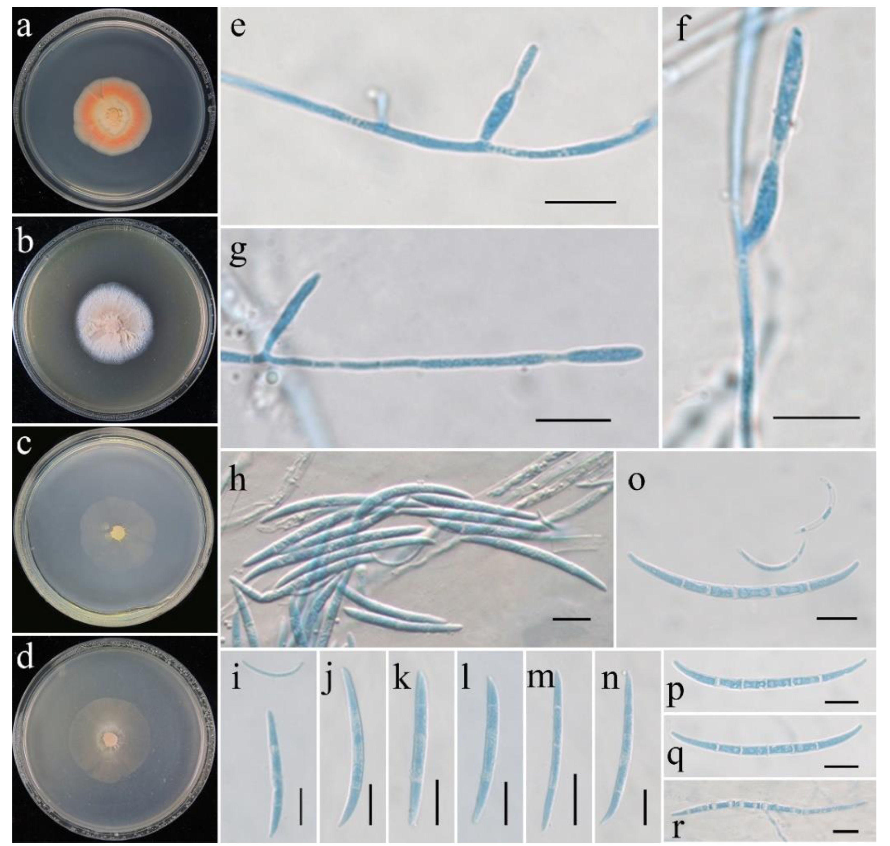
Fusicolla gigas C. Liu, Z.Q. Zeng & W.Y. Zhuang View in CoL , sp. nov. FIGURE 2 View FIGURE 2
Fungal Names: FN570867
Etymology:—Referring to the large-sized macroconidia produced by the fungus.
Typification:— China, Chongqing City, Wushan County, Hongchiba National Forest Park , 31°54′52′′ N, 109°08′56′′ E, in soil, 30 October 2020, Z.Q. Zeng, X.C. Wang, H.D. Zheng & C. Liu, CS25-8 ( CGMCC 3.20680 View Materials ) GoogleMaps .
Description: —Colonies on PDA reaching 36−39 mm diam. after 2 wk at 25 °C, slimy appearance due to abundant sporulation on medium surface, orange to pale yellow in center, pinkish orange at margin. Colonies on MEA reaching 35−39 mm diam. after 2 wk at 25 °C, surface floccose, with white aerial mycelium, light cinnamon in center. Colonies on SNA reaching 36−38 mm diam. after 2 wk at 25 °C, aerial mycelium absent, sporulation on center medium surface, light yellow to white. Colonies on OA reaching 39−40 mm diam. after 2 wk at 25 °C, aerial mycelium absent with slimy appearance due to abundant sporulation on medium surface, light yellow.
Asexual stage fusarium-like. Conidiophores arising directly from somatic hyphae, simple or rarely branched, monochasial, straight, hyaline, smooth-walled, aseptate or with basal septum, up to 42 µm long. Conidiogenous cells monophialidic or rarely polyphialidic, arising laterally from hyphae or in terminal pairs, cylindrical to subulate, 8−14 × 1.5−3 µm, thin- and smooth-walled. On PDA, macroconidia falcate, long fusiform, slightly narrowing towards the ends, acute, the apical cell often hooked with a more or less pointed tip, basal cell slightly pedicellate, hyaline, thin-and smooth-walled, (1−)3(−4)-septate, (25−)36−42(−53) × 2.5−3.5 µm (av. 39.5 × 2.8 µm) (n = 30), microconidia slightly curved to C-shaped, hyaline, smooth, aseptate, 11–18(−22) × 0.85–1.3 µm (av. 15.7 × 1.1 µm) (n = 20). On OA, macroconidia falcate, generally crescent to threadlike, (3–)4–9-septate, (32−)40–65(−80) × 2.3–3.8 µm (av. 53.2 × 2.9 µm) (n = 30), microconidia strongly curved, hyaline, smooth, 0−4(−5)-septate, (12−)15–24(−32) × 0.85–1.4(−2.2) µm (av. 19.3 × 1.2 µm) (n = 30).
Sexual stage not observed.
No known copyright restrictions apply. See Agosti, D., Egloff, W., 2009. Taxonomic information exchange and copyright: the Plazi approach. BMC Research Notes 2009, 2:53 for further explanation.
|
Kingdom |
|
|
Phylum |
|
|
Class |
|
|
Order |
|
|
Family |
|
|
Genus |