
Fridericia marginata, Schmelz, Rüdiger M. & Collado, Rut, 2013
|
publication ID |
https://doi.org/ 10.11646/zootaxa.3647.2.4 |
|
publication LSID |
lsid:zoobank.org:pub:33866E2B-6B0F-4124-A6A6-2B057E642149 |
|
DOI |
https://doi.org/10.5281/zenodo.5612044 |
|
persistent identifier |
https://treatment.plazi.org/id/301187BC-2C09-FFFF-88B0-FC2A485DFA5A |
|
treatment provided by |
Plazi |
|
scientific name |
Fridericia marginata |
| status |
sp. nov. |
Fridericia marginata View in CoL sp. nov.
( Figs 3 View FIGURE 3 A–D, 5D, Table 3 View TABLE 3 )
Holotype. MNHML MB29-000312, adult spcm, stained whole mount. Portugal, Coimbra, in soil from the experimental field area of the Coimbra Higher School of Agriculture (ESAC), meadow site ( Table 2 View TABLE 2 ); II 2012.
Paratypes. 15 spms. MNHML MB29-000313 –315, stained whole mounts, 2 adults, 1 subadult. ZMH OL 14523: 4 juvenile spms, stained whole mounts. ZMH OL 14524, fixed in Bouin's fluid, preserved in 70% ethanol: 8 spms. ZMH OL 14525, fixed in 70% ethanol, preserved in 100% ethanol: 5 spms.
Other material. 37 spms investigated in vivo, preserved in collective sample vials, in the authors' collection.
Etymology. Named for the ventro-lateral margins of the clitellum, consisting only of granulocytes.
Diagnosis. Less than 40 segments, max. 4 chaetae per bundle, clitellum saddle-shaped with ventral margins consisting of granulocytes only, coelomo-mucocytes with refractile vesicles, nephridia present at 10/11, chylus cells post-clitellar, no seminal vesicle, sperm heads 90 μm, sperm funnel small, spermathecae joint entally, without ectal gland, two stalked diverticula with ciliated subchamber, ampulla and diverticula elongate.
Description. Body colour opaque grey-white, often with a yellowish tinge (viv), slow body movements. Length c. 7–8 mm (viv), diameter c. 0.3 mm (viv). Segments 35–39. Chaetae max. 4 per bundle, formula 3,4 – 4,3,2: (3),4 – 4,3,2. Ventrally 2 per bundle from XXV to posterior end. Inner chaetae shorter than outer. Chaetae in caudal segments largest, 65–70: 5.5 μm, size of largest anterior chaetae c. 55: 4.5 μm. Epidermal gland cells scarce, pale, one row at chaetal level, cells quadrangular or without clear outline. Body wall c. 20 μm thick with longitudinal muscle layer thickest (c. 15 μm), cuticle thin (<1 μm). Preclitellar septa 4/5–9/10 thickened, thinner at 10/11 ff.
Brain rounded posteriorly, lateral sides parallel. Pharyngeal glands all united dorsally, dorsal lobes slightly increasing in size from IV to VI, ventral lobes strongly increasing in size from IV to VI. Oesophageal appendages elongate with 2–3 short or elongate branches in distal third. Chylus cells in XIII–XIV, 1.5–2 segment lengths, canals not widened into lacunae. Dorsal blood vessel from XVI–XVII. Pars tumida of midgut in XXVI–XXIX, extending over 2–4 segments, not distinguished in all specimens. Preclitellar nephridia 5 pairs, 6/7–10/11, length ratio anteseptale: postseptale 2: 3, medial to subterminal rise of efferent duct, terminal vesicle absent. Postclitellar nephridia with subterminal to terminal rise of efferent duct and elongate postseptale. Coelomo-mucocytes broadly oval, length 25–35 μm, with refractile vesicles at periphery in one incomplete row, vesicle diameter c. 1 μm, matrix transparent; cells very numerous, filling all spaces, making body opaque with grey-yellow tinge (viv); lenticytes minute, length c. 6–8 μm, sparse.
Clitellum saddle-shaped, prominent, cells in c. 35 dense rows with granulocytes and hyalocytes alternating; mid-ventral aclitellar field as wide as distance between bursal slits; hyalocytes absent at ventro-lateral borders and in a semi-circular field laterally of and around bursal slits; here only granulocytes in indefinite rows or in reticulate arrangement. (Single hyalocytes near ventral border between male and female pores observed in one specimen). Seminal vesicle absent; occasionally dense masses of developing sperm in anterior half of XI, and septum 10/11 bulged forward dorsally, unpaired. Spermatozoa> 200 μm long, heads c. 90 μm, not numerous on top of sperm funnel ( Fig. 3 View FIGURE 3 C). Sperm funnel comparatively small, hardly visible among coelomocytes, developing sperm, and oogonia (viv), about half as long as body diameter, tapering proximally, c. 2.5x as long as wide (e.g. 140: 60 μm), collar not wider than funnel body, not folded outwards. Vas deferens in dense irregular coils ventro-laterally, 8 μm wide. Male copulatory organ elevating body suface even when completely retracted (viv), transverse copulatory muscles strongly developed. Male glands broadly oval in ventral view, flat in side view, 1.5x as long as wide (e.g. 120 μm long, 80 μm wide, 50 μm high) bursal slits longitudinal, bursa thick-walled, with small and flat modiolus (comp. Schmelz 2003: 49) around ectal opening of vas deferens (the primary male pore); bursa plus male gland almost spherical. Subneural glands and other accessory glands absent. Spermathecae: no ectal gland; ectal duct about as long as body diameter, slightly widened in distal part, tapering again near ectal pore, ental bulb not projecting into ampullar lumen, only slightly wider than ectal duct, ectal duct canal straight throughout. Ampullae 1.5– 2 x as long as wide, tapering entad, ental tips fused, creating a common V-shaped lumen. Ampullar walls 4–6 μm thick, uniform, no histological differences observed, with smooth outer and inner surface, inner surface occasionally wavy, as a transitory state during contraction. Diverticula inserting on opposite sides of ental bulb at base of ampulla and projecting obliquely ectad, distance (= maximum width of spermatheca) c. 80 μm, c. 1/3 body diameter. Each diverticulum 2x as long as wide, diameter c. 20 μm, lumen subdivided into peripheral spermcontaining chamber and ciliated sub-chamber; both chambers connected by a porus; peripheral chamber wider than long, with dense sperm roll circulating; sub-chamber longer than wide, with ciliar movement, widely connected with ampullar lumen. No ciliar movement in ampulla. One mature oocyte at a time, extending over c. 2 segment lengths.
Remarks. The new species owes its name to the saddle-shaped clitellum with ventro-lateral margins consisting only of granulocytes, a character new in Fridericia , but common in some other genera, e.g. Guaranidrilus , Hemienchytraeus , Achaeta , Marionina . In the other Fridericia species with saddle-shaped clitellum the ventro-lateral borders do to our knowledge not differ from the rest and are composed of granulocytes and hyalocytes. However, not all species of the genus have been examined for that character. A further peculiarity is the slenderness of ampulla and diverticula, both about twice as long as wide.
Using the tabular comparison of Fridericia species with two spermathecal diverticula in Dózsa-Farkas (2009), F. marginata sp. nov. belongs to a group of 10 species characterized by proximally fused spermathecae. None of these species has the character combination of max. 4 chaetae per bundle, saddle-shaped clitellum, and stalked spermathecal diverticula. Using the key to Friderica species in Schmelz (2003), F. marginata would key out together with F. gamotheca Issel, 1905 . This species has also coelomocytes with few refractile vesicles, as F. marginata (Rota 1995, Schmelz pers. obs.). It differs from F. marginata in larger body size and higher segment number (37–50 according to Rota 1995), in a girdle-shaped clitellum according to Schmelz and Collado (2010), and in a complete fusion of the two spermathecal ampullae. A variant of F. gamotheca described from Morocco (Dózsa-Farkas 1989) and of uncertain taxonomic status (Schmelz 2003) is more in the size range of F. m a rg i na ta (length 5–7 mm, 28–33 segments), but differs in a girdle-shaped clitellum, coelomocytes without refractile vesicles and in the presence of a spermathecal ectal gland, among other characters. F. gamotheca is a problematic species, see F. roembkei sp. nov., remarks.
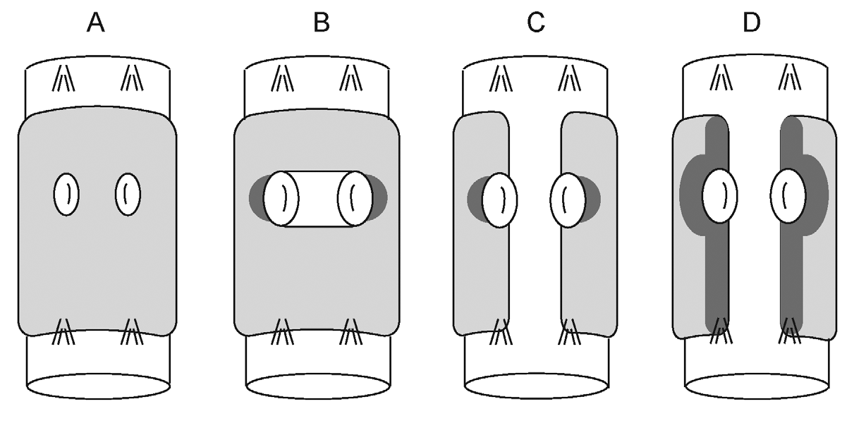
F. m a rg i n a t a and F. roembkei spp. nov. are quite similar at first sight, but they are easily distinguished by the coelomocytes—with refractile vesicles in F. m a rg i n a t a and without them in F. roembkei —and by the ventral pattern of the clitellum, see Figure 5 View FIGURE 5 . Further differences are more difficult to observe and may not be evident unless specimens of both taxa are available for comparison (in the following, character states of F. marginata first): spermathecal diverticula and ampulla elongate/not elongate; spermathecal ectal duct not longer/longer than body diameter; yellow epidermal gland cells absent/often present; pharyngeal glands in VI united/separate dorsally ( Table 2 View TABLE 2 ).

No known copyright restrictions apply. See Agosti, D., Egloff, W., 2009. Taxonomic information exchange and copyright: the Plazi approach. BMC Research Notes 2009, 2:53 for further explanation.