
Felisacus dauloi Woodward, 1958
|
publication ID |
https://doi.org/ 10.1206/0003-0090-403.1.1 |
|
persistent identifier |
https://treatment.plazi.org/id/296A879F-5655-751A-5D75-FB27FED60E38 |
|
treatment provided by |
Carolina |
|
scientific name |
Felisacus dauloi Woodward |
| status |
|
Felisacus dauloi Woodward View in CoL
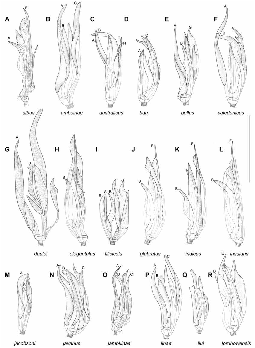
Figures 4 View FIGURE 4 , 8G View FIGURE 8 , 11X, Y View FIGURE 11 , 14K View FIGURE 14 , 18 View FIGURE 18 Felisacus dauloi Woodward, 1958: 236 (original description).
DIAGNOSIS: Recognized by the following combination of characters: body mostly whitish yellow or yellow without brown markings, with reddish tinge and yellow or red cuneus (fig. 4); antennal segment I cylindrical (as in Namyatova et al., 2016: fig. 8A), transverse depression on head extending present dorsally and laterally, vertex upraised (as in Namyatova et al., 2016: fig. 6D); relatively large body 3.8–4 in male and 4.3– 4.5 in female; cuneus ca. 3× as long as base; medial part of right paramere only slightly wider than basal part, shorter than basal and apical parts combined; outer margin of right paramere almost straight (fig. 11X); apical part of left paramere not widened, ca. 3× as long as wide (fig. 11Y); six spicules vesical spicules, including spicules A and B (fig. 8G).
REDESCRIPTION: Male. Total length 3.8–4.0. COLORATION (fig. 4): Head: Mostly yellow with reddish tinge and marking; longitudinal sulcus sometimes yellow brown, tubercle around antennal fossae, maxillary plate and buccula sometimes whitish yellow; clypeus yellow with longitudinal red stripe; mandibular plate red or yellow with reddish marking. Eye brown to dark brown, often with reddish margins. Labium: Yellow, segment III with pale brown or red stripe ventrally. Antenna: Segment I yellow to reddish brown, darkened apically; segment II reddish brown. Thorax: Pronotum yellow with pale brown anterior margin, often with longitudinal red stripe laterally; mesoscutum yellow; scutellum yellow, sometimes with red longitudinal stripe; thoracic pleura yellow with reddish areas, sometimes mostly red; scent gland evaporative area whitish often with reddish tinge apically. Hemelytron: Mostly colorless and translucent; inner part of clavus sometimes opaque, whitish yellow to yellow, with margins yellow to pale brown or reddish; corium with area along inner margin of corium pale brown or red, narrow and short, not reaching R+M; embolium sometimes yellow, with yellow to pale brown or red apex and margins; cuneus with margins and often apex yellow or red, sometimes mostly yellow; membrane with grayish tinge, with membrane cell yellow to pale brown or red. Legs: Coxae whitish yellow; femora whitish yellow basally and yellow to red apically; tibiae whitish yellow to yellow, yellow or reddish basally; tarsi pale brown, sometimes tarsal segment I whitish yellow. Abdomen: Ventral and lateral sides of pregenital segments whitish yellow, dorsal surface of pregenital segments and entire genital capsule red. SURFACE AND VESTITURE: Corium smooth, with shallow and scarce punctures. Dorsum, antennal segment I and femora clothed with setae mostly as long as or somewhat shorter than antennal segment II diameter; abdomen clothed with suberect mostly short simple setae. STRUCTURE AND MEASURE- MENTS: Body ca. 4.4–4.7× as long as pronotum width. Head: Depression, delimiting occipital region, present dorsally and laterally (as in Namyatova et al., 2016: fig. 4E); distance between depression and pronotum distinctly shorter than eye diameter; longitudinal sulcus on dorsal surface of head longer than eye diameter; distance from eye to pronotum as long as eye diameter, not swollen laterally (as in Namyatova et al., 2016: fig. 4E); vertex ca. 1.4–1.7× as wide as eye; vertex raised (as in Namyatova et al., 2016: fig. 6D); buccula ca. 0.2–0.25× as long as clypeus. Labium: Almost reaching posterior part of mesosternum; segments I and II strongly reduced, combined shorter than half of segment III; segment I shorter than wide (as in Namyatova et al., 2016: figs. 6E, 9C); segment II slightly longer than wide, its dorsal surface elongate posteriorly; segment III ca. as long as ventral side of head length; segment IV ca. 1.5× as long as segment III. Antenna: Segment I cylindrical (as in Namyatova et al., 2016: fig. 8A), ca. 1.7– 1.8× as long as head width, ca. 1.1× as long as pronotum width; segment II ca. 2.2× as long as width of head, ca. 1.4× as long as width of pronotum. Thorax: Anterior part of pronotum slightly shorter than posterior part; collar delimited; posterior part of pronotum slightly upraised; posterior margin of pronotum concave; pronotum ca. 1.2–1.3× as wide as long and ca. 1.4–1.7× as wide as head; mesoscutum slightly exposed. Hemelytron: Area along inner margin of corium flat; inner margin of cuneus convex (as in Namyatova et al., 2016: fig. 13E), medial margin of cuneus ca. 3× as long as base. Genitalia: Genital capsule (fig. 14K) slightly longer than wide; ventral wall twice as long as dorsal wall, with posterior margin of ventral wall semioval, smooth, without outgrowth(s), its apex inclined slightly leftward, not curved; sides of genital capsule not modified; right margin of paramere socket angulate, left margin rounded; distance between paramere sockets ca. 0.3× as long as base of genital capsule. Right paramere (fig. 11X) distinctly curved in apical half; apex straight dorsally; medial part only slighter wider than basal part, bearing setae, with outer margin straight and inner margin only slightly convex; outer angle distinct; inner angle rounded, without setae; basal part ca. 0.15–0.2× as long as rest of paramere. Left paramere (fig. 11Y) L-shaped; apical part not flattened, with toothlike outgrowth on posterior side medially (as in fig. 11G) and without outgrowth on dorsal surface; middle part widened, without swelling or outgrowth; setae only on middle part near outer margin. Aedeagus conjunctiva weakly sclerotized, secondary gonopore placed at base of vesica in repose; sclerotized part of ductus seminis around secondary gonopore shorter than wide; vesica with six spicules, including spicules A and B (fig. 8G).
Female. Total length 4.3–4.5. COLORATION (fig. 4): Head: Similar to male, sometimes pale brown posteriorly; mandibular plate yellow, yellow with reddish tinge or red. Labium: As in male. Antenna: Similar to male, segment I uniformly yellow or pale brown, yellow at base, segments III–IV brown to dark brown. Thorax: Similar to male, but mesoscutum and scutellum yellow, thoracic pleura sometimes with pale brown markings. Hemelytron: As in male. Legs: Similar to male, tarsi pale brown, sometimes segment I and II whitish to yellow. Abdomen: Ventral side and partly lateral side of pregenital segments and segment VIII whitish yellow, upper part of lateral side and dorsal surface of segments II–VIII and entire segment IX red. SURFACE AND VESTITURE: As in male. STRUCTURE AND MEASUREMENTS: Structure as in male; body ca. 4.2–4.6× as long as pronotum width; vertex ca. 1.6–1.8× as wide as eye diameter; antennal segment I ca. 1.5–1.8× as long as head width, ca. 0.9–1.1× as long as pronotum width; segment II ca. 1.9–2.2× as long as head width, ca. 1.2–1.4× as long as pronotum width; antennal segment III slightly longer than segment II; segment IV ca. 0.3× as long as segment III; pronotum ca. 1.2–1.3× as wide as long and ca. 1.6–1.8× as wide as head. Genitalia (as in Namyatova et al., 2016: fig. 23F, G): Dorsal labiate plate wider than distance between apodemes of second valvula; mostly smooth, without distinct striations, with semicircular sclerite and distinct sclerotized rings laterally; lateral oviducts placed almost medially, very close to each other, spermathecal gland placed between lateral oviducts; dorsal labiate plate with distinct tubercles, without membranous lobe medially.
DISTRIBUTION: Papua New Guinea (fig. 18).
HOST PLANT: Ferns in wet tropics ( Woodward, 1958).
REMARKS: Antennal segments III–IV in males were lost, in females only parts of antennal segment IV remain.
DISCUSSION: Felisacus dauloi was described by Woodward (1958) from a single female. One of us (A.A.N.) examined the holotype, preserved in the Queensland Museum. We have some other nontype specimens from BPBM, collected also in Papua New Guinea, and we found that they are very similar to the type of F. dauloi and treat them as belonging to that species.
Felisacus dauloi is similar to F. filicicola , F. ochraceus , and F. tanna in coloration and external morphology (figs. 4–7). Felisacus filicicola differs from F. dauloi in the mostly yellow or reddish-yellow cuneus and the presence of seven vesical spicules, including spicules A, B, E, and G (fig. 8I). Felisacus ochraceus differs in the cuneus uniformly yellow or with yellow or red inner part, the medial part of the right paramere is as wide as the basal part, and shorter than the basal and apical parts combined, the outer margin of the right paramere is distinctly concave (fig. 13C), and there are five vesical spicules, including spicules A, B, C, and H (fig. 9F). Felisacus tanna differs from F. dauloi in the apex of the cuneus colorless often with a yellow tinge and is never red, the middle part of the right paramere is broad, twice as wide as the basal part (fig. 13T), and the vesica has five spicules, including spicules A, B, and E (fig. 9M).
MATERIAL EXAMINED: Holotype: PAPUA NEW GUINEA: Eastern Highlands: Daulo Pass, 20 Aug 1956 – 22 Aug 1956, T.E. Woodward, 1♀ (00201839) ( QM). Additional material: PAPUA NEW GUINEA: Morobe Province: Kaisenik Rd, Wau Subdistrict, 7.38012 ° S 146.77817 ° E, 1100 m, 02 Mar 1978, W.C. Gagne, Pteris moluccanus (Pteridaceae) , 4♀ (00042366, 00042367, 00042277, 00042278) Pteris moluccana (Pteridaceae) , 33 (00042364, 00042365, 00043882), 1♀ (00043881) ( BPBM). Northern Province: 9–10 km WSW of Tufi, SE Cape Nelson, 9.1 ° S 149.3 ° E, 585 m, 08 Sep 1982 – 13 Sep 1982, G.A. Samuelson, 13 (00043880) ( BPBM).


| QM |
Queensland Museum |
| BPBM |
Bishop Museum |
No known copyright restrictions apply. See Agosti, D., Egloff, W., 2009. Taxonomic information exchange and copyright: the Plazi approach. BMC Research Notes 2009, 2:53 for further explanation.