
Feia nympha Smith, 1959
|
publication ID |
https://doi.org/ 10.11646/zootaxa.4097.3.3 |
|
publication LSID |
lsid:zoobank.org:pub:1F51C4F3-58E4-469A-B42F-FF4029660464 |
|
DOI |
https://doi.org/10.5281/zenodo.5619258 |
|
persistent identifier |
https://treatment.plazi.org/id/A46E7679-FFA6-8668-4DA4-1AD9FB8A5D9B |
|
treatment provided by |
Plazi |
|
scientific name |
Feia nympha Smith, 1959 |
| status |
|
Feia nympha Smith, 1959 View in CoL
( Figures 3 View FIGURE 3 & 4 View FIGURE 4 )
Feia nympha Smith, 1959: 206 View in CoL (Pinda, Mozambique).— Bogorodsky et al. (2010): 118 ( Sudan, based on juvenile).
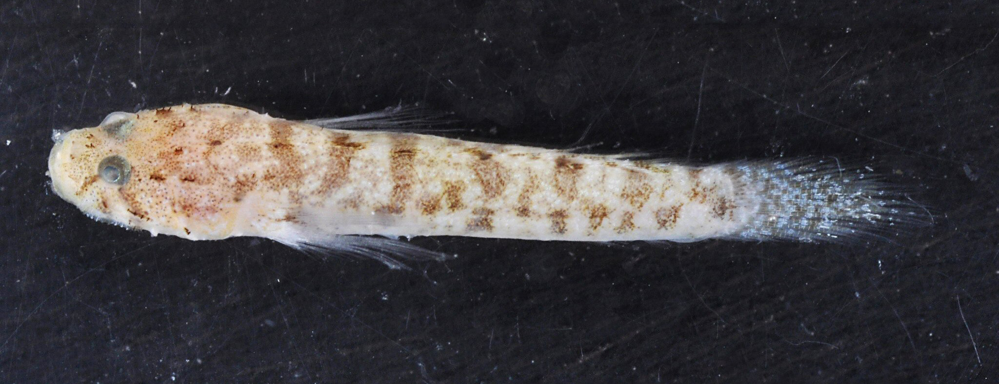
Material examined. SMF 35754 (field number KAU13-12), female, 15.4+ 4.9 mm, 14 June 2013, Red Sea, Saudi Arabia, 30 km south of Al Wajh, 26°03'30.36" N, 36°38'34.98" E, base of coral block, 3−4 m, coll. S.V. Bogorodsky.
Diagnosis. Pectoral-fin rays 14−15. Pelvic frenum weak (frenum height in midline 1/4 of pelvic-spine length). Scales small, cycloid, rarely extending anterior to below origin of second dorsal fin. Longitudinal scale series 7−25. Cheek and preopercle with low raised ridges. Gill opening restricted to just anterior to lower pectoral-fin base. Head and body with brown to light brown mottling and narrow irregular bars.
Brief description (based on the specimen SMF 35754, Fig. 3 View FIGURE 3 ). Head slightly depressed, profile rounded; eyes dorsolateral; anterior nostril a long tube without process from rim, reaching upper lip, posterior nostril difficult to detect; cheek and preopercle with low raised ridges; gill opening restricted to just anterior to lower pectoral-fin base.
Fins. Dorsal-fin rays VI + I,9; anal-fin rays I,9; pectoral-fin rays 14; caudal fin with 17 segmented rays, 13 branched; pelvic fins I,5. Pelvic fins fused, forming complete disc; pelvic frenum weak, height in midline 1/4 of pelvic-spine length.
Squamation. Scales small, all cycloid, present only posteriorly on body, reduced to patch on caudal peduncle, extending anterior to below second dorsal fin (a few scales along lateral midline on right side), rest of body naked. Longitudinal scale series 7 on left side, 10 on right.
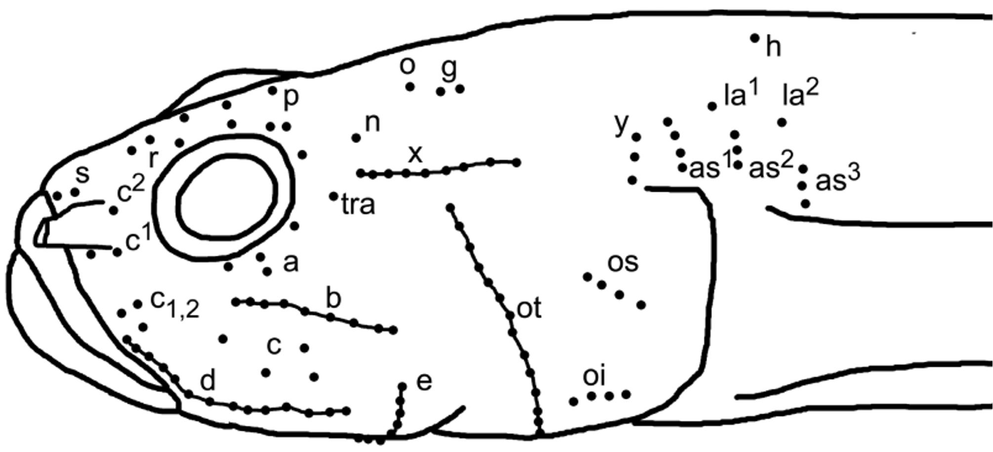
Cephalic sensory systems ( Fig. 4 View FIGURE 4 ). No head canals present. Rows and number of sensory papillae in rows reduced. In some rows papillae connected by membrane forming ridge. Rows of head sensory papillae were counted on the left side: (1) preorbital: internal upper row r as two papillae, external row s with just two papillae anteriorly close to upper lip. Lateral series c in three: superior (c2) as one papilla above anterior nostril; middle c1 two papillae below anterior nostril; inferior row c1,2 as three papillae above upper lip; (2) suborbital: row a as four papillae below posterior part of eye; row b (9) present on posterior cheek and preopercle bellow row a as longitudinal row with elongate papillae connected in ridge by membrane; row c as four scattered papillae between rows b and d, row d (14) as longitudinal row with elongate papillae connected in ridge by membrane above posterior upper lip and along lower cheek; (3) preoperculo-mandibular: external row e not divided (26) and with posterior part well in front of rear edge of preoperculum, internal row i not clearly divided (14), row f (11) two short rows converging posteriorly to form V-shaped pattern; (4) oculoscapular: anterior transverse series tra as single papilla behind eye, longitudinal row x (9) with elongate papillae connected in ridge by membrane, transversal row y (3) behind it. Axillary rows as1 (4), as2 (3), as3 (3), la1 (1) and la2 (1) present; (5) opercular: transverse row ot (17) with elongate papillae connected in ridge by membrane; superior longitudinal row os (4); inferior longitudinal row oi (4); (6) anterior dorsal: row n a single papilla behind upper edge of eye, row o a single papilla and row g two papillae in the central predorsal area and row h a single papilla in posterior predorsal area; (7) interorbital: interorbital area with longitudinal rows p (5).
Coloration of freshly collected material ( Fig. 3 View FIGURE 3 ). Head and body tan with brown to light brown mottling and narrow bars. Dorsal and lateral surface of head with numerous small melanophores, some sensory papillae rows more intensively pigmented, predorsal mostly mottled. Body with seven narrow brown saddles across back, first in front of first dorsal fin, last on top of caudal peduncle; saddles more or less joining seven brown bars on upper part of body including bar at base of caudal fin, bars interrupted into spots along mid-side of body. Head with short oblique brownish bar extending anteroventrally from eye to lips, another oblique brownish bar extending ventroposteriorly from eye to posterior part of cheek. Each dorsal fin with two proximal brown spots extending from the dorsal saddles, rest of fins mostly translucent. Pelvic and anal fins translucent. Caudal fin with brown pigment forming irregular vertical bars. Pectoral fins translucent with brown spot basally on upper rays.
Distribution and habitat. Feia nympha is a widespread species known from the Red Sea, east coast of Africa and islands of the western Indian Ocean to French Polynesia, in the western Pacific north to Japan, south to Queensland, Australia. But due to cryptic habitat it is rare in collections. The adult female was collected from an isolated reef of a large bank in Al Wajh area, Saudi Arabia. It was found at base of reef crest at depth of 3-4 m, on shallow slope to 6 m, near deeper sand and coral patches.
Remarks. At present the genus Feia includes four species: F. no t a Gill & Mooi, 1999 known from the Western Australia, F. nympha , F. r a nt a Winterbottom, 2003 known from Vietnam only, and F. dabra Winterbottom, 2005 known from Palau only. The present material from the Red Sea, Saudi Arabia, matches the data in the brief description of F. nympha ( Bogorodsky et al., 2010) based on a juvenile (8.5 mm SL) from Sudan. It also almost completely matches the species redescription given by Lachner & McKinney (1979) and the set of characters provided by Winterbottom (2005, Table I) for the four species. Considering the limited data taken from the small Sudan specimen, a detailed study of adult Red Sea material and comparison with the Pacific and Indian Ocean material of F. n y m p ha was needed ( Lachner & McKinney, 1979). The only differences were found in squamation, being reduced to a patch on the caudal peduncle in the present specimen, characterized in having a longitudinal scale count of just 7 on the left side and 10 on the right versus 15 scales in juvenile from Sudan ( Bogorodsky et al., 2010), 12−16 in specimens from Japan ( Ikeda et al., 2000), and 14-25 in the data given by Winterbottom (2003, 2005), as well as some cephalic sensory system characteristics. It is hard to follow the cephalic sensory system description in Lachner & McKinney (1979) since they used their own terminology. However, the illustration (Fig. 11 in Lachner & McKinney 1979) shows the same sensory papillae rows as in present specimen except for the rows h, la1 and la2 that are apparently missing. These rows are all in the posterior predorsal area; maybe they were overlooked by the authors or the rows were not visible due to local skin surface damage.
| SMF |
Forschungsinstitut und Natur-Museum Senckenberg |
No known copyright restrictions apply. See Agosti, D., Egloff, W., 2009. Taxonomic information exchange and copyright: the Plazi approach. BMC Research Notes 2009, 2:53 for further explanation.
|
Kingdom |
|
|
Phylum |
|
|
Class |
|
|
Order |
|
|
Family |
|
|
Genus |
Feia nympha Smith, 1959
| Kovačić, Marcelo, Bogorodsky, Sergey V. & Mal, Ahmad O. 2016 |
Feia nympha
| Smith 1959: 206 |
| Bogorodsky et al. (2010) : 118 |