
Exechonella kleemanni, Cáceres-Chamizo & Sanner & Tilbrook & Ostrovsky, 2017
|
publication ID |
https://doi.org/ 10.11646/zootaxa.4305.1.1 |
|
publication LSID |
lsid:zoobank.org:pub:1192C3A0-5CCB-4A86-903C-A2B82906A5F9 |
|
DOI |
https://doi.org/10.5281/zenodo.6017354 |
|
persistent identifier |
https://treatment.plazi.org/id/CF0AB852-FFC5-E916-FF03-FA3F91EEE52C |
|
treatment provided by |
Plazi |
|
scientific name |
Exechonella kleemanni |
| status |
sp. nov. |
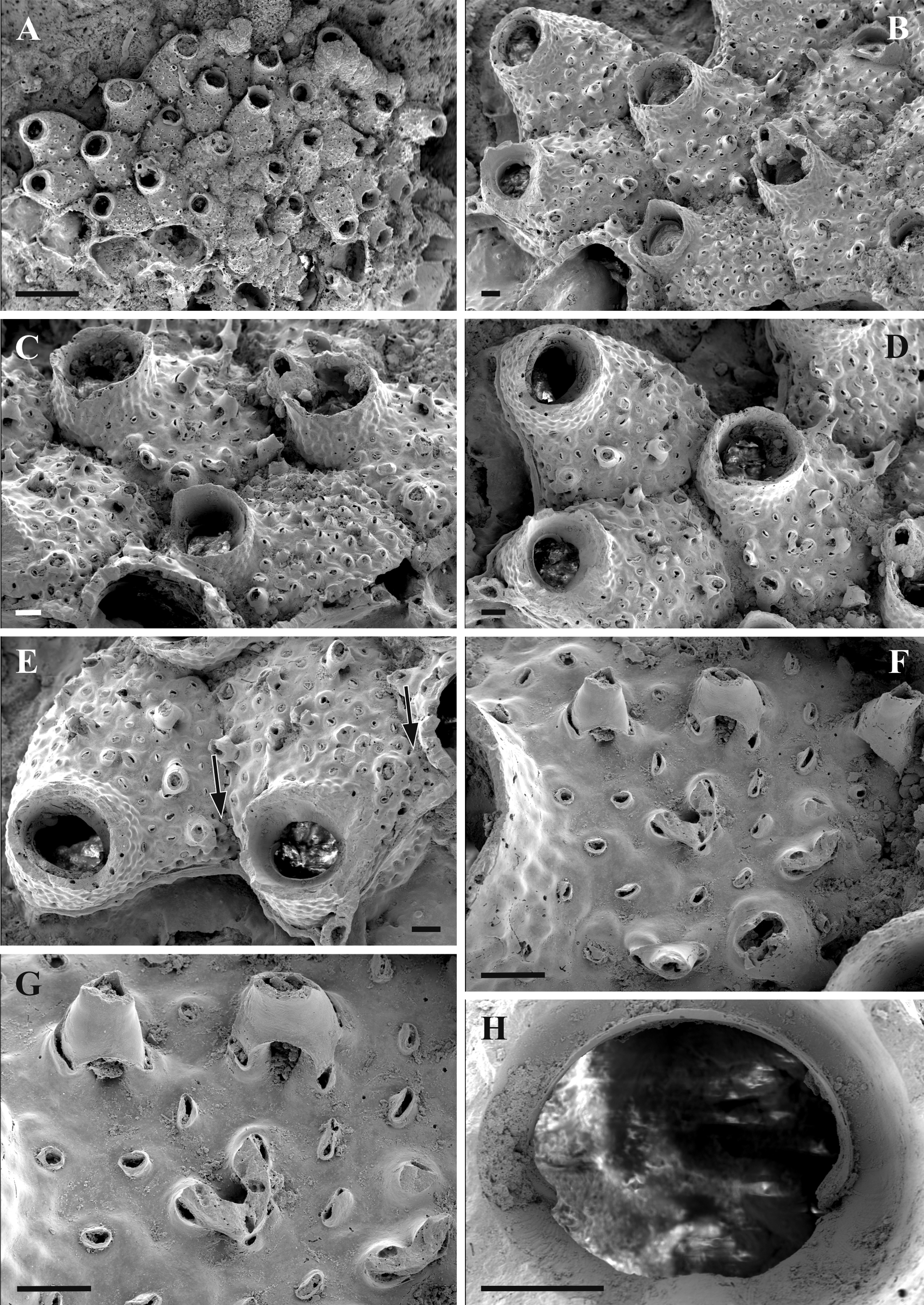
Exechonella kleemanni n. sp.
( Fig. 25 View FIGURE 25 , Table 24)
Material examined. Holotype: DPUV 2012-0004-0001 , on coral rubble (mounted on the SEM stub and coated with gold). Red Sea , the Northern Bay of Safaga, 23 July 1987.
Etymology. Named after marine biologist Dr. Karl Kleemann who collected this species, and who kindly donated to us various bryozoan colonies that he found on corals.
Description. Colony encrusting, unilaminar, multiserial. Autozooids convex, oval or pentagonal in shape, separated by deep grooves. Primary orifice oval, wider than long, poster (one-third) slightly narrower than the anter (two-thirds). Anter wall underlain by an inner lamina, which ends in drop-like condyles pointed to the orifice midline or downward. Orifice with proximal shelf (a distalmost part of the zooidal frontal shield proximally surrounded by a wall of the peristome) that has slight convex area or very low triangular projection in its central part. Areas to the left and to the right from this convex area/projection are wrinkled. Peristome tubular, slightly flared, with longitudinal grooves on its internal surface and pustulose externally. Frontal shield slightly pustulose being perforated by 31–56 foramina having a shape of short cylindrical or flattened tubes with gymnocystal internal wall surrounding its lumen and small, predominantly slit-like opening. Base of many or sometimes most of these tubes is surrounded by a low conical elevation while some are situated in a small depression. Most autozooids have 4–8 long, spire-like, thick-walled hollow processes which formation involves the fusion of two, three or, rarely, four foraminal tubes, whose openings are distinguished near the process base. Processes randomly distributed across the frontal shield with some having lateral position. Small oval marginal pores well seen around zooidal periphery. Vertical zooidal walls narrow, represented by multiporous mural septula with 1–2 rows of communication pores. Avicularia are not recognizable during external examination, but they might be present underneath of 1–2 lateralmost processes formed close to adventitious kenozooids with 5–6 pores. Ancestrula is unknown.
Northern Bay of Safaga, Red Sea
m±sd r n AzL 1048.7±97.4 930–1220 16 AzW 783.3±112.2 630–960 16 OrL 194.3±7.9 190–210 8 OrW 240±26.5 200–270 8 FoN 46±8.8 31–56 13 PeL 187.5±47.9 150–250 5 PeW 427.5±20.6 400–450 5 Remarks. Exechonella kleemanni n. sp., is characterized by its frontal shield with numerous foramina as short, mostly flattened tubes with small slit-like openings and spire-like, thick-walled hollow processes. The primary orifice oval with proximal shelf that is slightly convex or having a low triangular projection in its central part. Condyles are drop-like.
While having a number of similarities, the studied colony of E. kleemanni n. sp. differs from E. verrucosa by the (1) primary orifice shape that is subcircular in the latter species and oval with slightly convex proximal edge or small projection in the former, (2) condyles that are triangular and directed towards the centre of the orifice, being situated almost in middle of the orifice lateral sides in E. verrucosa , and drop-like, directed proximally, and being situated in the proximal third of the orifice lateral side in E. kleemanni n. sp., (3) peristome that is short, flared and with long spike-like projections on its rim in the former species, and longer, cylindrical and just slightly flared, without projections in the latter (though all peristomes are damaged in our material). Further differences include (4) shape of frontal foramina, with a small opening in a depression surrounded by a wide flat rim in E. verrucosa and with a slit-like opening on the top of flattened cylindrical tube in E. kleemanni n. sp., (5) shape of zooids that are rather flat with a clear outline of the marginal pores in E. verrucosa , and convex with marginal pores hardly seen in E. kleemanni n. sp. Finally, (6) avicularia with a characteristic nipple area are present in 1–2 larger lateralmost foramina in the former species whereas they were not detected during external examination, but they might be present underneath of 1–2 lateralmost projections in the latter.
Winston and Heimberg (1986) described and illustrated a similar species (as Coleopora verrucosa Canu & Bassler, 1927 ) with long conical processes associated with foramina from Komodo Island, Indonesia. Their specimen, however, differs from E. kleemanni n. sp. by the frontal shield evenly covered by the small foramina with slit-like opening (often slightly curved, with very narrow rim) each placed in the small depression. Also the shape of the peristome is slightly flared with a proximal process on its rim in the species from Komodo, and more tubular in E. kleemanni n. sp.
Recently , we examined two more specimens possessing the long spike-like projections associated with foramina of the frontal shield, one from Philippines ( USNM 7915 View Materials , Albatross Station D. 5751, depth 24 fathoms) and the other from the Great Barrier Reef (James Cook University Marine Biological Expedition, St. 986, 20 July 1987, kept at the Natural History Museum, London). The Australian specimen is reminiscent E. kleemanni n. sp. by the shape of its tubular peristome and tube-like foramina, but its preservation is too poor for a definite conclusion.
In contrast, the specimen from the Philippines described in this paper as E. rimopora n. sp. strongly reminiscent the aforementioned specimen from Komodo described by Winston and Heimberg (1986) as Coleopora verrucosa (see below).
Distribution. E. kleemanni n. sp. was found only in the Red Sea, Northern Bay of Safaga.
| USNM |
Smithsonian Institution, National Museum of Natural History |
No known copyright restrictions apply. See Agosti, D., Egloff, W., 2009. Taxonomic information exchange and copyright: the Plazi approach. BMC Research Notes 2009, 2:53 for further explanation.