
Exechonella ampullacea Hayward & Ryland, 1995
|
publication ID |
https://doi.org/ 10.11646/zootaxa.4305.1.1 |
|
publication LSID |
lsid:zoobank.org:pub:1192C3A0-5CCB-4A86-903C-A2B82906A5F9 |
|
DOI |
https://doi.org/10.5281/zenodo.6017304 |
|
persistent identifier |
https://treatment.plazi.org/id/CF0AB852-FFF9-E921-FF03-FF2B960EE794 |
|
treatment provided by |
Plazi |
|
scientific name |
Exechonella ampullacea Hayward & Ryland, 1995 |
| status |
|
Exechonella ampullacea Hayward & Ryland, 1995 View in CoL
( Fig. 1 View FIGURE 1 , Table 1)
Exechonella ampullacea: Hayward & Ryland 1995 View in CoL , p. 547, fig. 7e.
Exechonella ampullacea: Tilbrook & Gordon 2016 View in CoL , p. 597, fig. 3d.
? Exechonella tuberculata: Harmer 1957 View in CoL , p. 653–654 (in part), pl. 54, fig. 14.? Exechonella tuberculata: Gordon 1984 View in CoL , p. 70, pl. 23, fig. d.
Material examined. Holotype: QM G304975. Coral Sea , Great Barrier Reef , Heron Island , about 20 feet depth along the reef edge, 21 April, 1972. Paratype: QM G304977. Coral Sea , Great Barrier Reef, Heron Island, about 20 feet depth along the reef edge, 21 April, 1972.
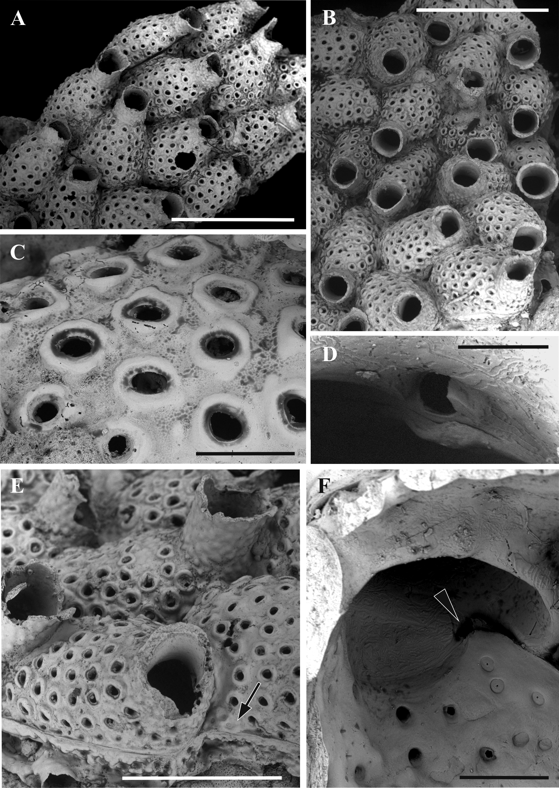
Description. Colonies encrusting, unilaminar, multiserial. Autozooids “bottle-like: convex, oval-elongated, separated by deep grooves and pits in the ‘corners’ between zooids. Primary orifice oval, wider than long, anter wall underlain by an inner lamina (only visible in oblique view) ending in distolateral condyles seen as narrow elongated plates with rounded distal part. Condyles are associated with a small opening (‘pocket’) of unknown function. Long tubular peristome is pustulose externally and with longitudinal grooves on its internal surface (in upper half), the rim is flared. Frontal shield pustulose, with 16‒41 rounded or oval foramina. The lumen of each foramen has smooth gymnocystal walls, whereas an area around it (often seen as a slightly elevated wide ring) has an inner wall surface. The proximal part of the frontal shield is ‘reduced’ in some zooids making a sort of ‘gaps’ in the ‘corners’ between zooids. Marginal pores small and rounded, with centrally perforated cuticular plate, predominantly seen in zooids on the periphery of the colony. The distalmost part of zooid below peristome can bear up to two rows of such pores. No avicularia. Adventitious kenozooids with a few pores, each having centrally perforated cuticular plate. Vertical zooidal walls narrow, represented by multiporous mural septula with communication pores arranged in two rows. Ancestrula unknown.
Heron Island, Great Barrier Reef
m±sd r n AzL 785±64 667–889 15 AzW 441±65 354–606 15 FoN 31±6.5 16–41 16 FoD 61±5.3 51–71 29 OD 30±7.6 20–40 29 Remarks. All species of Exechonella ampullacea -complex are characterized by very similar zooidal morphology having the bottle-like cystids, with long tubular peristomes that often prevent an observation of the inner lamina and the condyles. The main differences between the species are explained by the size, shape and, sometimes, number of the frontal foramina as well as presence and shape of projections associated with them (see below). E. ampullacea differs from the other species of this complex by the absence of any kind of frontal projections. In contrast, E. tuberculata and E. erinacea have massive conical (spike-like) projections developed in association with all foramina. The rest of the species have less developed processes in association with some foramina.
Harmer (1957) described under the name E. tuberculata two specimens from New Guinea that clearly represent two different species, as was noted by Tilbrook (2006) who earlier had the opportunity to examine most of Harmer’s material. Whereas the first, depicted on pl. 54, fig. 13, has numerous pointed processes associated with frontal foramina and thus might belong to E. erinacea , another, shown on pl. 54, fig. 14, is similar to E. ampullacea although its foramina look slightly smaller and not as numerous as in the holotype. Also, an elevated foraminal rim was not shown in the Harmer’s depiction. While both these specimens clearly belong to the species of the E. ampullacea species-complex, their restudy is required.
Gordon (1984) described E. tuberculata from Kermadec Ridge, New Zealand, that fairly corresponds to E. ampullacea by its general zooidal morphology and, in particular, by the absence of the frontal processes associated with foramina.
It should also be noticed that Cook and Bock (2004), when illustrating E. tuberculata , used a specimen from the Bass Strait. Earlier Cook (1967) made sketches of zooids from the colonies (as E. tuberculata ) collected in the type locality ( Port Phillip Heads , Victoria ), but they are too schematic for any definite conclusion. Six type specimens in two wooden slides with glass covers are kept in the Museum Victoria, Melbourne. Both slides contain three specimens of various sizes each, first labeled Lagenipora tuberculata McG, P. P.H., 65927, H627, F45627 View Materials , and the second slide labeled F45627 View Materials which was mentioned as a number of the holotype by Cook and Bock (2004). Photos of the slides with the specimens seen through the glass covers were kindly provided by the Invertebrate Palaeontology collection manager Dr R. Schmidt. Re-examination of this material that is presumably very fragile (P.E. Bock, pers. comm., 2016) as well as other specimens attributed to E. tuberculata is necessary to make further progress.
Another species belonging to the “group [that] includes … E. tuberculata and E. ampullacea was described by Cook and Bock (2004, p. 267) as Exechonella sp. cf. discoidea Canu & Bassler, 1929 . While it undoubtedly belongs to E. ampullacea species-complex, it is clearly not Actisecos discoidea ( Canu & Bassler, 1929) as will be shown later, and more similar to E. anuhaensis Tilbrook, 2006 .
Distribution. Exechonella ampullacea has been found at the Heron Island, Great Barrier Reef, Coral Sea, south Pacific Ocean, and Pulau Jong, Jurong Island, Singapore.
No known copyright restrictions apply. See Agosti, D., Egloff, W., 2009. Taxonomic information exchange and copyright: the Plazi approach. BMC Research Notes 2009, 2:53 for further explanation.
|
Kingdom |
|
|
Phylum |
|
|
Class |
|
|
Order |
|
|
Family |
|
|
Genus |
Exechonella ampullacea Hayward & Ryland, 1995
| Cáceres-Chamizo, Julia P., Sanner, Joann, Tilbrook, Kevin J. & Ostrovsky, Andrew N. 2017 |
Exechonella ampullacea:
| Tilbrook & Gordon 2016 |
Exechonella ampullacea
| : Hayward & Ryland 1995 |
Exechonella tuberculata
| : Gordon 1984 |
Exechonella tuberculata
| : Harmer 1957 |