
Endonura saleri, Fanciulli, Pietro Paolo & Dallai, Romano, 2008
|
publication ID |
https://doi.org/ 10.5281/zenodo.180806 |
|
DOI |
https://doi.org/10.5281/zenodo.5621698 |
|
persistent identifier |
https://treatment.plazi.org/id/4E4E526E-1E2C-215C-FF2F-FA05B1696761 |
|
treatment provided by |
Plazi |
|
scientific name |
Endonura saleri |
| status |
sp. nov. |
Endonura saleri sp. nov.
Material examined. Type material. Holotype (female) and 6 paratypes (5 females; 1 male) on slides; 12 individuals preserved in alcohol. Material was collected in the Cansiglio forest, in a site along the slopes of Mt. Pizzoc, 24.xi.1979. Samples consisted of leaves and humus from a beech wood. Holotype and paratypes are conserved in the collembolan collection of the Department of Evolutionary Biology of the University of Siena.
Description. body length up to 1,3 mm. Habitus as described by Cassagnau (1979) with abdominal segment VI well visible in dorsal view. Colour of the body white in alcohol, pigment present only in the ocular plate, near the two eyes. Dorsal tubercles well developed; head with 12 differentiated tubercles ( Tab. 1A View TABLE 1 A ; Fig. 22 View FIGURES 21 – 26 ); clypeal and antenno-frontal tubercles well separated each other ( Fig. 22 View FIGURES 21 – 26 ). Dorsal chaetotaxy constituted by long (Ml), short (Ms), and very short (Mss) macrosetae, mesosetae (me), microsetae (mi) and setae sensualis (S). Ml, Ms and Mss usually thickened, sheated, smooth and not pointed ( Fig. 21 and 26 View FIGURES 21 – 26 ). On the head, the ocular tubercle has 3 setae (Ml, Ms, me); antenno-frontal and clypeal tubercles with setae A, B, C, D, F and G; seta D is a free microseta that sometimes appears as a mesoseta; setae E and O absent, ( Tab. 1A View TABLE 1 A ; Figs 21, 22 View FIGURES 21 – 26 ).
Dorsal internal tubercle (Di) on the head is very simple while the external one (De) has a normal appearance. Di with one Ms and one Mss, De with one Ml and one Mss. Dorso-lateral tubercle with five setae (two Ml, one me and two mi), lateral and sub-ocular tubercle usually with 9 setae (three Ml, one Ms, five mi) ( Tab. 1A View TABLE 1 A ; Figs 22 View FIGURES 21 – 26 , 29 View FIGURES 27 – 30 ). Ventral chaetotaxy of the head and labium as in Fig. 23 View FIGURES 21 – 26 ; labrum as in Fig. 24 View FIGURES 21 – 26 . Apical vesicle of the IV antenna simple ( Fig. 27 View FIGURES 27 – 30 ); antennal organ III consists of two curved sensilla into a cuticular pocket and flanked by two cylindrical sensilla; ant. IV with 8 cylindrical sensilla ( Fig. 27 View FIGURES 27 – 30 ); ant. I and II with 7 and 11 setae respectively.
Chaetotaxy of thorax (Th.) and abdomen (Abd.) as in Table 1B View TABLE 1 A and Fig. 21 View FIGURES 21 – 26 . On Th. I tubercle Di not differentiated; Th. II–III with seta Di1 free; Th. III–Abd. III with seta De4 free, that usually appears as a Mss, or sometimes as a mi. Lateral tubercle of the Abd. III and IV with 4 and 6 setae, respectively. Abd. V with 3+3 setae on the fused Di tubercles (1 Ml, 1Ms and 1mi), De+Dl+L with 8 setae (7 + 1S) ( Fig. 30 View FIGURES 27 – 30 ). Ventral chaetotaxy as in Fig. 25 View FIGURES 21 – 26 and Table 1B View TABLE 1 A . Ventral tube with 4 setae. Chaetotaxy of the legs as described in table 1B; claws without inner tooth, setae B4 and B5 pointed and moderately long (B5>B4) ( Fig. 28 View FIGURES 27 – 30 ).
Etymology. The species derives its names from Zuan Saler, the first “Captain of the wood” nominated by the Venetian Doge Francesco Donato in 1404 with the appointment of preserving the forest of Cansiglio.
Discussion. Endonura saleri sp. nov. belongs to the group of species having two pigmented eyes and lacking both body pigmentation and seta O in the antenno-frontal tubercle of the head. The group includes several species; among them E. tartaginensis Deharveng, 1980 , E. pejai Deharveng, 1980 , E. ludovicae ( Denis, 1947) and E. transcaucasica ( Stach, 1951) . Endonura saleri sp. nov. and E. tartaginensis differ for the number of setae on the lateral tubercle of abd. III (3 in E. tartaginensis , 4 in E. saleri sp. nov.), those on the lateral tubercle (De+Dl+L) of Abd V (6 in E. tartaginensis vs 8 in E. saleri sp. nov.) and, in addition, E. tartaginensis show well differentiated Di tubercle on Th. I that is absent in the new species. E. saleri sp. nov. can be easily separated from E. pejai , E. ludovicae and E. transcaucasica by some features of the dorsal chaetotaxy and the shape and extension of the dorsal tubercles. In particular, E. pejai shows the complete fusion of clypeal and antenno-frontal tubercles, that are, instead, well separated in the new species; E. ludovicae have complete cryptopygy of Abd. VI while, on the contrary, Abd. VI is well visible in dorsal view in E. saleri sp. nov. E. transcaucasica shows well differentiated dorsal internal tubercles both on the head and on thorax I; Di tubercles on the head of E. saleri sp. nov. are only partially developed, while on thorax I Di are not developed at all.
Terga Legs
Di De Dl L Scx Cx Tr Fe T Th. I 1 2 1 – 3 6 13 19 Th. II 3 2+S 3+ S 3 2 8 6 12 19 Th. III 3 3+S 3+ S 3 2 8 6 11 18
Sterna
Abd. I 2 3+ S 2 3 Ventral tube: 4
Abd. II 2 3+ S 2 3 Ve: 5
Abd. III 2 3+ S 2 4 Ve: 4–5 Fu: 5 Abd. IV 2 2+ S 3 6 Ve: 8 Vl: 4 Abd. V 3 +3 7+S Ag: 3 Vl: 1 Abd. VI 7 Ve: 12 An: 2 mi Recently some new or newly redescribed species of Endonura have been reported ( Smolis et al. 2007, Smolis 2006, Smolis & Kaprus’ 2003, Pomorski & SkarŻyński 2000) from material collected in central-east Europe or the eastern part of the Mediterranean basin. Most of them display patterns of body pigmentation ( E. taurica , E. quadriseta , E. gracilirostris , E. dentifera , E. gladirostris ) ( Smolis et al. 2007, Smolis & Kaprus’ 2003) that make them easy to distinguish from E. saleri sp. nov.; others are unpigmented, such as E. carpatica Smolis, 2006 , from the Polish Carpathians, and E. urotuberculata Pomorski & SkarŻyński, 2000 , from Bulgaria. Both these species, however, have no eyes, so their diagnosis in comparison to E. saleri sp. nov. is quite simple. What appears of particular interest is the redescription of some syntypes of E. centaurea ( Cassagnau & Peja, 1979) by Pomorski and SkarŻyński (2000). Cassagnau and Peja (1979) described E. centaurea as a blind species even though they observed the presence, in the anterior part of the ocular tubercles, of a large cuticular grain that could be envised as a “vestigial eye”. Pomorski and SkarŻyński (2000) clearly ascertained the presence of two distinct eyes that, together with the unpigmented body and the lack of the O seta on the frontal tubercle, allowed the inclusion of E. centaurea in the same group as the new species. E. centaurea appears closely related to E. pejai ; both these species can be separated from E. saleri sp. nov. by differences in the distribution of dorsal tubercles (especially on the head) and some dorsal setae such as those on the lateral tubercle of abd III (three in E. centaurea , four in E. saleri sp. nov.) and Abd. V (6/ 7 in E. centaurea , 8 in E. saleri sp. nov.). However, as a general conclusion, several species of Endonura need to be revised in the light of new and numerous characters, in order to define their real taxonomic position inside the genus.
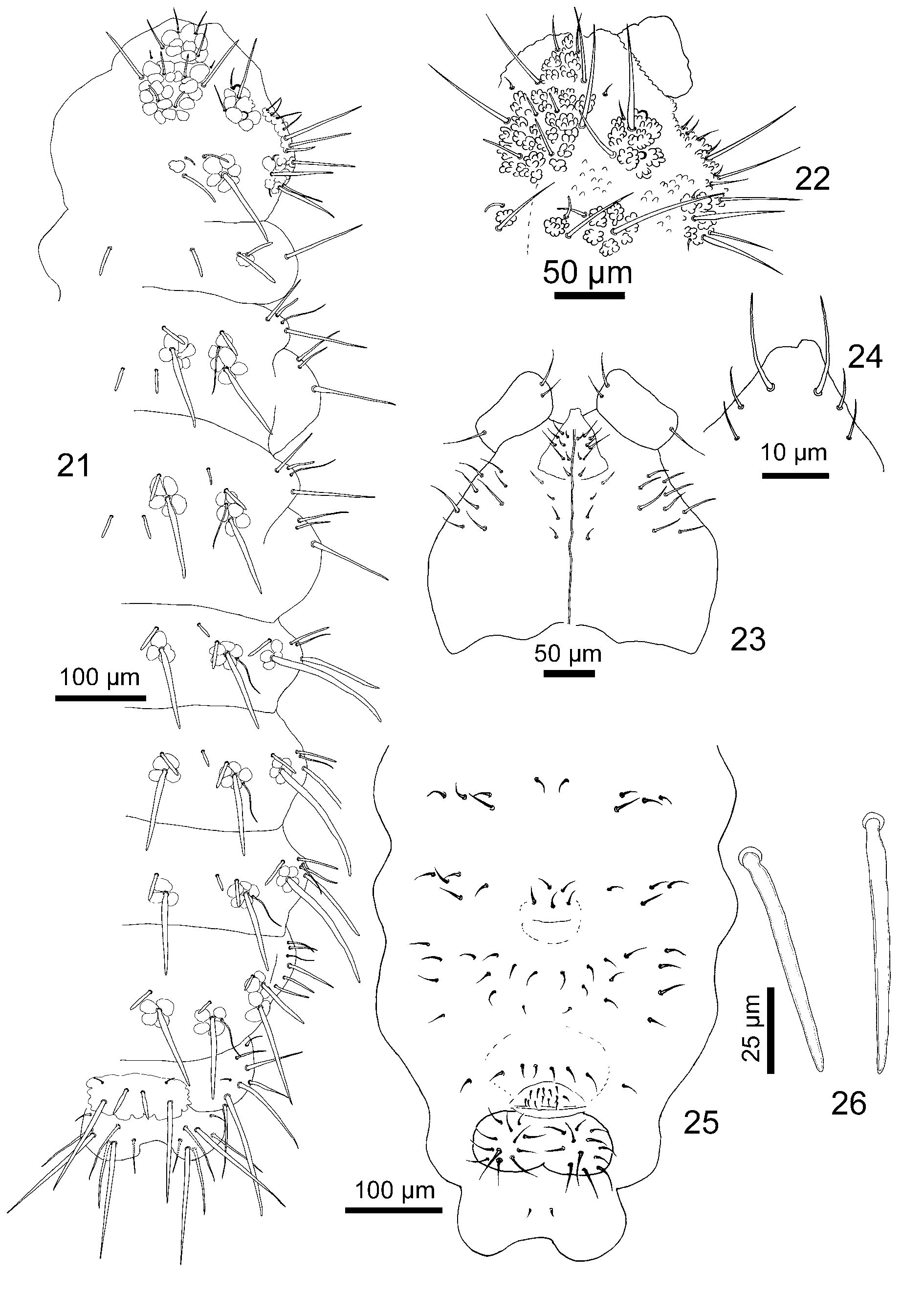
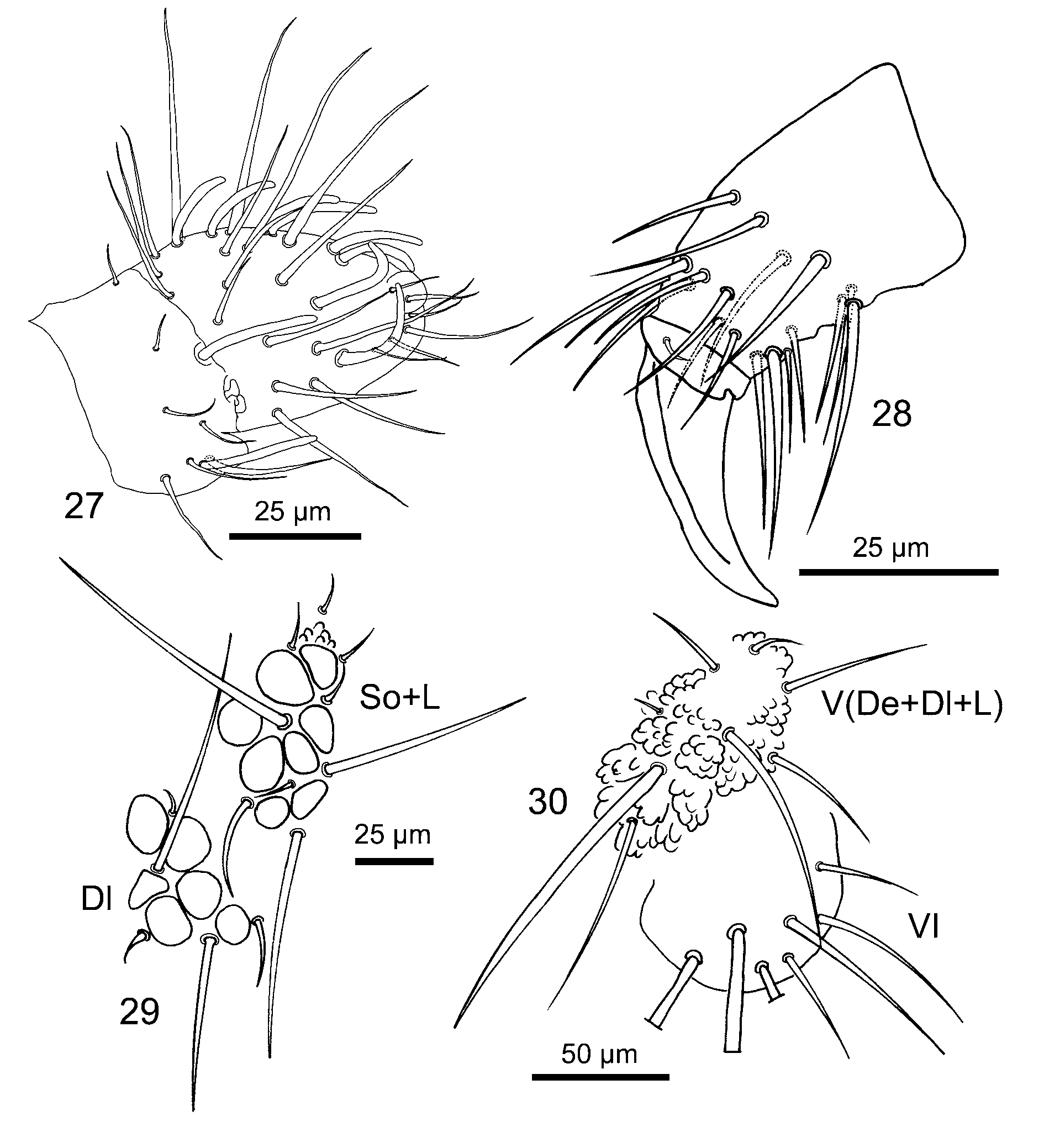
TABLE 1 A. Endonura saleri sp. nov. Tubercles and chaetotaxy of the head.
| Tubercle Number of setae | Types of setae | Names of setae |
|---|---|---|
| Clypeal (Cl) 4 | Ml, Ms. | F, G. |
| Antenno-frontal (Af) 8 | Mc, Ml, Ms, me. | A, B, C, D. |
| Ocular (Oc) 3 | Ms, Ml, me. | Ocp, Ocm, Oca. |
| Dorsal internal (Di) 2 | Ms, Mss. | Di1, Di2. |
| Dorsal external (De) 2 | Ml, Ms. | De1, De2. |
| Dorso-lateral (Dl) 5 | 2 ML, 1 me, 2 mi | Dl1, Dl5, Dl4, Dl2, Dl6 |
| Lateral-Suboc. (L+So) 9 | 3Ml, 1Ms, 5me | L1, L4, So1, L2, L3, So3–6 |
No known copyright restrictions apply. See Agosti, D., Egloff, W., 2009. Taxonomic information exchange and copyright: the Plazi approach. BMC Research Notes 2009, 2:53 for further explanation.