
Ecclisomyia maculosa Banks 1907
|
publication ID |
https://doi.org/ 10.11646/zootaxa.4413.2.1 |
|
publication LSID |
lsid:zoobank.org:pub:495CE6EB-4A83-4A05-8E42-A92889F1C1C4 |
|
DOI |
https://doi.org/10.5281/zenodo.5975597 |
|
persistent identifier |
https://treatment.plazi.org/id/652FF863-7631-433A-FF79-FCAC1AA15341 |
|
treatment provided by |
Plazi |
|
scientific name |
Ecclisomyia maculosa Banks 1907 |
| status |
|
Ecclisomyia maculosa Banks 1907 View in CoL
Figs. 22a–22c View FIGURES22 ; 23a–23c; 24a–24c; 25a–25c; 26a, 26b; 27a, 27b; 28a, 28b; 29a–29d; 39; 41; 42; 43c.
Ecclisomyia maculos a Banks 1907, 123, fig. 18, female, Boulder, Colorado, July 31, 1904 (Oslar), (description); Ross (1938, 31, 57, figs. 50, 50a, designation of Lectotype female, Boulder, Colorado and Allotype male, Slate Creek, Summit County, Colorado); Denning (1948, 18, figs. 3a, b, description male and female, bionomics); Ross (1950, 423, figs. 14, 14a, b. male genitalia, distribution); Denning (1951, 161, distribution); Nimmo (1971, 61, figs. 128a, b, 188–193, 598, description of male and female genitalia, bionomics, distribution); Anderson (1976, 80, distribution); Newell & Minshall (1977, 255, distribution); Swegman & Ferrington (1980, 289, distribution); Herrmann et al. (1986, distribution); Baumann & Unzicker (1981, 27, distribution).
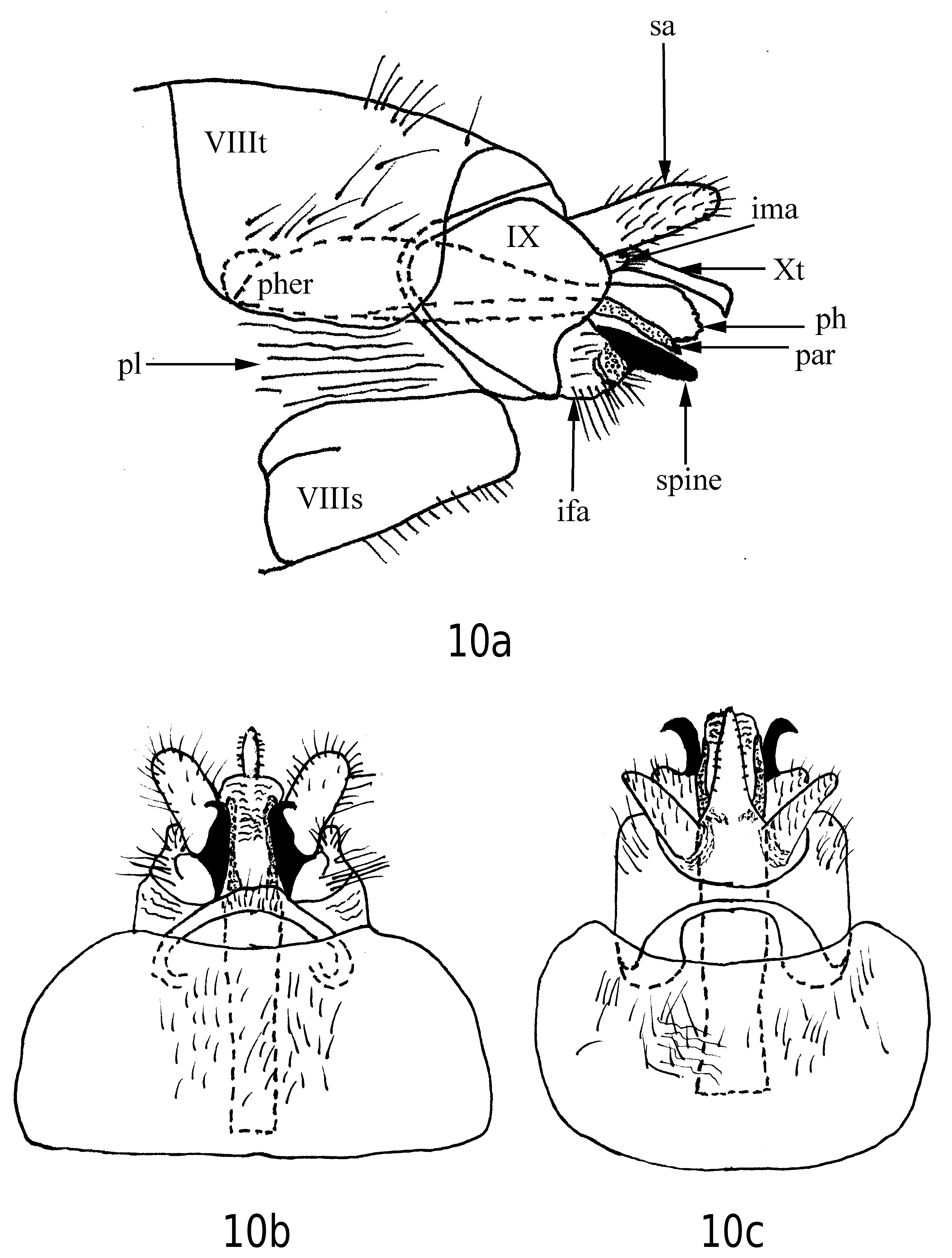
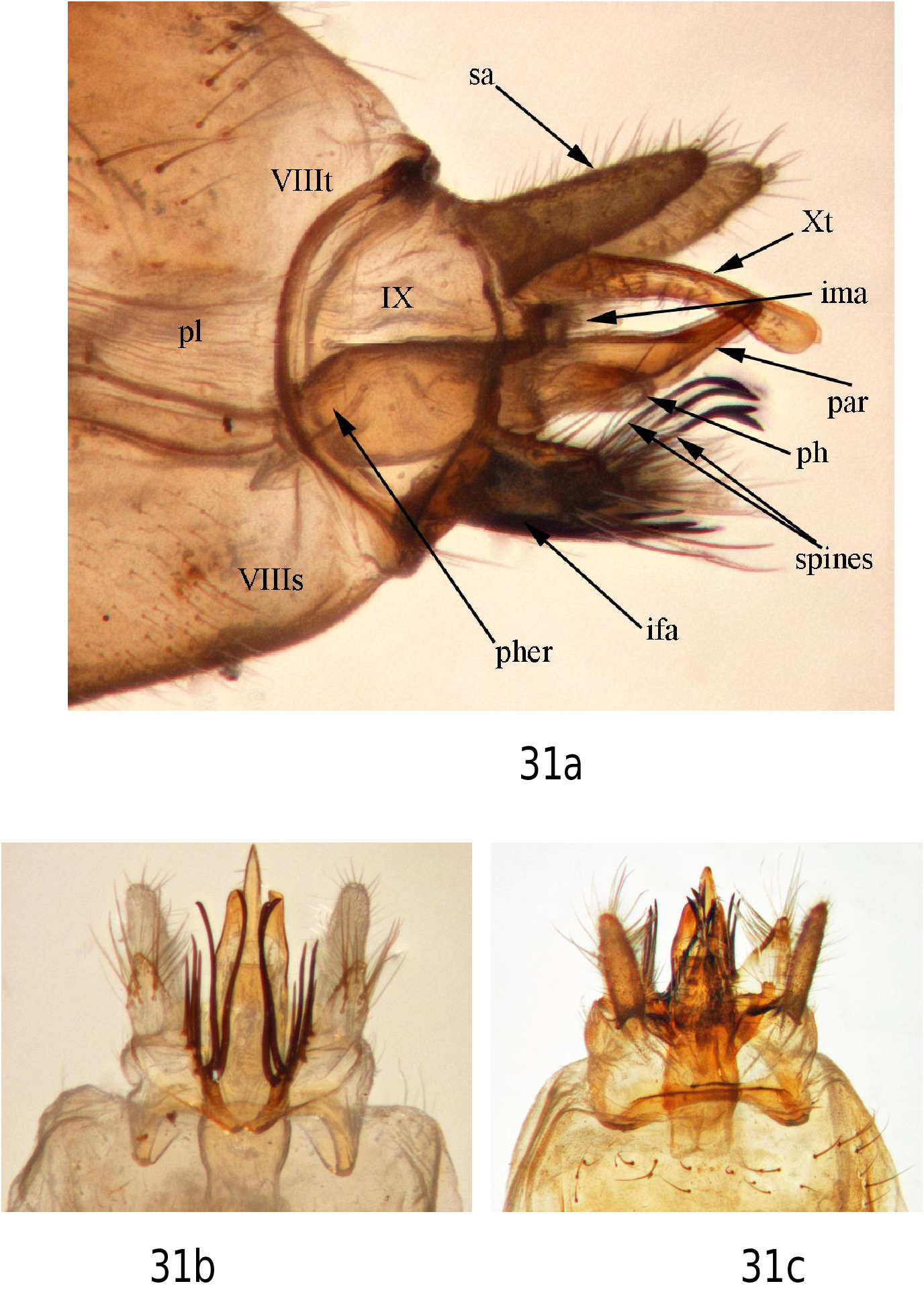
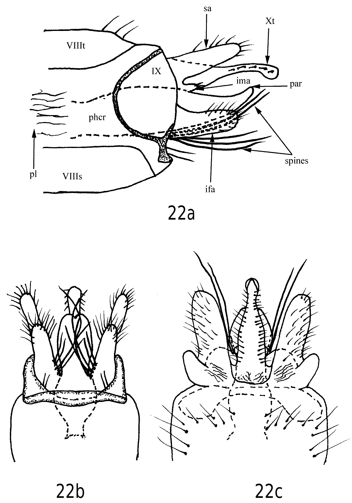
Male diagnosis. Each inferior appendages has 2 large, black, stout spines arising from its base ( Figs. 22b View FIGURES22 , 23b View FIGURES23 ), separating E. maculosa from E. bilera , which lacks spines on the inferior appendages, and from E. conspersa which has a single black, stout spine on each inferior appendage ( Fig. 10a View FIGURES 10 ). The inferior appendages, viewed laterally, extend further caudad than the triangular inferior appendages of E. simulata . The shallow U- to V-shaped notch at the distal ends of the fused parameres of E. maculosa ( Figs. 22b View FIGURES22 , 23b View FIGURES23 ) distinguishes it from E. simulata which has a deep U- to V-shaped notch at the distal end of the fused parameres ( Figs. 30b View FIGURES 30 , 31b View FIGURES 31 ).
Male description. See Banks (1907); Denning (1948); and Nimmo (1971). Supplemental description follows: Length 8–11 mm (N = 29). Head, thorax, legs brown. Wings brown, hyaline with scattered pale areas, giving wings spotted appearance. Ocelli midway between eyes and midline. Antennae with about 42–45 segments, dorsal surface of each segment with 4 stout black to reddish spines, except segment 1 (scape) and segment 2 with none. Antennal segments with numerous fine black setae; scape large, pedicel short, third antennal segment 2 X length of pedicel. Tergum VIII with row of transparent to reddish-brown setae across posterior area of tergum ( Fig. 23c View FIGURES23 ). Sternum VIII brown, with long reddish setae on distal ventral margin and distal lateral surfaces; in lateral view narrow, longer than wide ( Figs. 22a View FIGURES22 , 23a View FIGURES23 ). Pleura well developed, membranous. Tergum IX nearly semicircular in lateral view, longest mesolaterally, longitudinally short dorsal and ventrally; dorsal, ventral, and anterior margins heavily sclerotized ( Figs 22a View FIGURES22 , 23a View FIGURES23 ); left and right lateral portions connected dorsally and ventrally with transverse sclerotized bridges (straps) ( Figs. 22b, 22c View FIGURES22 , 23b, 23c View FIGURES23 ). Superior appendages divergent, nearly parallel-sided in lateral and dorsal views, rounded distally; with hair-like setae along lateral and distal margins ( Figs. 22a View FIGURES22 , 23a View FIGURES23 ). Intermediate appendage ventrad of tergum X sclerotized; distal margin with several elongate setae ( Figs. 22a View FIGURES22 , 23a View FIGURES23 ). Tergum X with short spike-like setae along lateral margins ( Figs. 22b, 22c View FIGURES22 ); apicolateral margins flanged ventrally; in dorsal view shaped somewhat like a bowling pin ( Fig. 22c View FIGURES22 ). Apices of parameres fused, with shallow U-shaped notch apically forming 2 short prong-like caudal extensions ( Figs. 22b View FIGURES22 , 23b View FIGURES23 ). Phallocrypt narrower anteriorly and caudally, wider mesally in lateral view ( Figs. 22a View FIGURES22 , 23a View FIGURES23 ). Inferior appendages divergent, elongate, apices rounded; in ventral view lateral margins nearly parallel; distal areas with numerous long reddish setae ( Figs 22a, 22b View FIGURES22 , 23a View FIGURES23 ); each with 2 large stout black to reddish-brown spines arising from base and extending caudad ( Figs. 22b View FIGURES22 , 23b View FIGURES23 ); 3 shorter, stout reddish to black spines along posterior mesal margin lateral of these 2 stout spines.
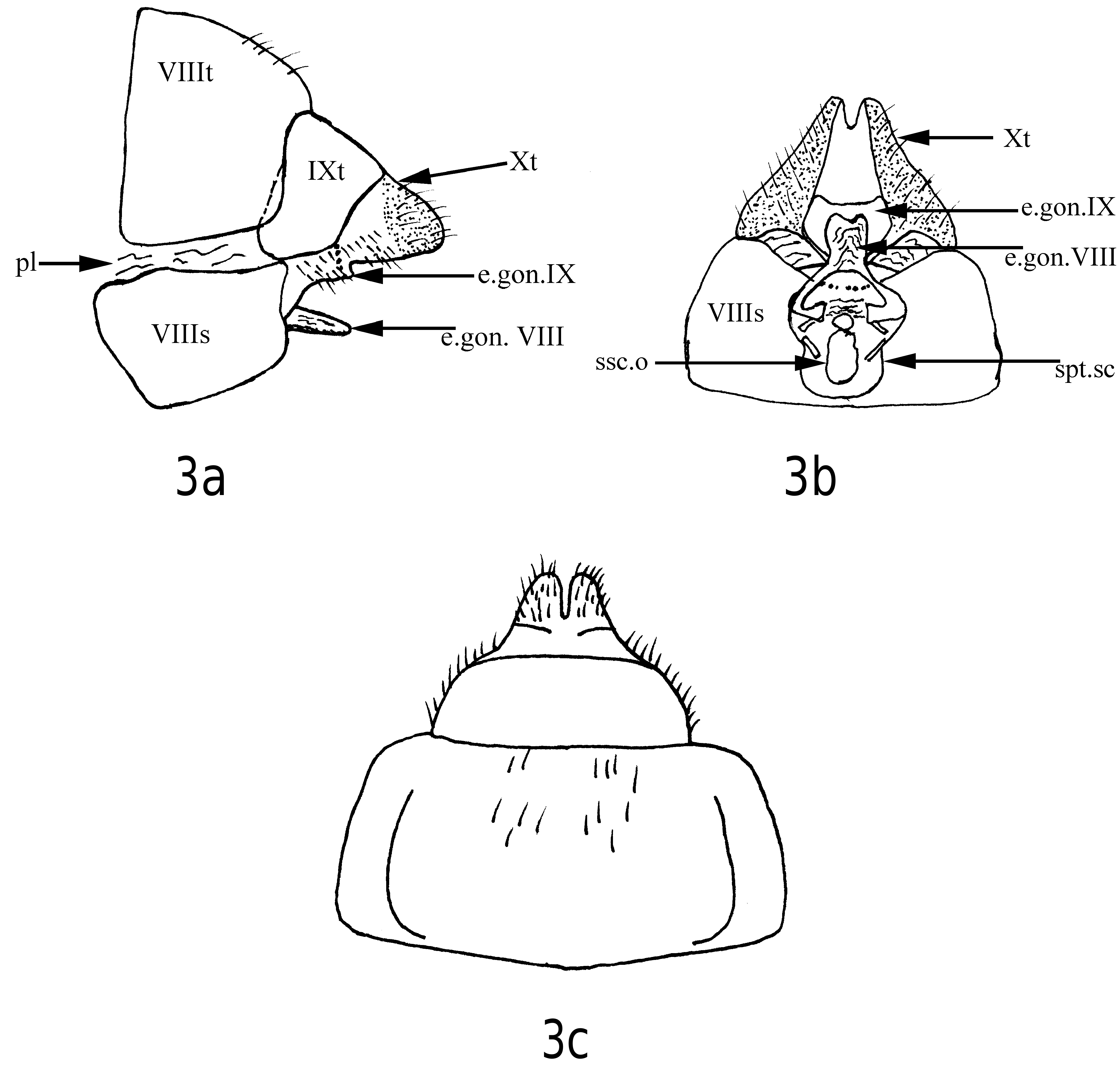
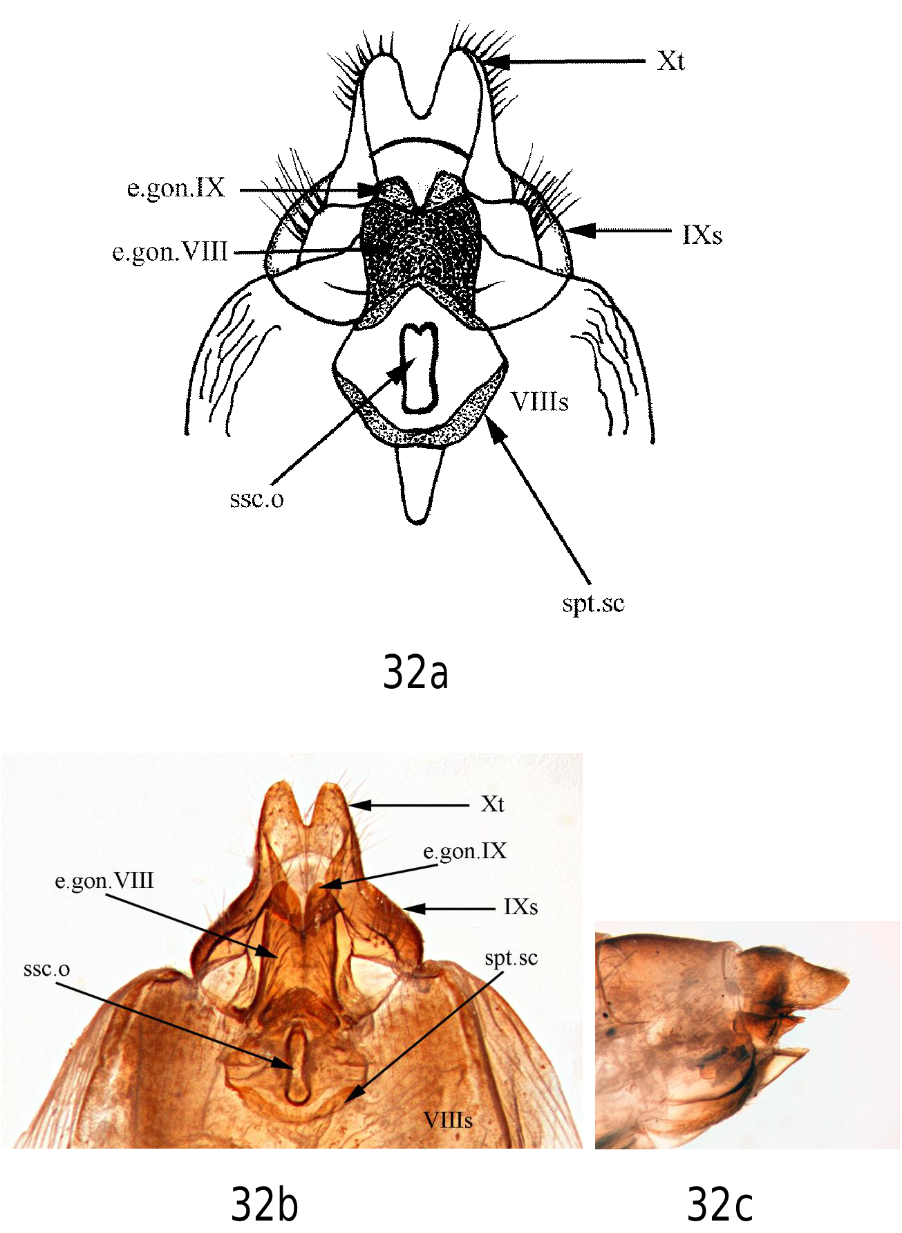
Female diagnosis. The distal margin of tergum X has a V-shaped incision extending about 1/2–3/4 the length of the segment in dorsal view ( Figs. 24b View FIGURES24 , 25b View FIGURES 25 ), whereas the distal margin of tergum X of E. simulata has a Ushaped incision that divides the tergum into 2 lobes, the incision about 3/4 the length of the tergum. The external part of gonopod VIII in both species is large; however, in E. maculosa it is nearly as wide as long ( Figs. 24c View FIGURES24 , 25b, 25c View FIGURES 25 ) whereas in E. simulata it is longer than wide ( Fig. 32a, 32b View FIGURES 32 ); the caudal margin may be convex ( Fig. 24c View FIGURES24 ), straight ( Fig. 25c View FIGURES 25 ) or with a V-shaped concavity ( Fig. 25b View FIGURES 25 ). The external gonopod of segment IX sclerotized, caudal margin with deep V-shaped declivity extending posterad about 2 X its width beyond the caudal margin of the extended gonopod of segment VIII ( Figs. 24c View FIGURES24 , 25b, 25b View FIGURES 25 ); in E. simulata it is wide, not as deeply cleft and only extends 1X its width posterad beyond the external part of gonopod VIII ( Figs 32a, 32b View FIGURES 32 ).The external part of gonopod VIII is massive ( Fig. 25b View FIGURES 25 ) whereas it is short and narrow in E. bilera ( Fig. 3b View FIGURES 3 ). The external part of gonopod IX has a deep mesal division ( Fig. 24c View FIGURES24 ); in E. bilera it is broadly concave as viewed from a ventral aspect ( Figs. 3b View FIGURES 3 , 4a View FIGURES 4 ). The spermathecal sclerite is not as long as in E. conspersa ; the spermathecal sclerite opening is relatively longer than in E. bilera .
Female description. See Denning (1948); Nimmo (1971). Supplemental description follows: Length 8–10 mm (N = 14). Head, thorax, legs reddish brown. Antennae each with approximately 38–42 segments; antennal segments with numerous translucent setae and short spines as in males, but less visible. Tergum X fused with tergum IX ( Fig. 24a View FIGURES24 ); distal margin of tergum X with V-shaped incision dividing tergum into 2 lobes for about 1/ 2–3/4 length of tergum. External part of gonopod VIII large, extending beyond genital opening; in ventral aspect lateral margins straight, caudal margin nearly straight ( Fig. 25c View FIGURES 25 ) to moderately convex ( Fig. 24c View FIGURES24 ), or may have a Vshaped mesal indentation ( Fig. 25b View FIGURES 25 ). External gonopod of segment IX sclerotized, narrow forming dorsal surface to vaginal opening; caudal margin with deep V-shaped declivity, extending posterad about 2 X its width beyond caudal margin of extended gonopod of segment VIII ( Figs. 24c View FIGURES24 , 25b, 25c View FIGURES 25 ). Spermathecal sclerite nearly as long as wide ( Figs. 24c View FIGURES24 , 25b View FIGURES 25 ), anterior margin rounded.
Pupal diagnosis. The E. maculosa pupa may be separated from that of E. bilera by the absence of dorsal gills on abdominal segment VIIa and presence on E. bilera . The E. maculosa pupa may be separated from that of E. conspersa by the absence of lateral line gills; they are present on abdominal segments IIp–IVa of E. conspersa ( Fig. 42 View FIGURE 42 ). The apical processes of E. maculosa each have 2 stout, long, black setae at the apex, whereas those of E. conspersa each have 3 stout, long, black setae. The E. maculosa pupa may be separated from that of E. simulata by the absence of single filament dorsal gills on abdominal segment IIa and presence on E. simulata ( Fig. 42 View FIGURE 42 ).
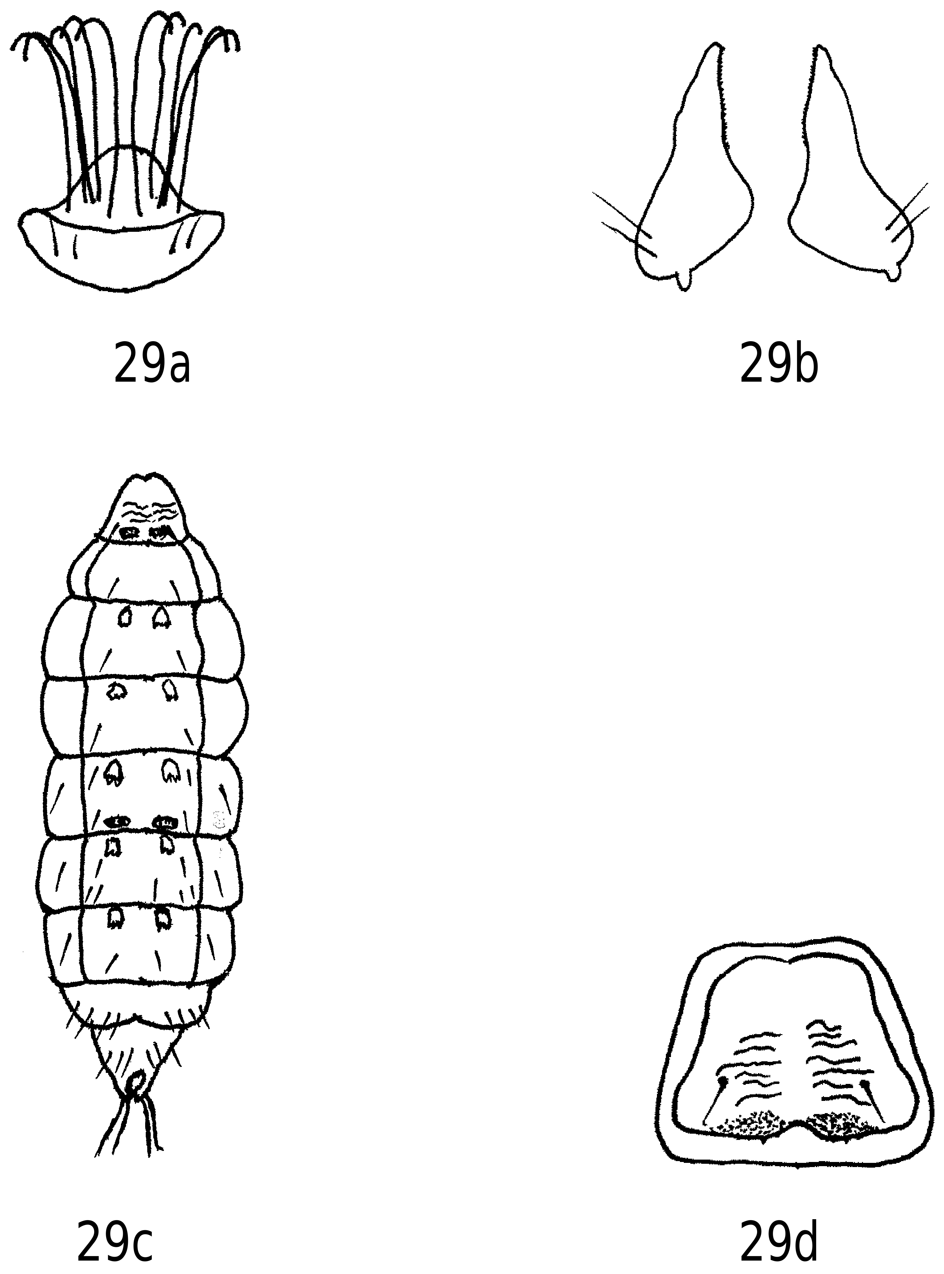
Pupal description. Length 8–10 mm (N = 8). Head: Light brown. Male antennae extending to or slightly caudad of abdominal apical processes; antennae in females extending to abdominal segment V. Labrum dorsal margin convex with 10 long black setae on dorsal surface, setae directed dorsad, distal end of each seta hooked ( Fig. 29a View FIGURES 29 ); posterior area lateral of meson with 2 short reddish setae. Mandibles black to reddish-brown, bases of mandibles broad, each tapering into single triangular apical tooth; mesal margin with minute serrations; ventrolateral surface with 2 long, black setae ( Fig. 29b View FIGURES 29 ).
Thorax: Pro-, meso-, and metanota light brown. Pro-, meso-, and metathoracic legs brown to yellow-brown. In mature pupa, tarsal and tibial segments with both single and paired reddish-black stout spines. Spines absent in immature pupa. Procoxae of both mature and immature pupae frequently each with 1 long black seta near lateral margin and 3–5 shorter black setae on ventral surface. Mesocoxae may each have 1 long black seta near lateral margin and 1–3 short black setae on ventral surface. Profemora each with single short black spine on ventral surface near distal margin. Mesotarsal segments 1–4 each with 2 lateral fringes of long reddish to opaque (whitish) silk-like hairs, directed posteriorly.
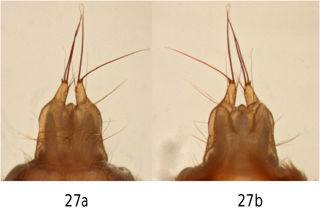
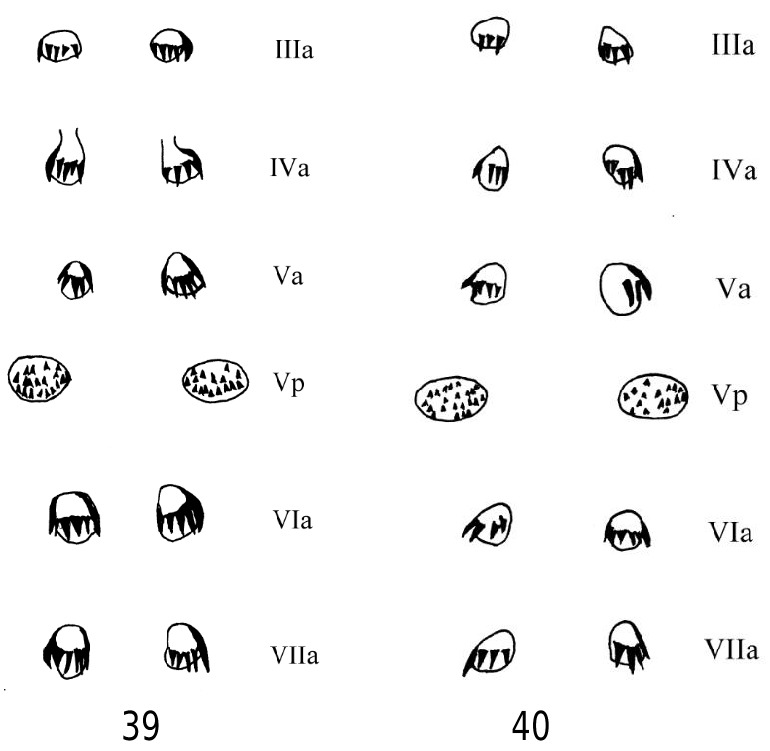
Abdomen: Lateral fringe on each side consisting of long feathery reddish hairs directed dorsad; originating from posterior area of abdominal segment V, with line of fringe arching ventrad, extending to anterior margin of abdominal segment VIII. Dorsal and ventral abdominal gills present, each single-filament; present dorsoposteriorly and ventroposteriorly on segments II–VII; present dorsoanteriorly and ventroanteriorly on segments III–VI; segments II and VII without gills dorsoanteriorly and ventroanteriorly; without lateral-line gills ( Fig. 42 View FIGURE 42 ). Dorsal process of abdominal segment I with 2 black sa 2 setae, each positioned lateral of meson; posterior margin sclerotized, light brown, forming pair of denticulate lobes lateral of meson, each lobe with 8–10 small spicules ( Fig. 29d View FIGURES 29 ); shape of dorsal process varying with maturity of pupa. Hook plates on segments III–VII ( Figs. 29c View FIGURES 29 , 39 View FIGURES 39, 40 , 41 View FIGURE 41 ). Pairs of hook plates present anteriorly on segments III–VII, posteriorly on segment V. Hook plate IIIa with 2–7 hooks; IVa with 2–8; Va with 3–8; Vp with 9–17; VIa with 2–6; and VIIa with 3–7. Hook plates reddish brown, hooks directed posterad on anterior hook plates of segments III-VII and anterad on circular to elliptical hook plates Vp. Dorsal abdominal segments each with 1 pair short black sa 2 setae on each of abdominal segments I–IV; 2–3 pairs on abdominal segments V and VII ( Fig. 29c View FIGURES 29 ); Dorsal abdominal segments VI sometimes with 1 pair short, black sa 2 setae; segments V–VII sometimes with 1 pair short, black sa1 setae. Dorsal abdominal segment VIII with 2 to 3 pairs of black to translucent setae lateral of meson. Distal area formed into 2 caudal lobes (apical processes); apical processes of male with light covering of spicules; spicules at apices of caudal lobes very difficult to discern with light microscope. Caudal lobes each with 2 long fine reddish-black to black setae on distal margin. Lobes of male and female slanted mesad; in ventral view lateral margins sinuate ( Figs. 26a View FIGURES 26 , 27a View FIGURES 27 , 28a).
Pupal case. Length 9–12 mm (N = 19). Width 2.5–3.0 mm (N = 6). Case long, narrow, constructed of small pebbles and grains of sand, with tight interstitial spaces, over an inner lining of silk surrounding pupa. Case nearly uniform in diameter, anterior end slightly wider than posterior end ( Fig. 43c View FIGURES 43 ). Ends of case rounded, each with small open area to allow for flow of water through case.
Larval diagnosis. Of the four Nearctic Ecclisomyia , only E. maculosa lacks a prosternal sclerite, or small sclerotized area on the venter of the prothorax. The parietals have distinct muscle scars ventral and posterior of the eyes. Ecclisomyia maculosa lacks both anterodorsal and anteroventral gills on abdominal segment VII, but gills are present in the other Nearctic Ecclisomyia ( Fig. 42 View FIGURE 42 ). In addition, the E. maculosa larva may be separated from that of E. simulata by the lack of dorsoanterior gills on abdominal segment II, and the presence of these gills on the E. simulata larva. The E. maculosa larva is without lateral line gills, whereas the E. conspersa larva has 2 gills with 1 dorsolateral gill and 1 ventrolateral gill on each side of abdominal segments II–IV ( Fig. 42 View FIGURE 42 ).
Larval description. Length (mature instar) 8–11 mm (N = 18). Head: Reddish brown. Parietals, genae and post genae glabrous, reddish brown to dark brown; area about coronal suture with faint yellow strip; muscle scars distinct posterior and lateral of eyes. Mandibles black, each with 4 small blunt apical teeth; posterolateral area with 1–2 short translucent setae; mesal margin with tuft of translucent feather-like setae ( Fig. 19a View FIGURES 19 ). Frontoclypeus smooth, without texture; posterior half without evident muscle scars, or muscle scars indistinct. Ventral apodome triangular, dark brown, 1.14–2.40 X longer than ventral ecdysical line. Early instar larva with ventral apodome shorter than ventral ecdysical line. Primary setae in positions 7 and 9–17 ( Fig. 19a View FIGURES 19 ). Primary setae in position 7 long, translucent; primary setae in position 9 black to reddish; primary setae in position 10 short, appressed, and transparent; primary setae in position 11 short, black to translucent; primary setae in position 12, short to long, black to transparent, fine, may be difficult to discern; primary setae in position 13 short, translucent, difficult to discern, may be missing; primary setae in position 14 long, black, and stout; primary setae in position 15 black; primary setae in position 16 appressed, translucent, hard to discern, may be absent; primary setae in position 17 black. Venter of head with primary setae in positions 8 and 18 ( Fig. 19b View FIGURES 19 ). Primary setae in position 8 short, may be translucent, difficult to discern; primary setae in position 18 translucent, short, difficult to discern, best seen in lateral view. Frontoclypeus smooth, without texture; wide anteriorly, narrow posteriorly, triangular; glabrous except for primary setae in positions 1–6 ( Fig. 19a View FIGURES 19 ). Primary setae in position 1 short or long, translucent, semiappressed; primary setae in positions 2 and 3 long and black, sometimes short and translucent; primary setae in position 4 appressed, short or long, black or translucent; primary setae in position 5 long, black, sometimes translucent, difficult to discern; primary setae in position 6 translucent, difficult to discern. Labrum brown to dark brown, sometimes brown posteriorly, yellowish anteriorly. Primary setae in positions 1–6 ( Fig. 19a View FIGURES 19 ); setae translucent, setal positions may be difficult to determine or setae absent. Primary setae in positions 1 and 2 translucent, short, best seen from ventral aspect. Cardo sclerites elongate, narrow, rectangular, and brown to dark brown. Submental sclerite with 1 short black seta set lateral of meson.
Thorax: Pro- and mesonota brown to reddish brown, nota with mesal yellow strip; metanotal sclerites brown. Anterior margins of pronota each with 10–13 long, black slender setae set equidistant from each other along anterior margin; short translucent setae positioned between these longer setae along anterior margin. Each half of pronotum with transverse line of 7–11 black ls setae posterior to mid-dorsal transverse groove; sa 2 setae short, black. Mesonotum with black ls setae set along anterior margin. Mesonotum with black setae in 3 distinct groups roughly corresponding to sa 1, sa 2, and sa 3 positions; muscle scars not evident or indistinct. Metanota sa 1 sclerites with 4–7 black ls setae, setal sockets prominent; metanotal sa 2 sclerites with 4–9 black ls setae; metanota sa 3 sclerites parenthesis-shaped, each with 6–17 ls black setae on anterior area of sclerites; metanotal sclerites in some specimens with setae translucent, not black. Prosternal horn short, sclerotized. Prosternal sclerite or sclerotized area absent. In prepupae examined, there is no prosternal sclerite or sclerotized area. Legs yellow–brown; foretrochantins dark brown; forming tent-like structure over procoxae. Procoxae with primary setae in positions 1 and 2; primary setae in position 2 black to translucent, setal sockets prominent ( Fig. 21a View FIGURES 21 ). Pro-, meso-, and metacoxae each with black primary setae in position 2; ventral margins with 4–7 black ls setae, sometimes short and translucent; dorsal margins of pro-, meso-, and metacoxae each with 15–17 long black setae, all coxal setae with prominent setal sockets ( Figs. 21a, 21b, 21c View FIGURES 21 ); some specimens with only 4–6 short translucent setae. Pro-, meso-, and metatrochanters each with black primary ls setae in positions 2, 3, and 5; seta in position 5 often absent or difficult to discern ( Figs. 21a, 21b, 21c View FIGURES 21 ). Ventral margins of pro- and mesotrochanters and pro- and mesofemora each with trochanteral brush, feather-like, translucent, extending to proximal ventral area of each femur ( Figs. 21a, 21b View FIGURES 21 ). Protrochanters each with single reddish elongate sl seta on ventral margin. Profemora with primary setae in position 2 and with 2 elongate long reddish-brown to translucent stout sl seta on ventral margin ( Fig. 21a View FIGURES 21 ). Meso- and metafemora each with black ls primary seta in positions 2, 3, and 4. Pro-, meso-, and metafemora with short reddish to translucent sl peg-like setae along ventral margins. Protibiae with primary setae in position 3. Pro- and mesopleura, and pro- and mesoepimera brown to dark brown.
Abdomen: Abdominal segment I with 4–5 black setae on dorsal hump; dorsal hump sometimes with sclerites; ventral of dorsal hump 4–13 black ls setae generally set on sclerotized areas, however setae sometimes with only basal sclerotization; these setae may also be short, reddish, without sclerotization. Dorsal hump sometimes with black ls setae lateral of meson, setal sockets prominent, setae possibly with sclerotized bases. Lateral humps each with 11 ls setae; dorsal areas may have 1–2 small sclerotized areas with 4–5 short black setae. Dorsal of lateral hump 6 black ls setae with sclerotized bases and prominent setal sockets. Each lateral hump with 1 long black seta ventrally. Ventral of lateral hump 3–9 ss to ls black setae sometimes with sclerotized bases; setal sockets prominent. Venter of abdominal segment I with 6–18 black ls setae forming an elliptical pattern; setal sockets prominent. Venter of abdominal segment I may have pair of sclerites or lightly sclerotized areas, each with 2–3 black setae, often with basal sclerotization. Ventral abdominal segments II–VIII glabrous. Dorsal and ventral abdominal gills present, each single-filament; present dorsoanteriorly and ventroanteriorly on segments III–VI; present dorsoposteriorly and ventroposteriorly on segments II–VII ( Fig. 42 View FIGURE 42 ). Dorsum of abdomen with short black primary sa 2 setae on abdominal segments II–VII; primary setae sometimes absent or difficult to discern, may be black or translucent. Dorsum of abdominal segments VII and VIII with short black setae adjacent to sa 2 setae. Tergum IX sclerite brown to yellow brown, elliptical, with 6 black, stout ls setae spaced equidistant along posterior margin; surface of sclerite with 5–6 shorter black setae. Venter of abdominal segment IX with 2–4 short black setae in sa 2 position. Lateral fringe of fine reddish hairs extending on each side from anterior of segment III to anterior of segment VIII. Chloride epithelia present on venter of abdominal segments III–VII; may not be evident in early instars. Anal prolegs each with lateral sclerite brown; narrowing ventrally and forming light brown sclerotized strap (band or projection) delineating sternum IX from sternum X; with 1–3 black ls seta on ventrocaudal margin; setal sockets prominent; with 1–6 black ls setae on dorsal surface and with basal tuft of 4–5 long brown to black setae, of which inner 2 setae longer than shorter outer setae. Sole plates brown to dark brown; heavily sclerotized, each without setae or only bearing single short black seta near distoventral margin. Caudal lobes glabrous except for minute sl reddish spicules on either side of anal opening. Anal hooks each bearing small accessory hook (denticule).
Distribution. Ecclisomyia maculosa is confined to the Intermountain Region and the Rocky Mountains, and has been recorded from Alberta, British Columbia, Colorado, Montana, and Wyoming ( Ross 1950). Newell & Minshall (1977) recorded E. maculosa from Idaho. The distribution record for E. maculosa reported for Oregon ( Denning 1951) is E. simulata , as reported by Ross (1950). I have examined specimens of E. maculosa from Alberta, British Columbia, Colorado, Idaho, Montana, and Wyoming. The specimens of E. maculosa from British Columbia have been from the southeastern Rocky Mountains.
Bionomics. In Colorado, E. maculosa appears to be primarily a subalpine to alpine species, frequently collected at altitudes of 2438–4314 m. Herrmann et al. (1986) reported an altitude range of 2558–3377 m. In Alberta, E. maculosa has been recorded from an altitudinal range of 1371–2134 m ( Nimmo 1971). This species has been collected from a shallow, narrow, swiftly flowing mountain stream ( Denning 1948). Nimmo (1971) noted that this species occurs in “small, riffled mountain creeks, running gently over fine gravel bottoms.” Ecclisomyia maculosa may be widely sympatric with E. conspersa . I have examined one female of E. maculosa and one male specimen of E. conspersa from Canyon Creek at the USFS trailhead west of Hamilton, Montana, and 1 male E. conspersa and 1 male E. maculosa from Tindall Meadows 12.9 km S of Landmark, Idaho. I am aware of only these two localities where both species are sympatric. Additional collecting may indicate other sites of sympatry for these two species.
Material Examined. CANADA: Alberta, sedge marsh, 1.6 km S Bow Pass, (Hwy. 93), Banff Nat’l. Park, 25- vii-1966, 1 M (APN) (SMEC); Lake Agnes, Lake Louise, Banff Nat’l. Park, 10-vii-1967, 1 M, 1F (APN) (SMEC); 10-viii-1967, 7 M, 1 F (APN) (SMEC); Lynx Creek, Forest Trail Rd., 14-vii-1967, 2 M (APN) (SMEC); Bow Pass, Banff. Nat’l. Park, 10-viii-1967, 1 M (APN) (SMEC); Snaring River, N of Jasper, (Hwy. 16), Jasper Nat’l. Park, 15-vii-1968, 1 M (APN) (SMEC); Poboktan Creek, (Hwy. 93), Jasper Nat’l. Park, 06-viii-1968, 1 M (APN) (SMEC); Yara Creek, Forest Trail Rd., 6.4 km N of Red Deer River Crossing, 13-ix-1968, 1 M (APN) (SMEC); Crowsnest Pass, 19-vii-2003, 3 M (PAO) (CSUC). British Columbia, Fairy Creek, Fernie, (Hwy. 3), Elk Valley, 12-viii-1967, 1 M (APN) (SMEC). COLORADO: Boulder Co., t ributary to N. Fork Boulder Creek, (Forest Rd. 298), 23-vii-2013, 1 F (CJV) (CSUC); Middle Fork Vrain River, Peaceful Valley, 30-vii-2013, 3 F (CJV) (CSUC); Como Creek, (County Forest Rd. 116 and Forest Rd. 248), 04-viii-2013, 1 M, 1 F (CJV) (CSUC); Middle Fork Vrain River, Peaceful Valley, N40.047 W105.877, 09-vii-2015, 1 M (T. McNary) (CSUC). Clear Creek Co., Quail Creek, nr. Jct. of Grizzly Gulch and Stevens Gulch Roads, 22-vii-2013, 2 M (S. Fitzgerald) (CSUC). Eagle Co., Beaver Creek, Beaver Lake Trail, 20-vii-2014, 3 M 6 F (S. Fitzgerald) (CSUC). Garfield Co., stream, below Slide, Mandell Lake, 01-viii-1982, 3 M P, 3 F P, 18 P (B. Brooks) (DERPC). Gilpin Co., Inlet stream, Crater Lake, 29- vii-1991, 2 F P, 4 P (D.E. Rathke) (DERPC). Grand Co., Tonahutu Creek, 19-vii-1988, 2 M, 4 F (BCK) (CSUC); Tonahutu Creek, above Big Meadows, 23-vii-1988, 7 M (M. Harris) (CSUC); Timber Creek, Back Country Cmpgd., 26-vii-1989, 1 M, 4 F (Barker and M. Harris) (CSUC); Colorado River, trailhead, Rocky Mt. Nat'l. Park, 25-vii-1997, 4 M, 7 F (PAO and E. Buckner) (CSUC). Lake Co., Lake Creek, 7.2 km below Independence Pass, (Hwy. 82), N39.192 W106.8968, 12-vi-2013, 3 L (BDH) (CSUC); N. Fork Lake Creek, off Rd. 826, 0.8 km S of Hwy. 82, 20-v-2013, 5 L (BDH) (CSUC); Busk Creek, Meadows, 0.9 km W Turquoise Lake, N39.444 W106.742, 01-vi-2013, 1 L (BDH) (CSUC); 16-vi-2013, 1 L (BDH) (CSUC). Larimer Co., Icy Brook above the Loch, Rocky Mt. Nat'l. Park, 03-viii-1986, 2 M, (BCK) (CSUC); 06-vii-1988, 3 M (BCK) (CSUC); Fall River, Endovalley Picnic Grounds, Rocky Mt. Nat'l. Park, 22-vii-1987, 2 M (BCK) (CSUC); Fall River 1.6 km E Hidden Valley Rd., Rocky Mt. Nat’l. Park, N40.4038 W105.6158, 16-vi-2007, 1 L (BDH) (CSUC); Spring Creek, Rocky Mt. Nat'l. Park, 29-vii-1989, 1 M (M. Harris) (CSUC); Spruce Creek, Rocky Mt. Nat'l. Park, 29-vii-1989, 1 M (M. Harris) (CSUC); Endovalley Picnic Area, Rocky Mt. Nat'l. Park, 13-vii-1993, 5 M (PAO) (CSUC); 07-viii-1995, 21 M, 1 F (D. Katz) (CSUC); Kern Lake Trailhead, Rocky Mt. Nat'l. Park, 22-vii-1998, 1 M (S. Simonson) (CSUC); S. Fork Poudre River, 03-viii-2003, 1 M (D. Finn) (CSUC). Saguache Co., Jones Creek, at Jones Creek Cmpgd., 12-vi- 2005, 3 M (BCK and R. Baumann) (CSUC). Summit Co., Boulder Creek, 3.2 km N of Rt. 9, 03-viii-1995, 1 M (BCK) (CSUC); 10 Mile Creek, nr. I-70 crossing, 01-viii-2014, 5 M, 3 F (S. Fitzgerald) (CSUC). IDAHO: Bear Lake Co., Eight Mile Creek, (Eight Mile Creek Rd.) 24 km S Soda Springs, 30-vi-2007, 1 L (M. Kippenhan) (CSUC). Lemhi Co., 11.3 km E Leadore, 26-vi-1965, 1 M (SDS and E.R. Logan) (DRGPC). Shoshone Co., Jct. of Boulder and Marble creeks, 19.3 km E Colder, 04-vii-1964, 8 L (SDS) (DRGPC). Valley Co., Tindall meadows 12.9 km S Landmark, 22-vii-1965, 1 M (SDS and E.R. Logan) (WFBM). MONTANA: Flat Head Co., Upper Kintla Cr., Glacier Nat’l. Park, 01-viii-1979, 3 M (J. Stanford) (FLBS); Upper Kintla Lake, Glacier Nat’l. Park, 01- viii-1979, 1 M (J. Stanford) (FLBS); Picnic Lakes, Jewel Basin, 07-vii-1987, 4 M (FLBS); Twin Springs, Nyack, 25-vii-2006 to 03-viii-2006, 9 M 44 F (M. Anderson) (FLBS); stream along Hwy. 2, at milemarker 193.5 (km 311.4), 03-vi-1996, 3 PP (DER) (DRGPC). Gallatin Co., Swann Creek, Swann Creek Rd., N45.3730 W111.1710, 06-viii-2015, 2 F (BCK and CJV) (CSUC). Ravilli Co., Canyon Creek, at USFS trailhead W of Hamilton, 30-vii- 2008, 1 F (R. Durfee) (CSUC); Daly Creek, 3.2 km upstream from Jct. with Skalkaho Creek, (Route 38), 05-viii- 2015 1 F (BCK, R. Durfee, and CJV) (CSUC). WYOMING: Albany Co., Meadow Creek, West Glacier Lake, Medicine Bow Nat'l. Forest, 15-vii-1987, 1 M (H. Copeland) (CSUC); West Glacier Lake, Medicine Bow Nat'l. Forest, 21-vii-1987, 4 F (BCK and W. Painter) (CSUC); Glacier Lakes, Medicine Bow Nat'l. Forest, 25-vii-1990, 9 M, 6 F (L. Bjostad, W. Painter, and D. Jewett) (CSUC). Fremont Co., McKenzie Highland Ranch NW of Dubois, 24-vi-1999, 1 M (K. Bagdonis) (CSUC). Johnson Co., Middle Fork Clear Creek, (Forest Service Rd. 323 off Hwy. 16) (Middle Fork Cmpgd.), 30-vii-2010, 2 M, 3 F (BCK) (CSUC). Teton Co., N. Fork Trail Creek, (Hwy. 22, E of Teton Pass), 09-vi-1997, 4 M, 2 M P, 1 F P, 7 P, 7 PP, 2 L (DER) (DERPC).


No known copyright restrictions apply. See Agosti, D., Egloff, W., 2009. Taxonomic information exchange and copyright: the Plazi approach. BMC Research Notes 2009, 2:53 for further explanation.