
Discophorellus transspinus, Zhang, Pei & Chen, Xiang-Sheng, 2011
|
publication ID |
https://doi.org/ 10.5281/zenodo.201241 |
|
DOI |
https://doi.org/10.5281/zenodo.6189156 |
|
persistent identifier |
https://treatment.plazi.org/id/D7125A27-FF96-274F-4EB6-F967353DF953 |
|
treatment provided by |
Plazi |
|
scientific name |
Discophorellus transspinus |
| status |
sp. nov. |
Discophorellus transspinus sp. nov.
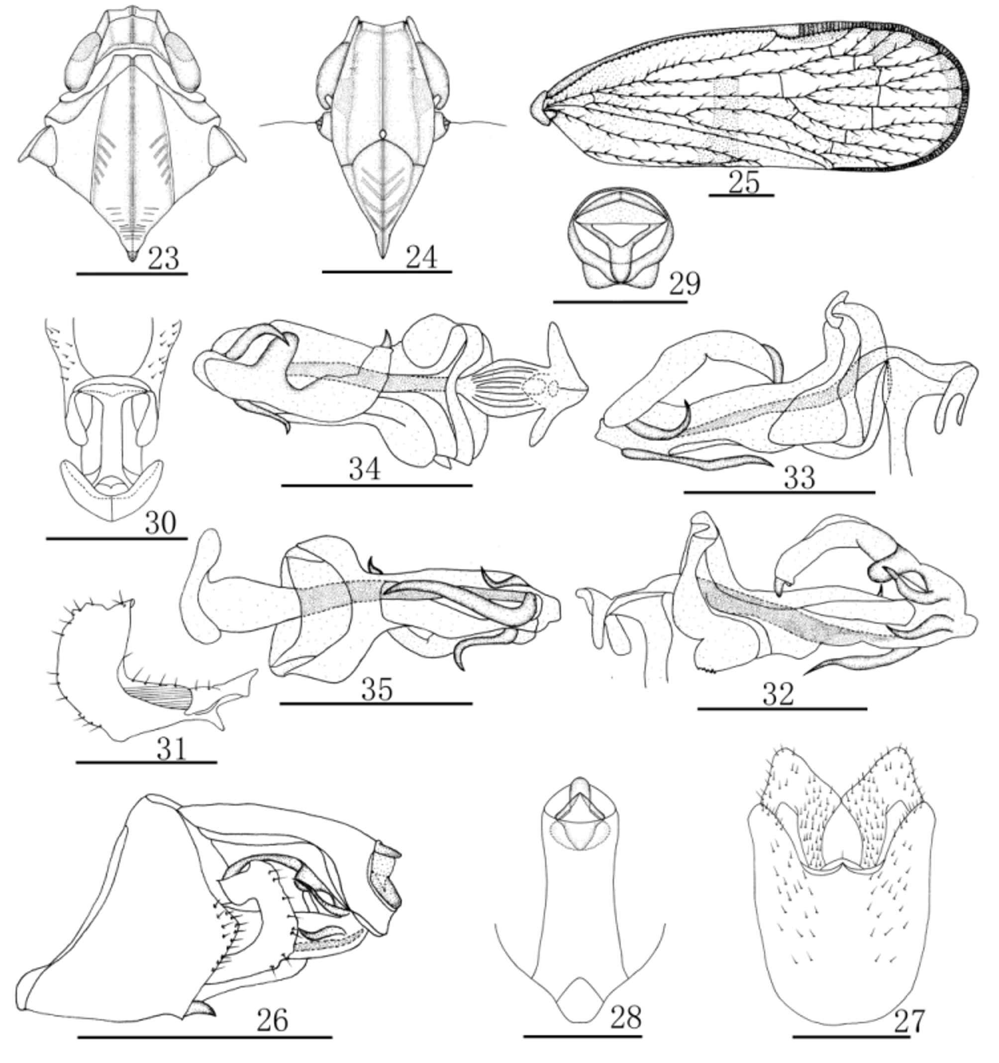
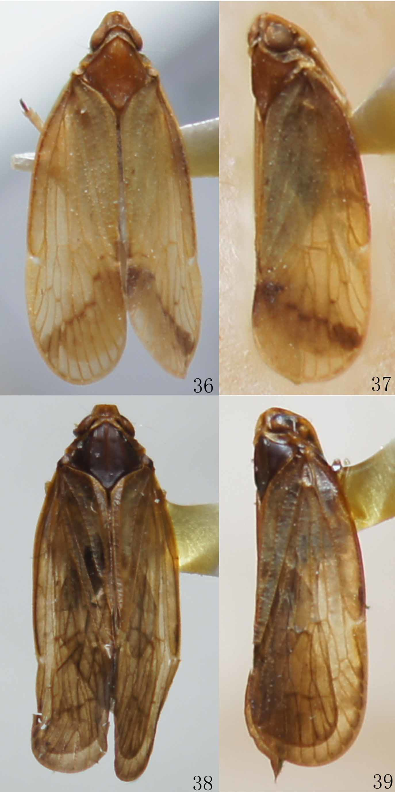
( Figs 23–35 View FIGURES 23 – 35 , 38, 39 View FIGURES 36 – 39 )
Description. Body length (from apex of vertex to tip of forewings): male 6.9–8.0 mm (n = 11), female 6.0– 8.5 mm (n = 22); forewing length: male 6.0–7.0 mm (n = 11), female 5.4–7.6 mm (n = 22).
Coloration. General color dark brown. Body covered with powdery wax. Eyes, anterior half blackish, posterior half brown. Median ocellus milk white, lateral ocellus pale yellow, semihyaline. Vertex dark brown, carinae black brown. Pronotum brown; Mesonotum black, carinae dark brown. Median area of frons black, lateral margin dark brown. Clypeus dark brown. Rostrum brown. Tegmina yellowish brown to dark brown, semihyaline, outer margin with spaced dimmed stains; veins yellowish brown (except C blackish brown), with concolorous tubercles; stigma blackish brown. Hind tibiae brown, lateral spines yellowish brown, apical spines black; hind tarsi with apical spines black and platellae yellowish brown. Abdomen black ventrally.
Head and Thorax. Eyes reniform, ventral margin concave above antennae; median ocellus slightly above intersection of frontoclypeal suture and median carina of frons. Vertex widening from subapical carina to both endpoints of lateral carinae as shown in Figs 23 View FIGURES 23 – 35 , 38 and 39 View FIGURES 36 – 39 ; 1.2 times wider than long; anterior margin with a middle cornuted process, posterior margin arc-shaped concave; area before subapical carina slightly hollowed, area behind subapical carina deeply hollowed, posterior margin forming a narrow footstep; both sides of median carina before subapical carina with arc-shaped scotches; subapical carina fusing with lateral carinae at apical seventh. Frons as shown in Figs 24 View FIGURES 23 – 35 , 1.4 times longer than wide; lateral carinae slightly S-like, elevated and lobate; anterior margin slightly concave into obtuse angle. Clypeus disc elevated, lateral side of median carina with oblique striations. Rostrum reaching hind coxae, apical segment 1.2 times longer than subapical segment. Pronotum as shown in Figs 23 View FIGURES 23 – 35 , 38 and 39 View FIGURES 36 – 39 , posterior margin deeply concave, forming obtuse angle; as long as vertex. Mesonotum 1.6 times longer than pronotum and vertex combined, inner sides of lateral carinae with oblique striations, median carina indistinct in posterior-median area, which bears transverse scotches. Forewings 2.7 times longer than wide, with setae on tubercles which situated along two sides of veins, CuP vein with tubercles but without setae; C with 45 tubercles; RP apically quadrifid, MA apically trifid, MP bifid; PCu+A1 relatively short; Sc+R and M fused out of superior angle of basal cell. Hind-tibiae with 2 lateral spines, 6 apical spines; chaetotaxy of hind tarsus 8–9/11, 2nd hind tarsus with 8 platellae.
Male Genitalia. Pygofer symmetrical; in lateral view, inner margin arc-shaped concave, dorsal margin tilted dorsad, lateral lobes symmetrical with medium parts caudally convex, triangular, with bristles; in ventral view, dorsal margin W-shaped, widening from base to apex. Medioventral process symmetrical, rounded in ventral view, with a seta at apex, 3.2 times wider than long, reaching to two ninths of length of lateral lobes; thumb shaped in lateral view, base covered by lateral lobes. Anal segment as shown in Figs 26, 28 and 29 View FIGURES 23 – 35 ; L-shaped in lateral view; symmetrical in caudad view, dorsal margin arched convex, ventral margin slightly concave into obtuse angle; 2.4 times longer than wide in dorsal view; closely connected with pygofer by two points; anal style short and stout, finger-like, not protruding anal segment. Genital styles as shown in Figs 26, 27 and 31 View FIGURES 23 – 35 ; in ventral view, symmetrical, hammer-like, with dense setae, internal processes broad, sub-angular, touching each other; in lateral view, ventral margin of basal part straight, apical part bending dorsad, dorsal margin strongly bent cephalad; closely connected with connective, unmovable. Aedeagus short and stout, as shown in Figs 26, 32–35 View FIGURES 23 – 35 , in total with four spines arising at apex of aedeagal shaft; two on left side, short and stout, both curved dorsocephalad; the other two on right side, one short and curved dorsocaudad, other one longer and directed ventrocephalad; both sides of periandrium with a hemispheric process. Connective slightly I-shaped as shown in Fig. 30 View FIGURES 23 – 35 , the width of aedeagal shaft 1.3 times as wide as the width of connective plus ventral arms. Flagellum semi-sclerotized, freely movable, arising slightly before apex of aedeagal shaft on the right side, generally curving left, basal third with a long and sub-oval process on left side, apex with a hornlike process.
Type material. Holotype: 3, Lijiaba (700m), Mayanghe National Natural Reserve, Yanhe County, Guizhou Province, China, 5–12 June 2007, X.-S. Chen. Paratypes: 4 3, 11 ƤƤ, same data as holotype; 2 ƤƤ, Xiannvdong (640m), Dashahe Provincial Natural Reserve, Daozhen County, Guizhou Province, China, 26 August 2004, X.-S. Chen; 4 3, 5 ƤƤ, Forest Park (1000m), Guiyang, Guizhou Province, China, 21 May 2006, P. Zhang; 2 ƤƤ, Taipingshan (520–859m), Liping County, Guizhou Province, China, 15–23 July 2006, Q.-Z. Song; 1 Ƥ, Taipingshan (520–859m), Liping County, Guizhou Province, China, 15–23 July 2006, Z.-G. Zhang; 1 3, Lijiaba (700m), Mayanghe National Natural Reserve,Yanhe County, Guizhou Province, China, 5–12 June 2007, H.-S. Deng; 1 3, 1 Ƥ, Maojia (600–900m), Mayanghe National Natural Reserve, Yanhe County, Guizhou Province, China, 5–12 June 2007, Z.-G. Zhang.
Distribution. Southwest China (Guizhou Province).
Remarks. This new species is similar in appearance to D. cehengensis sp. nov, but differs from the latter in the shape of the medioventral process, the degree of sclerotisation of the flagellum, the shape of the flagellum, the shape of the internal processes of the genital styles and the shape and direction of the spines of the aedeagus.
Etymology. The name is derived from the Latin words trans- (transverse) and spinus (spine), which refers to the long and sub-oval process at the basal third part of flagellum.
No known copyright restrictions apply. See Agosti, D., Egloff, W., 2009. Taxonomic information exchange and copyright: the Plazi approach. BMC Research Notes 2009, 2:53 for further explanation.