
Dinelytron grylloides Gray, 1835
|
publication ID |
https://doi.org/ 10.1016/j.jcz.2020.01.005 |
|
DOI |
https://doi.org/10.5281/zenodo.3716942 |
|
persistent identifier |
https://treatment.plazi.org/id/9D0A8794-FFDD-066C-05BC-65F9BCC2EC19 |
|
treatment provided by |
Plazi |
|
scientific name |
Dinelytron grylloides Gray, 1835 |
| status |
|
Dinelytron grylloides Gray, 1835 View in CoL
Fig. 11A-B View Fig .
Dinelytron grylloides Gray, 1835: 27 View in CoL ; Westwood 1859: 6; Kirby 1904: 408 (commentaries on holotype); Redtenbacher 1906: 150 (redescription); Rafael & Heleodoro 2017 (Brazilian catalog); Brock et al. 2019 (world catalog).
Examined material. “ FLORESTA da TIJUCA , D. Federal [Rio de Janeiro], BRASIL, iii - 1951, C. A. Campos Seabra [collector]” (1 _ MNRJ LOST IN THE BURN); “CEIOC 7645”, “Itstiaia [Itatiaia], E. [State] do Rio de Janeiro, Brasil, x - 1947 ”, “ Dinelytron sp, Conle O. det., XII. 2013 ” (1 _ CEIOC)/“ Espírito Santo, Brasil, J. Michaelis vend., 22.iv.1898 ”, “ Dinelytron grylloides Gray _, Jos. [ef] Redtenbacher determ. 1899, public. 1906-08, Bestimm. Vez. Nr. 386.” (1 _ ZMUH)/ “ Espírito Santo, Brasil”, “ Damasippus pulcher Redt., Det. REHN 19, Hebard cln.” (1 _ ANSP).
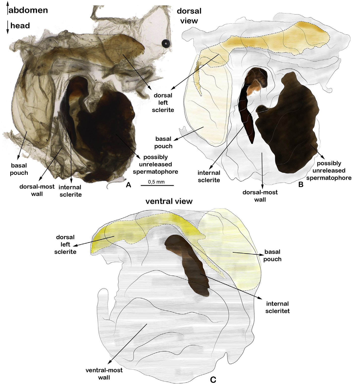
Diagnosis. Clypeus 5.1 times wider than high ( Fig. 11C, D View Fig ). Area between clypeus and labrum ellipsoid, conspicuous ( Fig. 11C, D View Fig ). Anterior femur five times longer than wide ( Fig. 11B View Fig ). Medial vein of tegmina bifurcated in Medial anterior and Medial posterior; Medial anterior bifurcated in Medial anterior 1 and 2 at approximately half tegmina length; Medial anterior 2 bifurcated in Medial anterior 2-1 and 2-2 right after previous bifurcation ( Fig. 11E View Fig ). Subgenital plate subellipsoid, 1.5 times longer than sternum 7, with widened and concave basal margin, convex lateral margin, apical margin medially deeply emarginated ( Fig. 12B View Fig ). Vomer subtriangular, not surpassing tergum 10; narrowing in medial third; lateral margin asymmetrical ( Fig. 12C View Fig ). Genitalia in dorsal view: basal pouch resembling a “D”. Tubes of dorsal left sclerite almost forming a right angle at the connection with basal pouch ( Fig. 13 View Fig ). Internal sclerite 1.1 times shorter than basal pouch, slim, slender ( Fig. 13 View Fig ).
Description _. General coloration light brown. Head. Frons with conspicuous frontal suture, forming a triangular sulcus, stained black ( Fig.11C View Fig ); coronal suture inconspicuous. Clypeus light brown, 5.1 times wider than high, with conspicuous semiellipsoid depression medially ( Fig.11C, D View Fig ). Area between clypeus and labrum ellipsoid, conspicuous ( Fig. 11C, D View Fig ). Labrum concolor with clypeus, asymmetric, with left half conspicuously longer than right half ( Fig. 11C, D View Fig ). Antenna with flagellomere 1 rectangular, two times longer than wide, 2.2 times longer than flagellomere 2; flagellomere 2 subquadrangular; flagellomere 3 subrectangular, 1.3 times wider than high, 1.2 times longer than flagellomere 2. Compound eye globose, light brown with black spots ( Fig. 11C View Fig ).
Thorax. Pro- and meso-notum rugose ( Fig. 11A View Fig ). Pronotum with conspicuous longitudinal medial sulcus ( Fig. 11A View Fig ). Mesonotum 1.7 times longer than pronotum, with longitudinal medial carina ( Fig. 11A View Fig ). Metanotum with longitudinal medial carina. Coxopleurite rugose, dark brown. Mesothoracic epimeron subtriangular, smooth; episternum rugose, with a sinuous longitudinal carina; white setae present at ventral margin ( Fig. 11B View Fig ). Metathoracic pleural region smooth, shiny, dark brown with small light brown spots ( Fig. 11B View Fig ). Probasisternum light brown, opaque, with black spot medially. Meso- and meta-basisternum shiny, dark brown, with concolor medial circular sclerite.
Legs. Anterior femur dorsally light brown, ventrally light yellow; mid and posterior femur idem but at anterior and posterior margins. Anterior femur five times longer than wide, anteriorly and posteriorly setose, with posterior margin slightly sinuous; dorsally with black spots and three parallel, longitudinal carinae ( Fig. 11B View Fig ); ventrally smooth. Anterior tibia anteriorly and posteriorly covered by setae, with three parallel, dorsally longitudinal carinae and black spots ( Fig. 11B View Fig ); ventrally smooth. Mid femur with granules and posteriorly longitudinal medial carina; posterodorsal margin sinuous, better observed dorsally; posteroventral margin with four spines. Posterior femur dorsally and ventrally covered by long setae; posteriorly with inconspicuous light brown spots, two inconspicuous parallel longitudinal carinae; posterodorsally sinuous, better observed dorsally ( Fig. 11B View Fig ); posteroventrally with six spines. Posterior tibia antero- and postero-dorsally with two conspicuous carinae.
Wings. Tegmina light brown, with white spots; radial vein light green, remaining veins light yellow ( Fig.11E View Fig ). Radial vein bifurcated in Radial anterior and posterior at apical third; Radial posterior bifurcated in Radial posterior 1 and 2 at tegmina apex ( Fig. 11E View Fig ). Medial vein bifurcated in Medial anterior and Medial posterior; Medial anterior bifurcated in Medial anterior 1 and 2 at approximately half tegmina length; Medial anterior 2 bifurcated in Medial anterior 2-1 and 2-2 right after previous bifurcation ( Fig. 11E View Fig ). Cubital vein slightly sinuous ( Fig. 11E View Fig ). Posterior wing with costal area white at proximal half, concolor with tegmina in distal half ( Fig. 11A View Fig ).
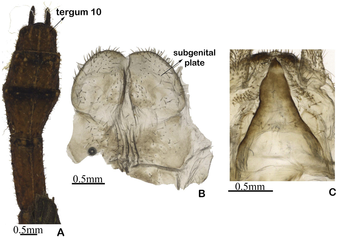
Abdomen. Abdominal terga dark brown. Terga 2-7 rectangular, decreasing gradually in length, longer than wide, with horizontal black spots at posterior margin and granules. Tergum 8 dorsally trapezoidal, with apical margin 1.5 times wider than basal margin ( Fig.12A View Fig ). Tergum 9 dorsally rectangular, wider than long ( Fig.12A View Fig ). Tergum 10 dorsally pentagonal with straight basal and lateral margin, apical margin slightly convex, forming a small apex medially ( Fig. 12A View Fig ). Cercus flattened laterally, black, densely covered by setae, with acute apex ( Fig.12A View Fig ). Abdominal sterna light brown. Sterna 1-6 rectangular, longer than wide, approximately of same length. Sterna 7-9 with small setae. Sternum 7 trapezoidal, with basal margin straight, 1.2 times wider than apical margin; lateral margin slightly curved posteriorly; apical margin straight. Sternum 8 subrectangular, 1.5 times wider than long, with basal margin sinuous, lateral margin convex, apical margin concave ( Fig. 12B View Fig ). Subgenital plate subellipsoid, 1.5 times longer than sternum 7, with widened and concave basal margin, lateral margin convex, medially deeply emarginated at apical margin ( Fig. 12B View Fig ). Vomer subtriangular, not surpassing tergum 10; narrowing in medial third; lateral margin asymmetrical ( Fig. 12C View Fig ).
Genitalia ( Fig. 13 View Fig ). Basal pouch resembling a “D”. Dorsal left sclerite attached to dorsal wall of genitalia; tubes of dorsal left sclerite almost forming a right angle at the connection with basal pouc. Internal sclerite 1.1 times shorter than basal pouch, slim, slender. Flagellum present. In Fig. 13A, B View Fig is displayed a dark mass at the left portion of dorsal lobe, resembling a sclerite. However, it is possibly a unreleased spermatophore formed prior to the specimens death.
Variations. Specimems from MNRJ and ZMUH have similar coloration. CEIOC specimen has a yellowish pronotum, tegmina greener and posterior legs and abdomen (dorsally) are light orange and dark brown respectively. The ANSP specimen has the entire body blackened and tegmina in dark tones of green.
Measurements (mm). Body length 37.0-38.5; dorsal head length 2.2; pronotum 2.3-2.7; mesonotum 3.2-3.6; anterior femur 7.8-8.0; anterior tibia 4-4.1; mid femur 5.5-5.7; mid tibia 2.5-2.7; posterior femur 8.2-8.5; posterior tibia 5.5-5.8.
Geographical records. Brazil, Espírito Santo; Rio de Janeiro: Itatiaia .
Remarks. As the type species of Dinelytron , this species is fundamental to the revision of the genus, as it provides a baseline for comparisons with other intrageneric species and serves as a reference for the differentiation of Dinelytron with other genera of Prisopodini . Westwood (1859) confirmed that the type specimen was lost, as it was included in the part of the entomological collection of the Museum of the Zoological Society which was sold.
The specimens were identified as Di. grylloides based on Redtenbacher (1906) description of the subgenital plate - “ Lamina subgenitalis _ apice profunde incisa ”, which can be translated freely as “male subgenital plate with deep incision at apex”. This is consistent with the subgenital plate observed in the designated specimens. Moreover, Redtenbacher identified the ZMUH specimen as Di. grylloides .

No known copyright restrictions apply. See Agosti, D., Egloff, W., 2009. Taxonomic information exchange and copyright: the Plazi approach. BMC Research Notes 2009, 2:53 for further explanation.
|
Kingdom |
|
|
Phylum |
|
|
Class |
|
|
Order |
|
|
Family |
|
|
Genus |
Dinelytron grylloides Gray, 1835
| Heleodoro, Raphael Aquino & Rafael, Jose Albertino 2020 |
Dinelytron grylloides
| Gray 1835: 27 |