
Cratera picuia, Lago-Barcia & Carbayo, 2018
|
publication ID |
https://doi.org/ 10.11646/zootaxa.4500.4.3 |
|
publication LSID |
lsid:zoobank.org:pub:70672C0A-EC78-40BA-85EE-6206184CE0F0 |
|
persistent identifier |
https://treatment.plazi.org/id/4F7187CF-C545-FFFC-D5A8-F0ECFECAF80D |
|
treatment provided by |
Felipe |
|
scientific name |
Cratera picuia |
| status |
sp. nov. |
Cratera picuia sp. n.
Synonymy
Geoplana hina ; Carbayo et al., 2013, non Cratera hina ( Marcus, 1951)
Type material. Holotype F1613 ( MZUSP PL 1008 ): Parque Nacional Saint-Hilaire / Lange , Matinhos, State of Paraná, Brazil (25.76437, -48.62266). F. Carbayo et al. coll., 10 January 2008, transverse sections of cephalic region on 11 slides; horizontal sections of ovaries region on 10 slides; transverse sections of prepharyngeal region on 9 slides; sagittal sections of pharynx and copulatory apparatus on 7 slides. GoogleMaps
Distribution. Only known from the type locality, Parque Nacional Saint-Hilaire/Lange, Matinhos/ PR, Brazil.
Etymology. The name picuia is a free composition of the Tupi (indigenous Brazilian language) words py (meaning inside) and cuia (meaning gourd, the dried shell of a cucurbit plant ( Tibiriçá, 1984)). It refers to the uncommon, inner location of the prostatic vesicle inside the penis bulb.
Diagnosis. Species of Cratera with dorsal color comprising a pair of pearl orange paramedian bands separated by a median ivory-colored band, and paired oyster-white marginal bands, each one longitudinally divided in half by black grey spots. Prostatic vesicle canalicular, located inside penis bulb. Ejaculatory duct not dilated distally.
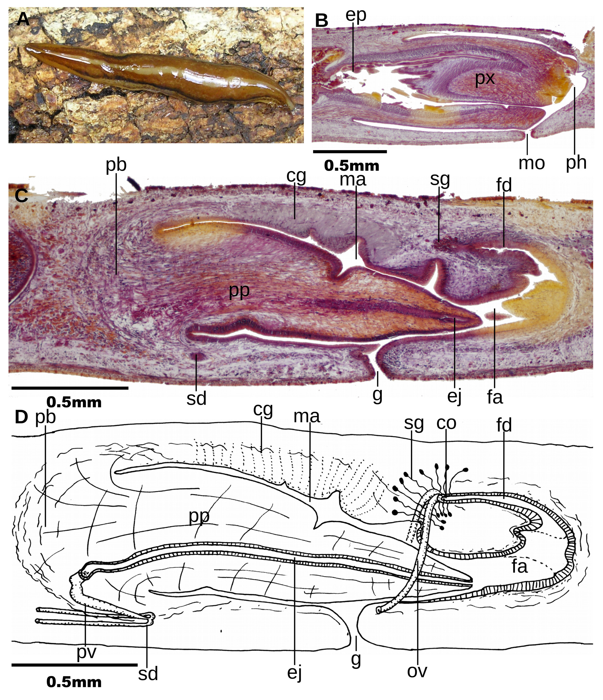
Description. External aspect. Live holotype with elongated body, with margins approximately parallel; anterior extremity of the body damaged during handling and lost ca. 0.5mm; posterior end damaged, but pointed. Dorsum convex, ventral side slightly convex. Dorsal color comprises a pair of pearl orange paramedian bands (1.5/ 7th) separated by a median ivory-colored band (1/7th of body width), and paired oyster white marginal bands (1/ 7th) each one longitudinally divided in half by black grey spots which become more abundant posteriorly so as to create an irregular longitudinal stripe ( Fig. 11A View FIGURE 11 ). Ventral body surface, white. After fixation, body became cream with the exception of the black grey lines, which became brown grey. Preserved holotype 28 mm long, and 3 mm wide. Eyes monolobulated, 50 µm in diameter. A ~ 0.5 mm long section of anterior apex of the body was lost; it is assumed they contour cephalic region. They spread over dorsum from anterior extremity, backwards. At the level of the pharynx they extend on a lateral band, on each side of the body, with 1/3 of the body width. From that region towards the posterior extremity they spread on a band progressively narrower. On black-brownish bands, small halos around each eye. Sensory pits are 30 µm deep invaginations arranged in a single row. It is assumed sensory pits contour anterior end. Behind, sensory pits extend backwards to at least a length equal to 36% of body length. Relative mouth:body length, 68% and relative gonopore:body length, 75%.
Internal morphology. Creeping sole occupies 88% ventral body width. Rhabditogen cells and erythrophil glands producing erythrophil fine granules pierce dorsal epidermis. Glandular margin comprised of two types of glands producing granules, erythrophil and xanthophil, respectively. Ventral epidermis pierced by glands producing erythrophil granules.
Cutaneous musculature with the usual three layers present in the subfamily Geoplaninae : subepithelial circular (one-fiber thick), diagonal with decussate bundles (5 µm thick) and an innermost longitudinal layer (27 µm thick dorsally, 47 µm ventrally), the latter is arranged in bundles. Cutaneous musculature as thick as 9% of body height. Parenchymal musculature composed of three muscular layers: a dorsal layer with diagonal decussate fibers (20-25 µm thick), a supraintestinal (70 µm thick) loose layers and a subintestinal transverse layer (50 µm thick). Ventral nerve plate present.
Relative position mouth:pharyngeal pouch length, of 72% ( Fig. 11B View FIGURE 11 ). Pharynx cylindrical, with its dorsal insertion placed backwards. Esophagus present, with 30% of pharynx length.
Close to the insertions of the pharynx, outer epithelium of the pharynx underlain by a thin layer (3 µm thick) of longitudinal muscle, followed by a thin layer of circular fibers (5 µm thick). Under epithelium of the distal section of the pharynx, the longitudinal layer seems to be absent. Inner pharynx musculature consisting of a subepithelial layer (2 µm thick) of longitudinal musculature, followed by a circular layer (80 um thick) and an outermost 10 um thick layer of longitudinal fibers.
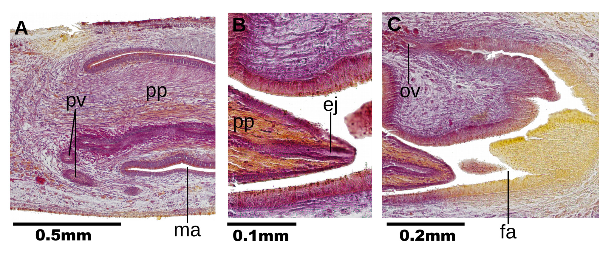
Testes dorsally located between intestinal branches. Testes are arranged in single-to-double row on each side of the body. They extend from 0.5 mm behind the level of the ovaries (equal to 30% of the body length) to 0.6 mm before the root of the pharynx (equal to 59% of the body length). Penis bulb consisting of packed muscle fibers variously oriented. This bulb extends from 0.3 millimeters anterior to penis papilla to envelope the anterior 0.25 mm of the male atrium. In their distal section, sperm ducts run medially to join below the level of the ventral insertion of the penis papilla ( Fig. 11C View FIGURE 11 ), and subsequently continuing with the intrabulbar prostatic vesicle ( Figs. 11D View FIGURE 11 and 12A View FIGURE 12 ). The prostatic vesicle is canalicular, with proximal portion running anteriorly, and distal, shorter portion running posteriorly. Prostatic vesicle lined by a columnar, ciliated epithelium, which is surrounded by a 10 µm thick circular muscle. Glands producing intense erythrophil granules open into prostatic vesicle. The anteriormost portion of the ejaculatory duct is out of the penis papilla and is slightly sinuous. Most of the ejaculatory duct runs straight through the ventral portion of the penis papilla. The ejaculatory duct is lined with a cuboidal, ciliated epithelium. It is surrounded by a 15 µm thick circular muscle. The lumen of ejaculatory duct is 10 µm in diameter proximally; very gradually it narrows to 5 µm distally before opening at the tip of the penis papilla ( Fig. 12B View FIGURE 12 ). There is no distal widening of the ejaculatory duct. Anterior 3/4th of this duct is pierced by very numerous glands producing erythrophil granules.
Penis papilla relatively conical, with its dorsal insertion slightly anterior than the ventral. It exceeds the gonopore level to occupy anterior half of the female atrium. Anteriorly, this papilla is lined with a columnar epithelium; gradually, this epithelium passes to squamous. Penis papilla epithelium is underlain by a circular muscle (18 µm thick), followed by a longitudinal muscle (4 µm thick). Most epithelium of penis papilla is pierced by glands producing xanthophil granules, but they are most abundant along distal half. Glands producing cyanophil granuels are scarce and restricted to the proximal, dorsal half of the epithelium. Male atrium not folded. It is lined with a cuboidal, non-ciliated epithelium, and is underlain by an 5-10 µm thick circular muscle followed by a 5 um thick longitudinal muscle. Roof of male atrium pierced by a large mass of glands producing cyanophil granules.
The ovaries are ovoid, 400 µm long and 200 µm wide. They are located at a distance from anterior end equal to 28% of body length. The ovovitelline ducts arise from the dorso-lateral aspect of the ovaries. Ovovitelline ducts rise laterally to the female atrium, and join to form the very short common glandular ovovitelline duct (50 µm) above female atrium. Numerous shell glands discharge into final portion of ovovitelline ducts. The common glandular ovovitelline duct joins the long female genital duct (400 µm) ( Fig. 12C View FIGURE 12 ). This duct is an anteriorly directed projection from the postero-dorsal region of female atrium. It is lined with a columnar, ciliated epithelium which is pierced by glands producing cyanophil granules. The glands piercing this epithelium are scarce but their bodies are abundant surrounding the epithelium.
Female atrium is ample, and not separated from the male atrium. A dorso-lateral fold narrows the lumen of the female atrium. Female:male atrial length is 8.5:10. This female atrium is lined with a nonciliated, columnar epithelium, which is 30 µm high. The female lining epithelium is surrounded by a 10–15 µm thick muscle layer of longitudinal and circular fibers intermingled. This epithelium is pierced by two types of glands, one producing eryhtrophil granules, another producing cyanophil granules.
Remarks. This species was confounded with Cratera hina ( Marcus, 1951) by Carbayo et al. (2013). The chromatic pattern of the dorsum of C. picuia sp. n. resembles that of C. hina but is not identical. Two circumstances led Carbayo et al. (2013) to misidentify the specimen (FC, pers. obs.): (a) the fact that intraspecific variation in the dorsal color pattern is not rare in geoplaninids [e.g. Cephaloflexa bergi ( Graff, 1899) , Obama josefi ( Carbayo & Leal-Zanchet, 2001) , O. nungara Carbayo et al., 2016 ]; and (b) the close resemblance of the shape of the internal organs of the new species, even the location of the prostatic vesicle, with that of the original description of C. hina . However, as shown above, in C. hina the prostatic vesicle is extrabulbar, whereas in C. picuia sp. n. the prostatic vesicle is intrabulbar. This latter trait is unique in Cratera and differentiates readily C. picuia sp. n. from all its congeners.
| MZUSP |
Museu de Zoologia da Universidade de Sao Paulo |
No known copyright restrictions apply. See Agosti, D., Egloff, W., 2009. Taxonomic information exchange and copyright: the Plazi approach. BMC Research Notes 2009, 2:53 for further explanation.
|
Kingdom |
|
|
Phylum |
|
|
Order |
|
|
Family |
|
|
Genus |
Cratera picuia
| Lago-Barcia, Domingo & Carbayo, Fernando 2018 |
Cratera hina ( Marcus, 1951 )
| Lago-Barcia & Carbayo 2018 |
Geoplana hina
| Marcus 1951 |