
Conura paraleucotela, Brotto & Tavares, 2021
|
publication ID |
https://doi.org/ 10.11646/zootaxa.4942.3.5 |
|
publication LSID |
lsid:zoobank.org:pub:FF2841C8-D952-44E0-83AC-75C3FA5953EC |
|
DOI |
https://doi.org/10.5281/zenodo.4619701 |
|
persistent identifier |
https://treatment.plazi.org/id/03A3AD23-625B-C664-FF29-7BDDFDE028E1 |
|
treatment provided by |
Plazi |
|
scientific name |
Conura paraleucotela |
| status |
sp. nov. |
Conura paraleucotela sp. nov.
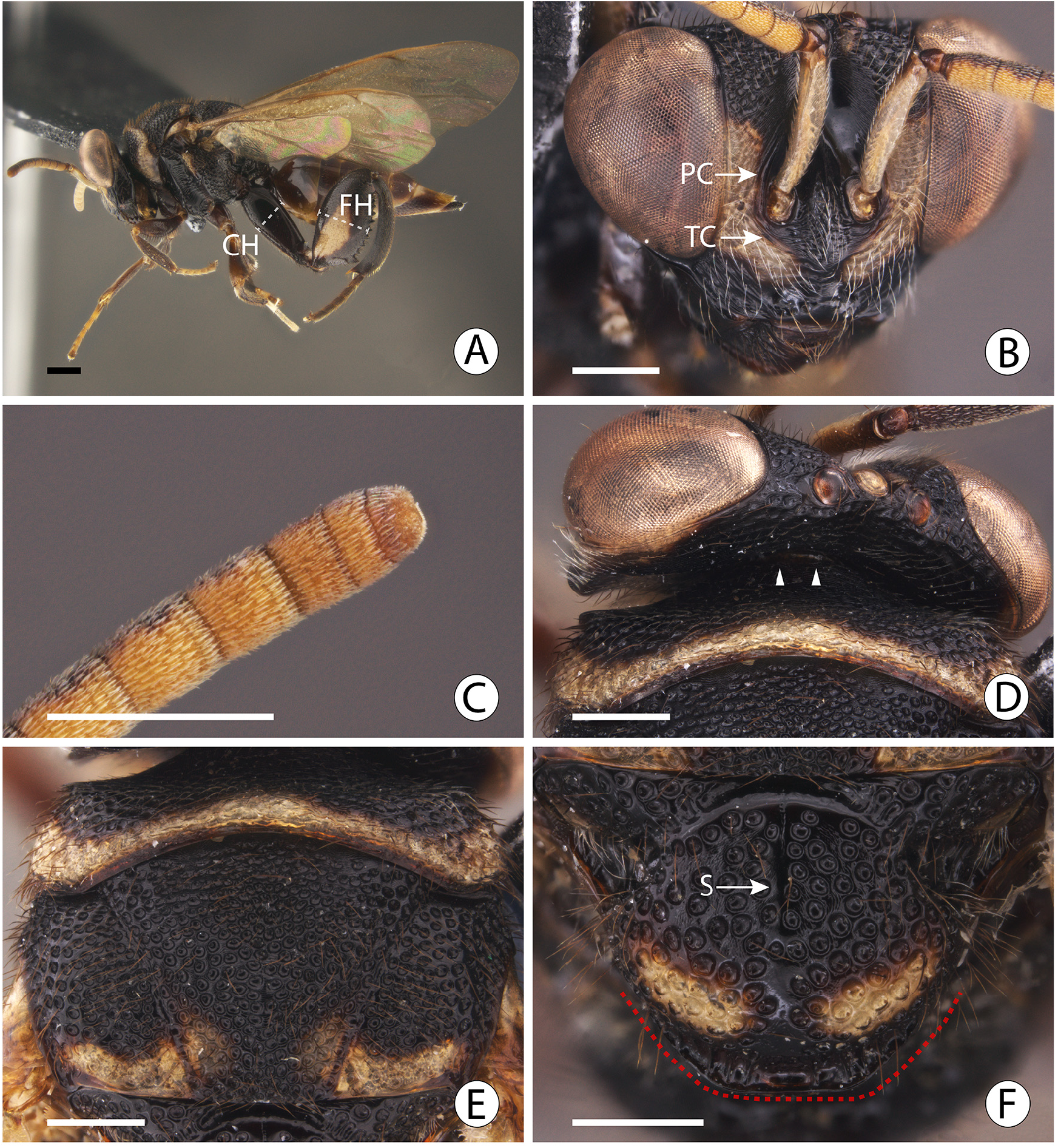
Figs 3 View FIGURE 3 A–F; 4A–D
Zoobank: urn:lsid:zoobank.org:act:F34A8B89-0F11-4682-BBB6-80DC4EADFD79
Etymology. From the Greek prefix παρά- (“ para ” meaning resembling, near), referring to the close similarity of the female of this species to those of C. leucotela and C. pseudoleucotela .
Description. FEMALE. Holotype: length 7.36 mm.
Color Body mainly black ( Fig. 3A View FIGURE 3 ), but antennal scape on dorsum, pedicel and anellus ( Fig. 3B View FIGURE 3 ), dorsum of Fu1 to clava ( Figs 3A, 3B View FIGURE 3 ), front and middle legs ( Fig. 3A View FIGURE 3 ), middle of metascutellum ( Fig. 4A View FIGURE 4 ), and gaster brown ( Figs 4C, D View FIGURE 4 ), and following yellow: submedian spots anterior to median ocellus ( Fig. 3B View FIGURE 3 ), venter of antennal scape, funiculars and clava, lower half of parascrobal area ( Fig. 3B View FIGURE 3 ); side of lower face dorsally ( Fig. 3B View FIGURE 3 ), upper part of gena ( Figs 3A, 3D View FIGURE 3 ), posterior transversal stripe on dorsum of pronotum ( Figs 3D, E View FIGURE 3 ), posterior margin of mesoscutum except medially ( Fig. 3E View FIGURE 3 ), tegula, posterior spot on axilla; large submedian spot on either side of mesoscutellum in posterior half ( Fig. 3F View FIGURE 3 ), sides of metascutellum, lateral panel of pronotum ( Fig. 3A View FIGURE 3 ), spot on mesepimeron ( Fig. 3A View FIGURE 3 ), inner face of protibia, protarsus, base of mesotibia, mesotarsus, spot on metacoxa ventrodistally, wide stripe on inner and outer faces of metafemur ( Fig. 3A View FIGURE 3 ), transverse band on gastral tergites, and base and apex of Gt7+8 ( Figs 3A View FIGURE 3 , 4D View FIGURE 4 ). Wings hyaline, veins dark brown.
Head. Clava with second segment slightly wider than the first segment so as to appear slightly swollen ( Fig. 3C View FIGURE 3 ); paratorular carina longer than greatest diameter of antennal foramen; lower face with conspicuous transverse carina below antennal foramen ( Fig. 3B View FIGURE 3 : TC).
Mesosoma. Pronotal anterior furrow delimited posteriorly by sharp margin ( Fig. 3D View FIGURE 3 , arrow heads); mesoscutum median area with regular umbilicate foveae, interstices not so narrow, giving sculpture a reticulate appearance ( Fig. 3E View FIGURE 3 ); mesoscutellum convex, basally with wide smooth and shiny transverse band ( Fig. 3F View FIGURE 3 ), with median nonfoveate strip over basal half ( Fig. 3F View FIGURE 3 : S), and diameter of foveae on disc 0.47–0.52× MOD, with interstice width usually greater than 0.5× diameter of foveae ( Fig. 3F View FIGURE 3 ); frenal carina almost straight posteriorly ( Fig. 3F View FIGURE 3 : red dashed line); metascutellum convex, smooth and shiny; propodeum oblique ( Figs 3A View FIGURE 3 , 4D View FIGURE 4 ), anterior costula conspicuous laterally, inconspicuous medially (( Fig. 4A View FIGURE 4 : ACP), median carina (anterior to posterior costula) 0.4× median length of propodeum ( Fig. 4A View FIGURE 4 : MCP), posterior costula conspicuous and limited to posterior half of propodeum ( Fig. 4A View FIGURE 4 : PCP), adpetiolar area with median and submedian carinae ( Fig. 4A View FIGURE 4 : AdA); metafemur with 10 teeth, basal tooth not followed by a minute tooth; metasternum concave, with median carina conspicuously raised as lamina ( Fig. 4B View FIGURE 4 : MCM).
Metasoma. Petiole visible dorsally, short, 0.60× as long as wide ( Figs 4C, D View FIGURE 4 ), basal lamina present dorsally and ventrally ( Fig. 4D View FIGURE 4 ), submedian carinae absent, one lateral carina present; Gs1 not projected forward, petiole attached to gastral base ( Fig. 4D View FIGURE 4 ); dorsally, Gt1–Gt5 about 0.6× as wide as long ( Fig. 4C View FIGURE 4 ); Gt6 with posterior margin slightly concave ( Fig. 4C View FIGURE 4 ); Gt7+8 about 0.3× as long as Gt1–Gt6 combined ( Fig. 4D View FIGURE 4 ).
MALE. Unknown.
Host. Unknown.
Distribution: BRAZIL (Amazonas: Manaus).
Remarks. The only known female of C. paraleucotela is easily distinguished from those of C. leucotela and C. pseudoleucotela by having the propodeum oblique ( Fig. 3A View FIGURE 3 , 4D View FIGURE 4 ), metasternum slightly concave ( Fig. 4B View FIGURE 4 ), petiole short, but dorsally visible ( Fig. 4C View FIGURE 4 ), Gs1 not projected anteriorly ( Fig. 4D View FIGURE 4 ), and Gt7+8 shorter, about 0.3× as long as Gt1–Gt6 combined ( Figs 4C, D View FIGURE 4 ).
Material Examined. HOLOTYPE ( INPA): female, labeled ‘BRA[ Brazil], AM [Amazonas], Manaus, ZF 3 [ Reserva Florestal ZF-3], Km 23, Res. 1112, 03. VI. 1986, Armad [ilha]. Malaise, Bert Klein col.’. The specimen has the right and left hind legs glued on a card.

| INPA |
Instituto Nacional de Pesquisas da Amazonia |
| AM |
Australian Museum |
| VI |
Mykotektet, National Veterinary Institute |
No known copyright restrictions apply. See Agosti, D., Egloff, W., 2009. Taxonomic information exchange and copyright: the Plazi approach. BMC Research Notes 2009, 2:53 for further explanation.