
Cnemidocarpa concha, Monniot, 2002
|
publication ID |
https://doi.org/ 10.1046/j.1096-3642.2002.00017.x |
|
persistent identifier |
https://treatment.plazi.org/id/436F3F3C-FFCC-FFB0-FF21-FB50FCAF1B2F |
|
treatment provided by |
Carolina |
|
scientific name |
Cnemidocarpa concha |
| status |
sp. nov. |
CNEMIDOCARPA CONCHA View in CoL SP. NOV. ( Fig. 22 View Figure 22 )
Part of Cnemidocarpa hemprichi: Michaelsen, 1919: 76 , pl. 1, fig. 9.
Material
Syntypes: MNHN S1 CNE 189 View Materials , Yemen: Socotra Island, coll. C. Monniot (1997) .
Other material examined: Djibouti, coll. F. Jousseaume & H. Coutière, 1897. Gulf of Suez, coll. R. Ph. Dollfus, 1929.
Description
The largest of nine specimens is 9 cm in length. The body is pear-shaped, the anterior part narrower than the posterior part that contains the digestive tract. The cloacal siphon opens in the middle of the dorsal side. The tunic is very hard, thick, of a brownish red colour, with large longitudinal swellings. There are few epibionts.
In formalin the body wall is pale in colour except for the siphonal rim which is darker. Its tissue is opaque and thick. The oral siphon is short. About 30 oral tentacles are irregularly distributed, in two orders of size.
The prepharyngeal band has two blades, the posterior one higher. The dorsal tubercle is large and occupies a large part of the deep V drawn by the prepharyngeal band. The aperture is convoluted.
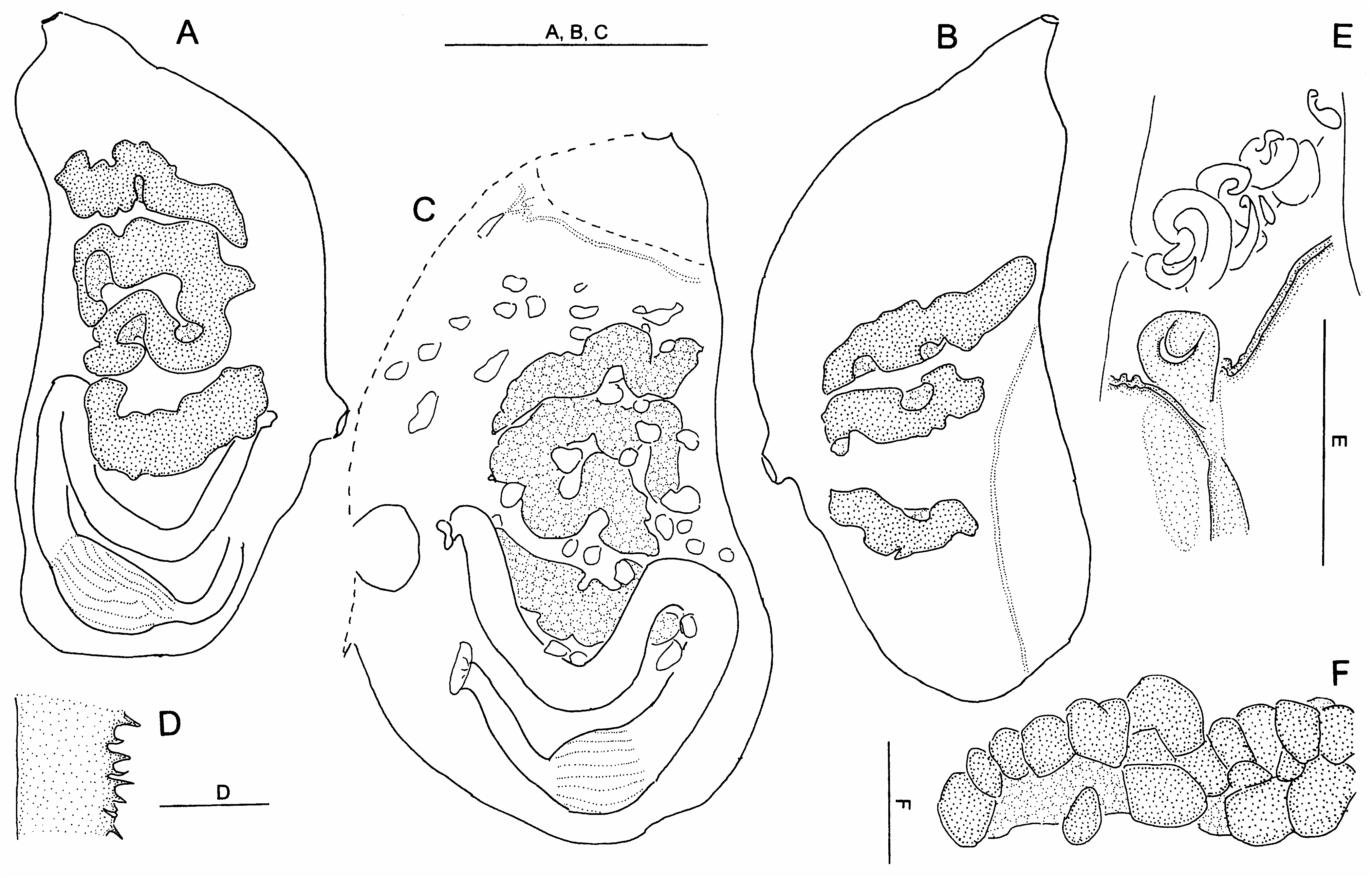
The four branchial folds on each side lie well apart from each other. An average of 30 longitudinal vessels were counted on the folds and ten between the folds. Between the folds there are six stigmata per mesh, cut by a parastigmatic vessel. The branchial sac extends posteriorly to the oesophageal opening. The dorsal lamina is a blade of equal height along its length. The endostyle is wide, curved along the posterior part of the branchial sac. Its edges fuse a little before the oesophagus entrance. Then it forms a closed appendix, coiled like a snail, protruding into the cloacal cavity against the oesophagus ( Fig. 22C View Figure 22 ).
The gut occupies the posterior quarter of the body, in a double loop ( Fig. 22A View Figure 22 ). The stomach is spindlelike, passing evenly into a narrower intestine that remains cylindrical along its whole length. There is no caecum. When the intestine reaches the oesophagus level, it curves in a rather long narrow loop ( Fig. 22A View Figure 22 ). The lobed anus is close to the cloacal aperture ( Fig. 22A View Figure 22 ).
The gonads are elongated, straight, parallel, and very numerous ( Fig. 22A,B View Figure 22 ). Few of them are ramified. The genital openings, at the apex of each gonad, are aligned along a wide curve around the cloacal aperture ( Fig. 22A View Figure 22 ). The gonads are included within the opaque internal layer of the body wall.
Numerous endocarps are distributed over the whole internal side of the body wall, both among the gonads and in the gut loop ( Fig. 22A,B View Figure 22 ). They are transparent vesicles of irregular shapes.
Remarks
This species differs from all described Cnemidocarpa species by its voluminous snail-like coiled postendostylar appendix. An uncoiled endostylar appendix of equivalent structure occurs in Ciona intestinalis ( MacDonald, 1859; Roule, 1884; Millar, 1953; Hoshino & Tokioka, 1967). This kind of structure has only been mentioned by Michaelsen (1919) in Cnemidocarpa hemprichi Hartmeyer, 1916 . I think that Michaelsen (1919) assembled under this name several similar species. Among them the largest specimens, with numerous radiating gonads, belong to the new species C. concha .
Cnemidocarpa concha differs from C. hemprichi by the large size of its specimens, its very thick body wall, and the convoluted aperture of its dorsal tubercle. The branchial sac, which has many more longitudinal vessels on and between the folds, may be taken to have the same structure, taking in account the larger size of the C. concha specimens. In C. concha the gonads are more numerous than in C. hemprichi , they are in long tubes, and regularly arranged with their genital papillae on a circle.
The geographical distribution of C. concha covers the Red Sea from Suez to Djibouti and extends to Socotra Island.
Etymology
From the Latin, concha = shell, to refer to the snaillike postendostylar appendix of this species.
CNEMIDOCARPA HEMPRICHI HARTMEYER, 1916 View in CoL ( Fig. 23 View Figure 23 )
Cnemidocarpa hemprichi Hartmeyer, 1916: 218 View in CoL , figs 6,7 – Red Sea.
Not Cnemidocarpa hemprichi: Michaelsen, 1919: 76 View in CoL ; not Monniot C., 1973: 51, fig. 1 = Cnemidocarpa schumacheri View in CoL sp. nov.
Cnemidocarpa madagascariensis Hartmeyer, 1916: 222 View in CoL , figs 8,9 – Madagascar; Vasseur, 1967: 116, pl. 5, figs 41–43 – Mauritius.
Material
Yemen: Socotra Island, 1 specimen, coll. C. Monniot, 1997 .
Description
The specimen measures 3 cm in length. The tunic is thin, soft but resistant, with a wrinkled surface without epibionts. The internal anatomy corresponds exactly to Hartmeyer’s description. The oral tentacles are numerous, in three orders of size. The dorsal tubercle opens in a C-shaped slit ( Fig. 23E View Figure 23 ). The dorsal lamina is high with a denticulate edge ( Fig. 23D View Figure 23 ). The four folds on each side of the branchial sac are clearly separated. The branchial formula on the right side is:

| MNHN |
Museum National d'Histoire Naturelle |
| R |
Departamento de Geologia, Universidad de Chile |
| V |
Royal British Columbia Museum - Herbarium |
No known copyright restrictions apply. See Agosti, D., Egloff, W., 2009. Taxonomic information exchange and copyright: the Plazi approach. BMC Research Notes 2009, 2:53 for further explanation.
|
Kingdom |
|
|
Phylum |
|
|
Class |
|
|
Order |
|
|
Family |
|
|
Genus |
Cnemidocarpa concha
| Monniot, Claude 2002 |
Cnemidocarpa hemprichi: Michaelsen, 1919: 76
| Michaelsen W 1919: 76 |
Cnemidocarpa hemprichi
| Hartmeyer R 1916: 218 |
Cnemidocarpa madagascariensis
| Vasseur P 1967: 116 |
| Hartmeyer R 1916: 222 |