
Chimarra fijiana, Johanson, Kjell Arne & Oláh, János, 2012
|
publication ID |
https://doi.org/ 10.5281/zenodo.210736 |
|
DOI |
https://doi.org/10.5281/zenodo.5664494 |
|
persistent identifier |
https://treatment.plazi.org/id/9F3E87DD-560E-FFF7-E89A-FDA0FE50FAA1 |
|
treatment provided by |
Plazi |
|
scientific name |
Chimarra fijiana |
| status |
sp. nov. |
Chimarra fijiana , new species
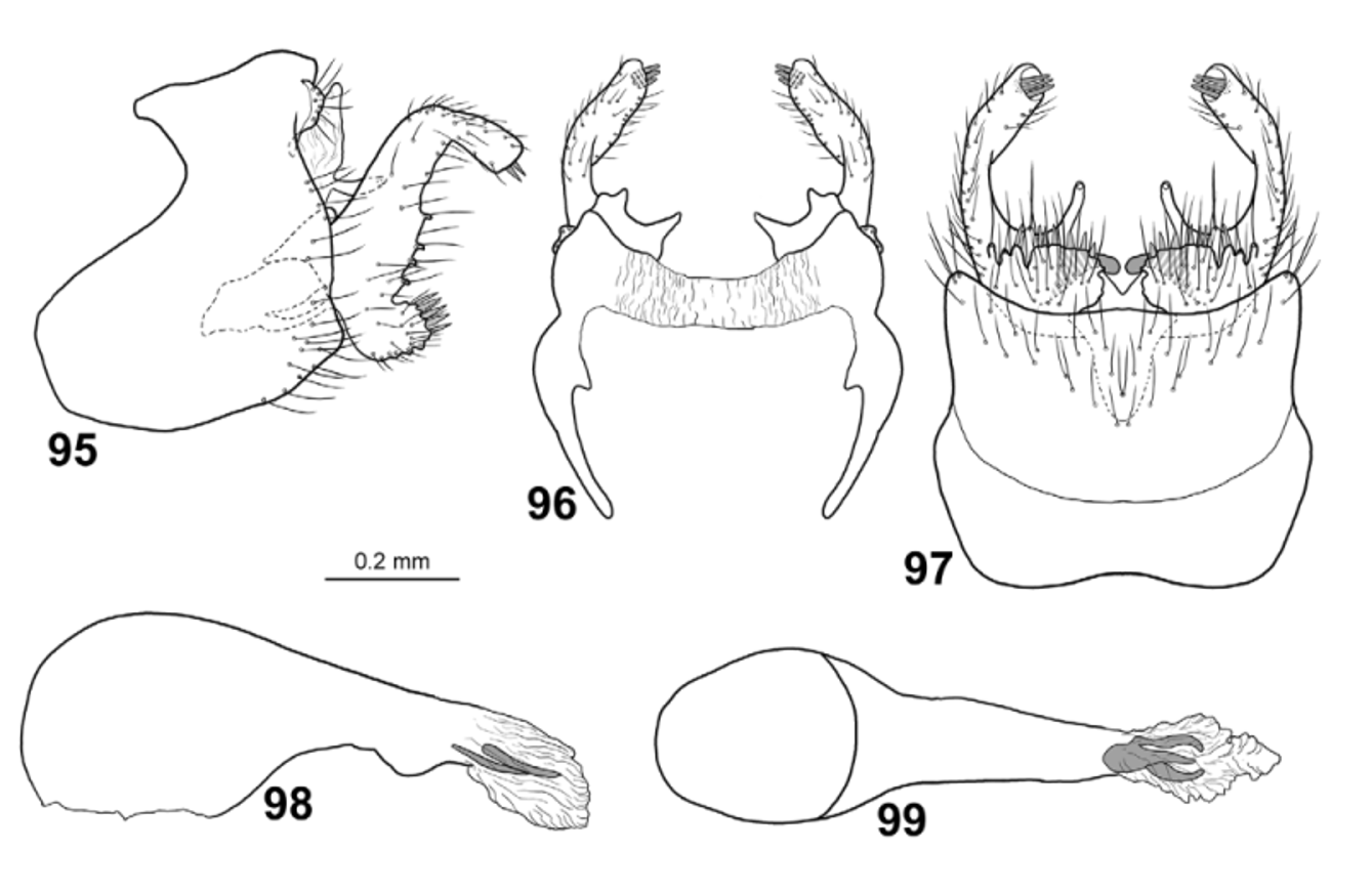
Figs. 18 View FIGURES 14 – 21 , 95–99 View FIGURES 95 – 99
The genitalia of C. fijiana are unique among the Fijian Chimarra in having both the ventral branch and the distal part of each gonopod angled posteriorly at 90°, and both apices of the gonopods with apical megasetae.
Male. Body dark pale yellowish-brown, dorsal part of meso and metathorax slightly darker than rest of body. Large dark area between lateral ocelli. Foreleg anterior claw as long as foreleg spur.
Wings ( Fig. 18 View FIGURES 14 – 21 ). Forewings 5.4 mm (n=1), brown; Forewings broad, ratio of length to breadth 3.4; R1 slightly curved before crossvein r; radial sector not produced anterad immediately before discoidal cell; discoidal cell originating slightly before mid-length of wing, about 3x longer than wide; median cell as long as discoidal cell; crossvein r originating from basal part of R2; fork I originating before crossvein s at distance equal to length of crossvein s; nygma located near base of fork II; fork III1 /4 as long as wing; fork V slightly shorter than fork II; Cu2 ending in wing margin close to A. Hind wings 4.3 mm (n=1), brown; ratio of length to breadth 3.0; margin weakly incurved at arculus, where Cu1 and Cu2 fused with margin; fork I sessile; fork III more than 2x longer than discoidal cell and1/4 as long as wing; fork V about as long as fork I; 1A+2A more than 3x longer than 1A.
Male genitalia ( Figs. 95–99 View FIGURES 95 – 99 ). Segment IX slightly taller than long; anterodorsal margins strongly concave in lateral view; ventral margins slightly convex; each posterior margin weakly produced posterad, starting immediately below each cercus, margin slightly undulating; dorsal part of segment IX produced anterad into triangular; ventral side of posterior 1/2 of segment IX covered by setae ( Fig. 97 View FIGURES 95 – 99 ). In dorsal view with pointed anterior lobes; anterodorsal plates widely separated by rectangular incision in dorsal view. In ventral view segment IX with shallowly concave anterior and posterior margins; posterior margin with very short central projection. Tergum X divided into dorsal and ventral branches. Dorsal branches of tergum X oriented vertically immediately posteriorly of segment IX, narrowing dorsally in lateral view; tapering apically in lateral view ( Fig. 95 View FIGURES 95 – 99 ); in dorsal view divided into pair of broad plates with pointed mesal and lateral corners ( Fig. 96 View FIGURES 95 – 99 ). Ventral branches of tergum X originating from lower part of dorsal branches, slender, finger-like, curved posterad in lateral and ventral view, with pair of sensillae located near apex of each branch. Cerci very short, wart-like; located dorsally on segment IX, near dorsal margin; covered by long setae. Gonopods shorter than segment IX, uniformly slender in lateral view; nearly Ushaped, with dorsal and ventral branch. Dorsal branch of each gonopod long, each with basal 2/3rds oriented nearly vertically, bent 90° posteriorly at apical 1/3rd; with 3 ventromesad orienting megasetae originating from ventromesal pocket at apex; ventral part of posterior margin of dorsal branch with large setal tubercles. Ventral branch of each gonopod strongly curved posterodorsad at base; with row of megasetae. Phallic apparatus slightly longer than rest of genitalia; phallotheca, in lateral and ventral view, with anterior part about 2x thicker than posterior part; posterior part narrowing distally in ventral view; posterior part with rounded ventral process in lateral view; apicoventral spine absent; phallotremal sclerite forming 3 longitudinal rays in ventral view; endothecal spines not observed.
Female. Unknown.
Holotype male: VITI LEVU: Vuda Prov., Koroyanitu Natural Heritage Park, Savuione Trail, Malaise trap, 12–19.x.2002, 17°40’S, 177°33’E, 450 m, M. Irwin, E. Schlinger & M. Tokota’a [loc#04] [ FNIC].
Paratypes: S ame data as holotype [loc#04] — 9 males [ NHRS]. Same data as holotype, except 21.ix–7.x.2002 [loc#04] — 2 males [ BPBM]. Vuda Prov., Koroyanitu Pk., 1 km E Abaca Vlg., Savuione Trail, Malaise trap, 26.x–5.xi.2002, 17°40’S, 177°33’E [17.3333°S, 177.55°E], 800 m, leg. M. Irwin, E. Schlinger & M. Tokota’a [loc#03] — 1 male [ BPBM]. Same data, except 12–19.xi.2002 [loc#03] — 1 male [ BPBM]. Same data, except 11–19.iii.2003, 17.667°S, 177.55°E [loc#03] — 1 male [ BPBM]. Same data, except 22.iv–5.v.2003 [loc#03] — 15 males [ BPBM]. Same data, except Kokabula Trail, 2–10.vi.2003 [loc#05] — 3 males [ NHRS]. Vuda Province, 1 km SW Vaturu Dam, Malaise trap 3, 2–14.vii.2004, 17.754°S, 177.665°E, 620 m, leg. E. I. Schlinger & M. Tokota’a [loc#17] — 1 male [ FNIC]. Sigatoka Prov., Sigatoka Sand Dunes National Park, Malaise trap in coastal forest, 22.ix–8.x.2002, 18°10’S, 177°30’E [18.1667°S, 177.5000°E], 30 m, leg. M. Irwin, E. Schlinger & M. Tokota’a [loc#18] — 1 male [ BPBM]. Naitasiri Prov., Bakobalevu Logging Road, Malaise trap, 17.iii–9.iv.2003, leg. E. Schlinger & M. Tokota’a [loc#19] — 1 male [ NHRS].
Etymology: Fijiana , named Fiji, the type country of the species.
Distribution: Viti Levu.

No known copyright restrictions apply. See Agosti, D., Egloff, W., 2009. Taxonomic information exchange and copyright: the Plazi approach. BMC Research Notes 2009, 2:53 for further explanation.