
Caledonispa sarasini ( Heller, 1916 )
|
publication ID |
https://doi.org/ 10.11646/zootaxa.4690.1.1 |
|
publication LSID |
lsid:zoobank.org:pub:18200D80-191F-4FEE-9B90-EAB43BEA218B |
|
persistent identifier |
https://treatment.plazi.org/id/03A1D663-8772-E474-FF7D-F9A10637794E |
|
treatment provided by |
Plazi |
|
scientific name |
Caledonispa sarasini ( Heller, 1916 ) |
| status |
|
Caledonispa sarasini ( Heller, 1916) View in CoL
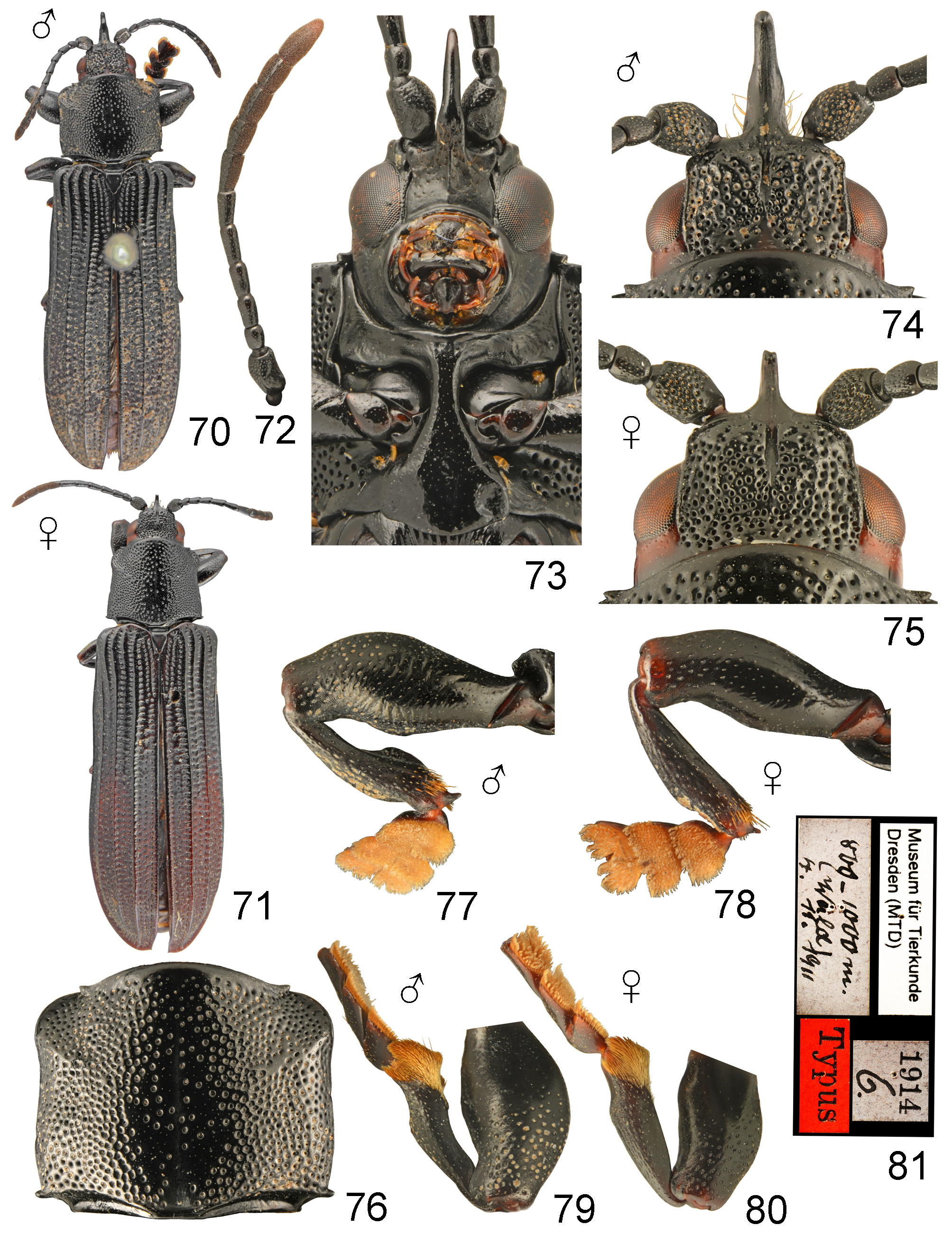
( Figs 70–81 View FIGURES 70–81 , 223)
Brontispa sarasini Heller, 1916: 306 (original description, incl. fig.); Spaeth 1936: 294 (noted); Risbec 1936: 185 (noted), 1944:
69 (noted); Maulik 1938: 50 (noted, not Brontispa View in CoL ). Brontispa Sarrasini View in CoL [sic!]: Lepesme 1947: 546 (noted, list of palm pests). Brontispa sarazini [sic!]: Risbec 1950: 379 (noted). Caldonispa sarasini: Uhmann 1952: 83 (transfer, lectotype designation), 1958: 206 (catalogue), 1964: 452 (catalogue); Gressitt 1957: 228 (faunistics, misidentification = C. freycinetiae ), 1960a: 24 (noted), 1960b: 107 (faunistics, biology); Maddison 1993: 84 (noted). Calenodispa sarrasini [sic!]: Mariau 1999: 232 (noted), 2001: 131 (noted).
Type locality. New Caledonia, Mt. Canala , 800–1000 m a.s.l. Description. Length 13.50–13.80 mm, width 4.00– 4.50 mm .
Almost whole body black, in some specimens posterior half of elytra with hardy perceptible ferrugineousbrown hue, head behind eye and coxae often ferrugineous-brown ( Figs 70, 71 View FIGURES 70–81 ). Abdomen completely black or sides of ventrites with small ferrugineous-brown patches. Legs and antennae black, tarsi sometimes ferrugineous-brown. Body glabrous except for yellowish hairs on frontoclypeus, short golden brown pubescence on distal antennomeres, golden orange pubescence on tarsal pads and apices of tibiae. Head 1.8 × as broad as long, interocular plate rectangular, slightly convex, especially in anterior part, at base distinctly impressed thus distinctly separated from vertex. Surface of interocular plate coarsely and densely punctate and with narrow, median sulcus along 3/4–4/5 length, sides and anterior corners of interocular plate narrowly margined, but corners obtuse; interantennal process in both sexes long, sexually dimorphic, strongly flattened laterally with thin sulcus along dorsum ( Figs 74, 75 View FIGURES 70–81 ). Frontoclypeus slightly longer than wide (excluding interantennal process), subacute apically, anterior corners forming elevated tubercles, area below tubercles flat, with several setose punctures, along middle runs well marked median keel ( Fig. 73 View FIGURES 70–81 ). Anterior margin of prosternum distinctly elevated, in middle with shallow emargination. Antennae 0.3 × as long as body, slightly compressed apically; antennomere I long but stout, approximately 1.2 × as long as broad; antennomere II very short, as long as broad, twice shorter than I; antennomere III approximately 1.4 × as long as II; antennomeres IV–VI and VIII approximately as long as III; antennomeres VII, IX, X approximately 1.2–1.3 × as long as III; antennomere XI 1.6 × as long as X, subangulate apically ( Fig. 72 View FIGURES 70–81 ). Pronotum approximately 1.1 × as broad as long, shallowly constricted in middle, anterior margin distinctly convex, basal margin shallowly bisinuate; anterior angles obtuse with small, sharp anterior tubercle, basal angles bearing small, elongate acute tooth; disc mostly flat, slightly impressed near anterior corners and with well developed median sulcus, shiny, punctation on sides of prothorax coarse and dense, punctures almost touching each other, on top of disc punctures sparse, especially along medial sulcus runs more or less impunctate area ( Fig. 76 View FIGURES 70–81 ). Elytra approximately 2.4 × as long as broad, subparallel basally and slightly broadened from base to middle and widest somewhat behind middle. Apex of elytra not emarginate, only sutural angle with small spine, lateral angle broadly rounded. Disc with 10–12 punctures in scutellar row, eight rows in humeral part but with numerous additional punctures below humeral callus, ten rows behind middle and 12 rows apically but slope with several additional punctures thus rows diffused among secondary punctation; intervals 4, 6, 8 narrowly costate on whole length, costae on intervals 4 and 6 flat anteriorly broadened and obtuse, other intervals flat. Ventral surface mostly shiny only abdomen more or less dull, pronotal hypomera moderately but densely punctate, punctures almost touching each other; prosternal process smooth laterally with minutely punctate intercoxal area and expanded apex; lateral plates of meso- and metaventrite smooth, central plate of mesoventrite with longitudinal striation, metaventrite laterally with coarse and dense punctures, rest of surface with distinct longitudinal and oblique striation; surface of first two and basal half of third ventrite more or less irregular, last two ventrites smooth. Legs stout, sexually dimorphic.
Sexual dimorphism distinct, in males interantennal process extremely long, twice as long as antennomere I and extending to ¾ length of antennomere III, strongly curved and reminiscent of rhinoceros horn ( Fig. 74 View FIGURES 70–81 ); in females process distinctly shorter, as long as or slightly shorter than antennomere I, hook shaped ( Fig. 75 View FIGURES 70–81 ). Femora in males strongly swollen, fore tibiae apically, on inner margin shallowly emarginate thus tibiae in ⅓ length before apex with distinct angulation ( Fig. 79 View FIGURES 70–81 ); mid tibiae in males explanate in apical ⅓ length, inner margin forming distinct plate, apex armed with large black spine ( Fig. 77 View FIGURES 70–81 ). In females femora only slightly swollen, fore tibiae on inner margin not emarginate, without angulation ( Fig. 80 View FIGURES 70–81 ), mid tibiae not explanate apically, without plate on inner margin, apically armed with small black spine ( Fig. 78 View FIGURES 70–81 ). Apex of abdominal ventrite deeply emarginate in males while shallowly emarginate in females.
Host plant. Pandanaceae : Pandanus sp. ( Gressitt 1960b). Based on additional specimens the species feeds also on Freycinetia sp. ( Pandanaceae ).
Remarks. Risbec (1936) mentioned the species in his work about parasites of the coconut trees and stated that it most likely feeds on some forest palms as it was described from mountains. Despite that, Lepesme (1947) suggested that the species is a pest of coconut tree ( Cocos nucifera ). Risbec (1950) suggested that the statement is erroneous. Nevertheless, Mariau (1999, 2001) again listed C. sarasini among pests of coconut tree. It is very unlikely that the species is actually associated with coconut trees as it occurs at high elevations and therefore we concur with Risbec and do not list Cocos nucifera among host plants.
Type material examined. Lectotype (designated by Uhmann 1952: 83): ♂ ‘ Drs. F. Sarasin & J. Roux | Neu- kaledonien | Mt. Kanala | [blue, p, cb] || 800-1000 m. | Wald | 4.11.1911 [hw on verso of previous label] || Typus [r, p, cb] || 1914 | 6. [w, p, cb] || Museum für Tierkunde | Dresden [w, p, cb]’ ( MTD) . Paralectotype: ♀ the same data as lectotype ( MTD) .
Additional material examined. NEW CALEDONIA: Aoupinie , top camp, 21°11′S, 165°18′E, 850 m, 23.i.2001 – 1.ii.2002, 2 ♀ (Malaise trap), Burwell & Monteith leg. ( QMBA) GoogleMaps ; Plateau de Dogny , 1030 m, 4.iii.1960, 2 ♂, 1 ♀ (ex Pandanus sp.), J. L. Gressitt leg. ( BPBM) , 1000 m, 22.iii.1963, 1 ♀ (ex Freycinetia sp.), R. Straatman leg. ( LS) .
No known copyright restrictions apply. See Agosti, D., Egloff, W., 2009. Taxonomic information exchange and copyright: the Plazi approach. BMC Research Notes 2009, 2:53 for further explanation.
|
Kingdom |
|
|
Phylum |
|
|
Class |
|
|
Order |
|
|
Family |
|
|
Genus |
Caledonispa sarasini ( Heller, 1916 )
| Borowiec, Lech, Świętojańska, Jolanta & Sekerka, Lukáš 2019 |
Brontispa sarasini
| Spaeth, F. 1936: 294 |
| Risbec, J. 1936: 185 |
| Heller, K. M. 1916: 306 |