
Bulbamphiascus incus, Gee, 2005
|
publication ID |
https://doi.org/ 10.1080/00222930500060397 |
|
persistent identifier |
https://treatment.plazi.org/id/03B28791-FA30-FFDF-FE47-8876FB33FFE2 |
|
treatment provided by |
Felipe |
|
scientific name |
Bulbamphiascus incus |
| status |
sp. nov. |
Bulbamphiascus incus sp. nov.
( Figures 1–6 View Figure 1 View Figure 2 View Figure 3 View Figure 4 View Figure 5 View Figure 6 )
Material examined
Holotype: adult „ (dissected on to four slides) from Loch Kishorn, NHM Reg. No. 2004.4118. Paratypes: 20 adult ♀♀ (two dissected each on to three slides and 18 spirit preserved) and 19 adult „„ (two dissected each on to three slides and 17 spirit preserved) from Loch Kishorn, NHM Reg. Nos 2004.4119–4157; seven adult ♀♀ and nine adult „„, spirit preserved from Loch Diabaig, NHM Reg. Nos 2004.4158–4173.
Description of female
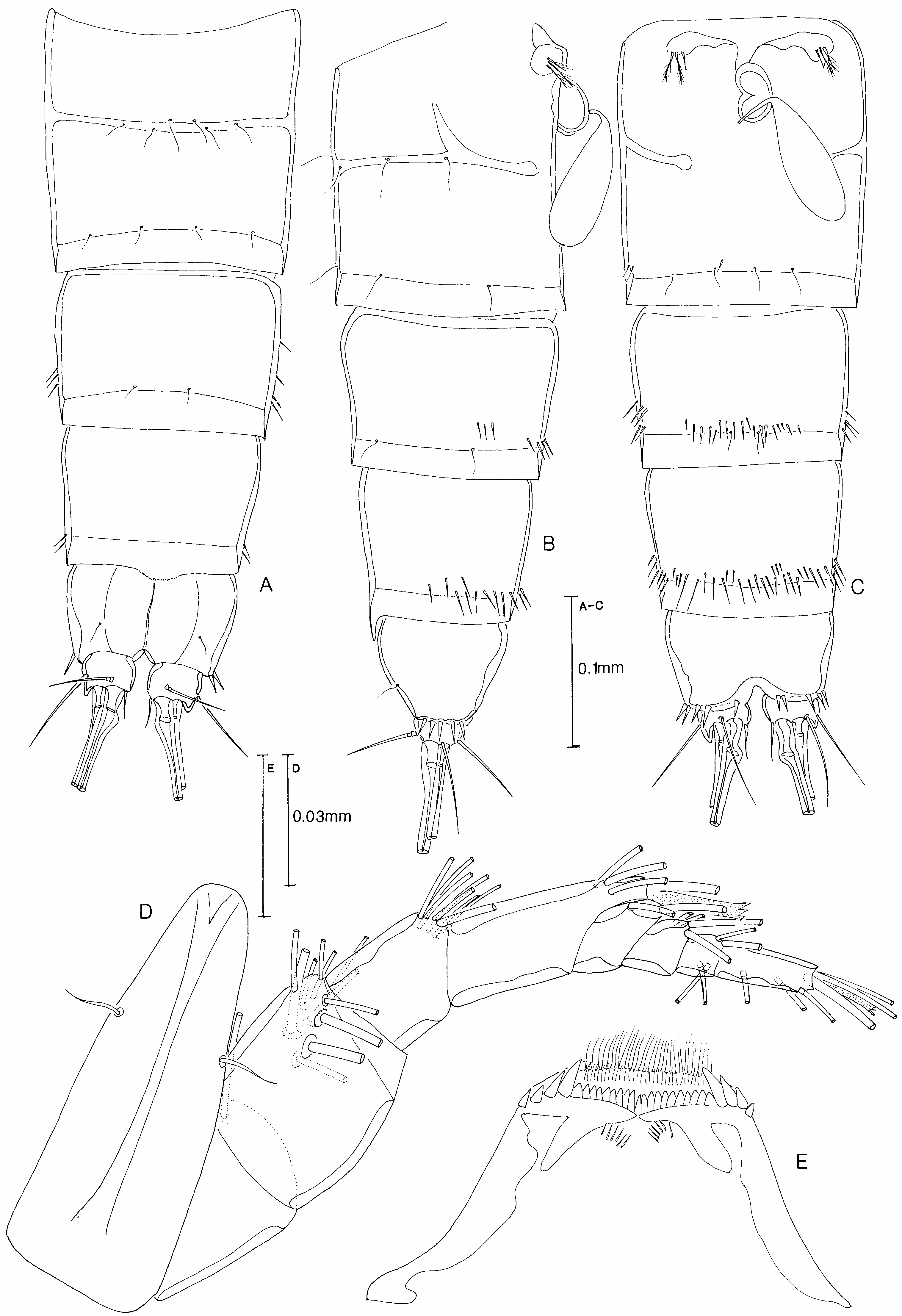
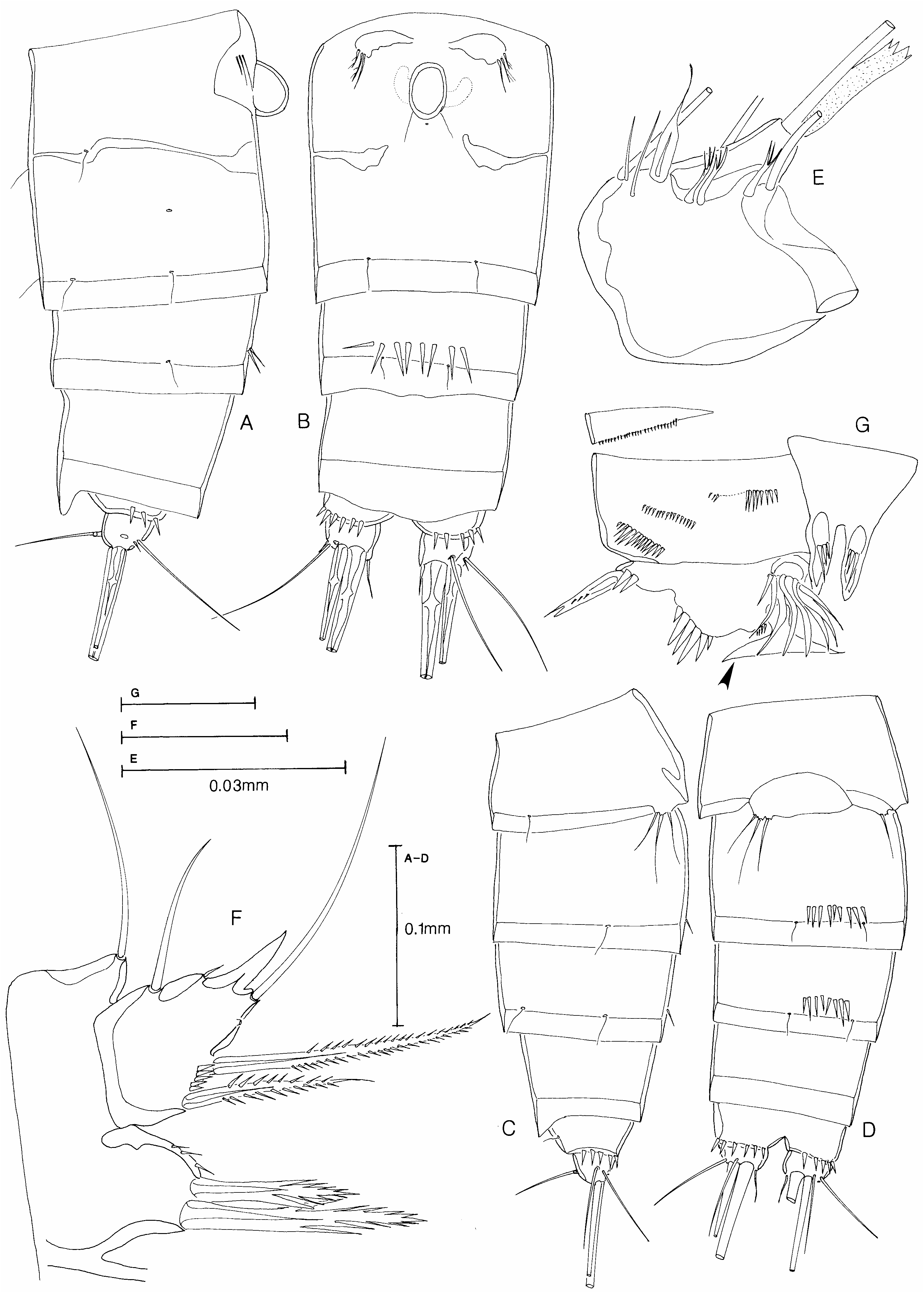
Body. Length 0.900 – 1.097 mm (mean 51.005 mm, n 510); sub-cylindrical, widest at posterior margin of cephalothorax, tapering gradually posteriorly. Rostrum ( Figure 1D View Figure 1 ) defined at base, elongate, triangular, extending beyond the second antennular segment, with a pair of small sensilla on lateral margins. Cephalothorax tapering anteriorly, as long as free prosomites. Genital double-somite ( Figure 1A–C View Figure 1 ) divided dorsally and laterally by subcuticular rib. Genital field ( Figure 1C View Figure 1 ) with separate genital apertures covered by vestigial P6, each bearing two pinnate and one smooth setae; copulatory pore situated medially, posterior to genital apertures, and obscured by large oval-shaped copulatory bulb; internal seminal receptacles kidney-shaped. Anal somite with small semicircular operculum near median dorsal anterior border and overlain by a minutely dentate pseudoperculum ( Figure 1A View Figure 1 ). Caudal rami ( Figures 1A–C View Figure 1 , 5F View Figure 5 ) broader than long in dorsal view with a slender tube pore on ventral posterior margin; seta I (antero-lateral accessory seta) small and naked, seta II (antero-lateral seta) and seta III (postero-ventral seta) long and smooth; terminal setae IV and V well developed with few short spinules in central region; in most specimens seta V swollen in the fracture region; seta VI (terminal accessory seta) short and smooth; seta VII (dorsal seta) triarticulate GoogleMaps .
Somatic ornamentation ( Figure 1A–C View Figure 1 ). Body surface appears smooth under light microscope, all somites except preanal furnished with numerous sensilla and pores. Prosome without spinule rows; urosomite 4 with a median ventral row of fine spinules and a ventro-lateral patch of fine spinules, variable in extent; preanal somite with a continuous row of spinules ventrally and ventro-laterally; anal somite with a lateral and ventral row of strong spinules round the base of caudal rami. Hyaline frills of urosomites with minutely dentate margin.
Antennule ( Figure 1D View Figure 1 ). Eight-segmented, segment 2 slightly swollen, with convex margins, segment 4 two times longer (on anterior margin) than broad; aesthetascs on fourth and distal segments. Setal formula as follows: 1-[1], 2-[11], 3-[8], 4-[3+(1+a)], 5-[2], 6-[4], 7- [4], 8-[4+(2+a)].
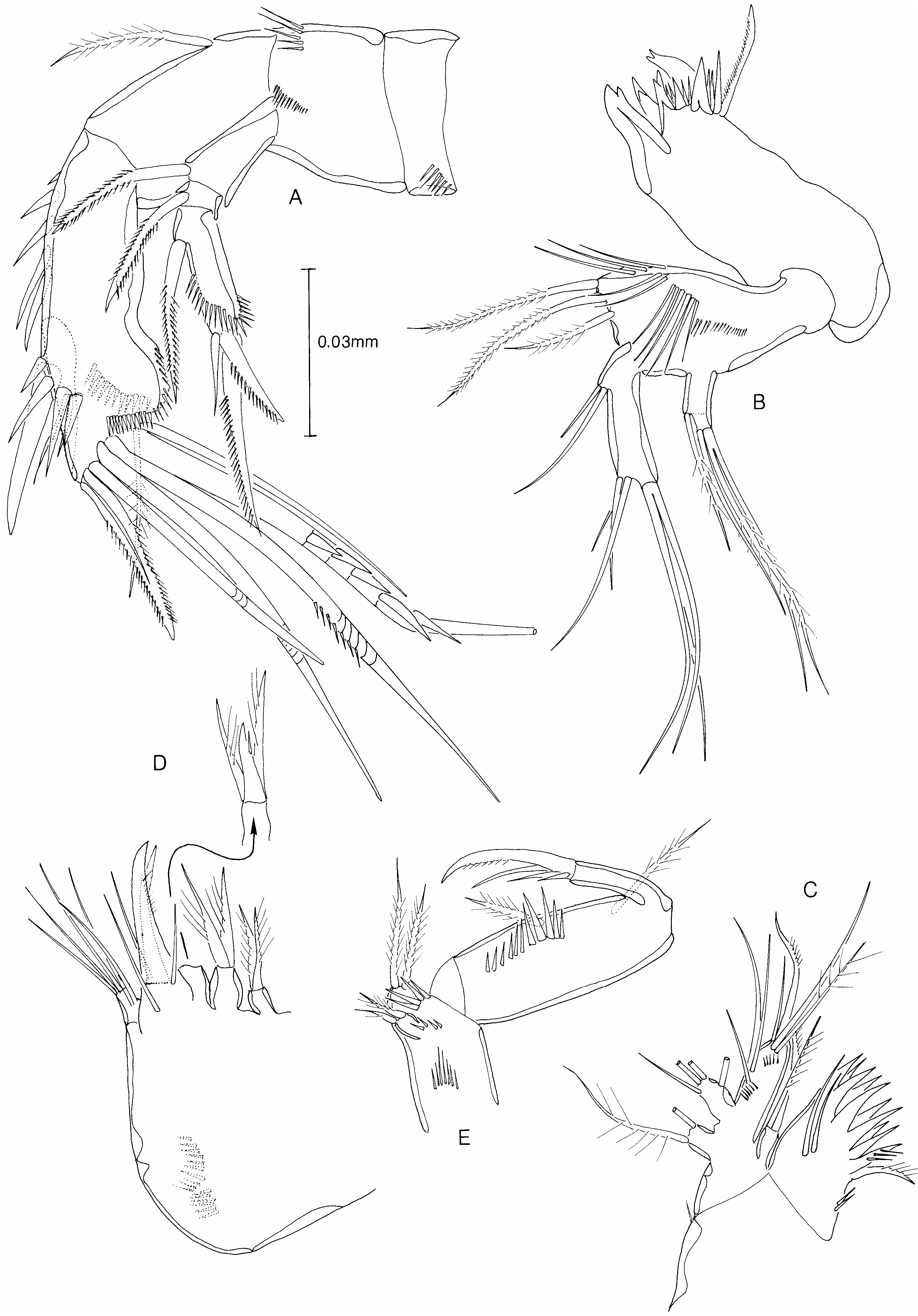
Antenna ( Figure 2A View Figure 2 ). Coxa well developed with row of setules. Allobasis with partial suture and row of spinules dorsally at base of exopod; one seta on abexopodal margin. Exopod three-segmented, proximal segment with one pinnate seta; middle segment short, with a pinnate seta; distal segment with an oblique row of strong spinules; with one pinnate seta on lateral margin and two stout pinnate spines and one naked seta on distal margin. Free endopod segment with two rows of strong spinules on outer margin, row of smaller spinules on distal and inner margin and on ventral face; lateral armature of two large pinnate spines and two setae; distal margin armed with one pinnate spine, four geniculate setae (two pinnate medially), one naked and one plumose seta.
Labrum ( Figure 1E View Figure 1 ). Posterior margin armed with lateral groups of four teeth, a median row of smaller teeth and two rows of setules.
Mandible ( Figure 2B View Figure 2 ). Coxa stout, gnathobase armed with a number of stout bicuspid teeth and a row of finer unicuspid teeth; two setae (one pinnate) at inner distal corner. Basis broad, with two rows of spinules on anterior face and three pinnate setae on distal margin. Exopod indistinctly two-segmented, proximal segment with one lateral pinnate seta; distal segment with three setae, two fused at base. Endopod large, one-segmented; with eight setae (two setae proximally, three setae subdistally and, on distal margin, three setae fused at base).
Maxillule ( Figure 2C View Figure 2 ). Arthrite of praecoxa with two setae on anterior face; distal margin with four pairs of recurved naked spines, and two pectinate spines. Coxa with two pinnate setae on distal margin. Basis with two rows of spinules on anterior face and distal margin; bearing seven elements (two naked setae and two pinnate spines distally and three naked setae subdistally). Exopod one-segmented with two plumose setae. Endopod onesegmented with four setae.
Maxilla ( Figure 2D View Figure 2 ). Syncoxa with two rows of spinules and three endites, proximal and middle endite armed with two, distal endite with three, pinnate spines. Allobasal endite with a large fused pectinate spine, a smaller articulating spine and three naked setae. Endopod one-segmented with five setae.
Maxilliped ( Figure 2E View Figure 2 ). Syncoxa with three surface rows of spinules and four pinnate setae (two on distal margin and two subdistally on a small peduncle). Basis with two pinnate setae on palmar margin and a sub-marginal row of spinules. Endopod one-segmented, with a terminal, partially pinnate, claw and three accessory setae.
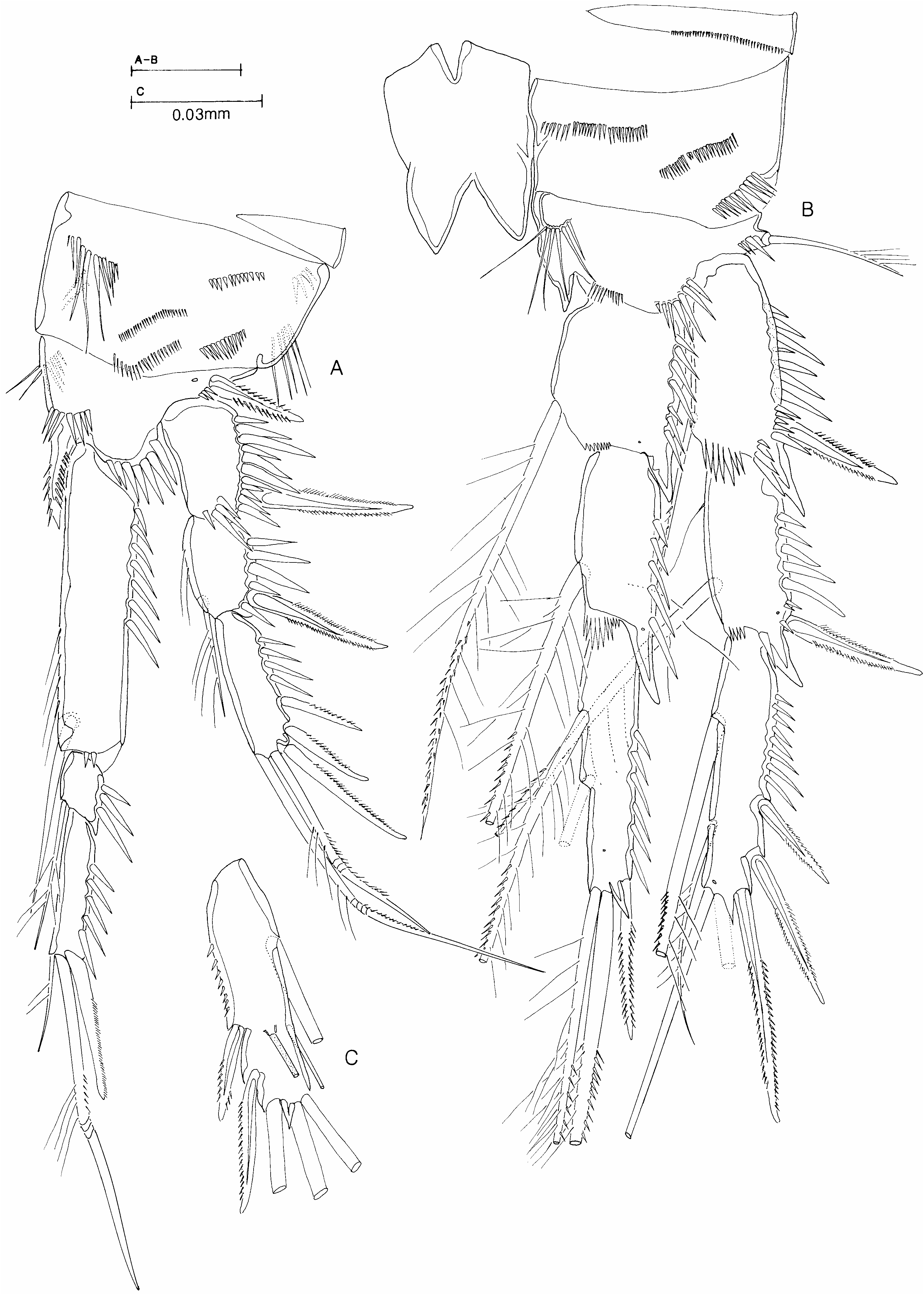
P1 ( Figure 3A View Figure 3 ). Intercoxal sclerite (not shown in figure) small, ovoid, without ornamentation. Praecoxa with a row of minute spinules along distal margin. Coxa with four rows of small spinules and one row of long spinules on anterior face and three rows of spinules on posterior face. Basis with rows of spinules on inner and median distal margin and at base of inner and outer pectinate spines. Exopod three-segmented, each segment with row of strong spinules on outer margin, exp 2 with row of setules and a plumose seta on inner margin, exp 3 with two geniculate setae on distal, and three spines on outer, margin. Endopod three-segmented; enp 1 longer than enp 2 and 3 combined, reaching nearly to distal margin of exp 3, row of strong spinules on outer margin, row of setules and a strong seta on inner margin; enp 2 less than half length of enp 3, with row of spinules on outer margin and a pinnate seta on inner margin; enp 3 with row of spinules on outer margin and, on distal margin, a small pinnate seta, a large geniculate seta and a spine.
P2–P4 ( Figures 3B View Figure 3 , 4A View Figure 4 ). Intercoxal sclerite strongly developed, almost square, sclerite of P2 with two rows of spinules. Protopod ornamented as for P1 except coxa with only three rows of spinules on anterior face and none on posterior face; basis without inner spine but with a distinct chitinous extension on inner distal margin. Rami three-segmented, equal in length in P2 and P3, endopod slightly shorter than exopod in P4; distal segment longest; all segments with row of strong spinules on outer margin; proximal two segments of both rami with spiniform extension of outer distal margin; inner distal seta on exp 3 weakly developed. Setal formula of swimming legs as follows:
Exopod Endopod
P1 0: 1: 023 1: 1: 021
P2 1: 1: 223 1: 2: 121 (1: 312)
P3 1: 1: 223 1: 1: 221
P4 1: 1: 323 1: 1: 121
Parentheses denote male condition.
P5 ( Figure 6A View Figure 6 ). Elements of each side not fused medially. Baseoendopod and exopod separate. Inner expansion of baseoendopod reaching about half length of exopod; with a few spinules on outer margin; armed with five pectinate or pinnate setae (three on inner and two on distal margin). Exopod about twice as long as broad, with few spinules on inner and outer margin; with six setae, proximal inner seta pinnate, distal inner seta and terminal seta naked, borne on a short peduncle, proximal and medial outer setae short and normal, distal outer seta markedly swollen at base.
Description of male
Similar to female except for urosome, antennule, P1 basis, P2 basis and endopod, P3 exp 3 and P5.
Body. Length 0.59–0.80 mm (mean 50.66 mm, n 516), urosomites 2 and 3 not fused. Genital somite ( Figure 5C View Figure 5 ) with vestigial P6 forming one fixed and one articulating plate each bearing three setae.
Somatic ornamentation ( Figure 5A–C View Figure 5 ). As in female except that a ventral and ventro-lateral row of spinules present on urosomite 3 as well as urosomites 4 and 5.
Antennule ( Figure 5D View Figure 5 ). Haplocer, 10-segmented with segment 4 a small segment overlaying the proximal portion of swollen segment 5; geniculation between segments 7 and 8 which both bear modified elements; aesthetascs on fifth and distal segments. Segment 5 with distinctly shaped seta (broad base and flagellate tip) near proximal margin (shown more clearly in Figure 7E View Figure 7 ). Setal formula as follows: 1-[1], 2-[11], 3-[8], 4-[2], 5-[7+(1+a)], 6- [2], 7-[3?], 8-[2?], 9-[4], 10-[5+(2+a)].
P1 ( Figure 5E View Figure 5 ). Basis with a single, long chitinous projection at inner proximal corner.
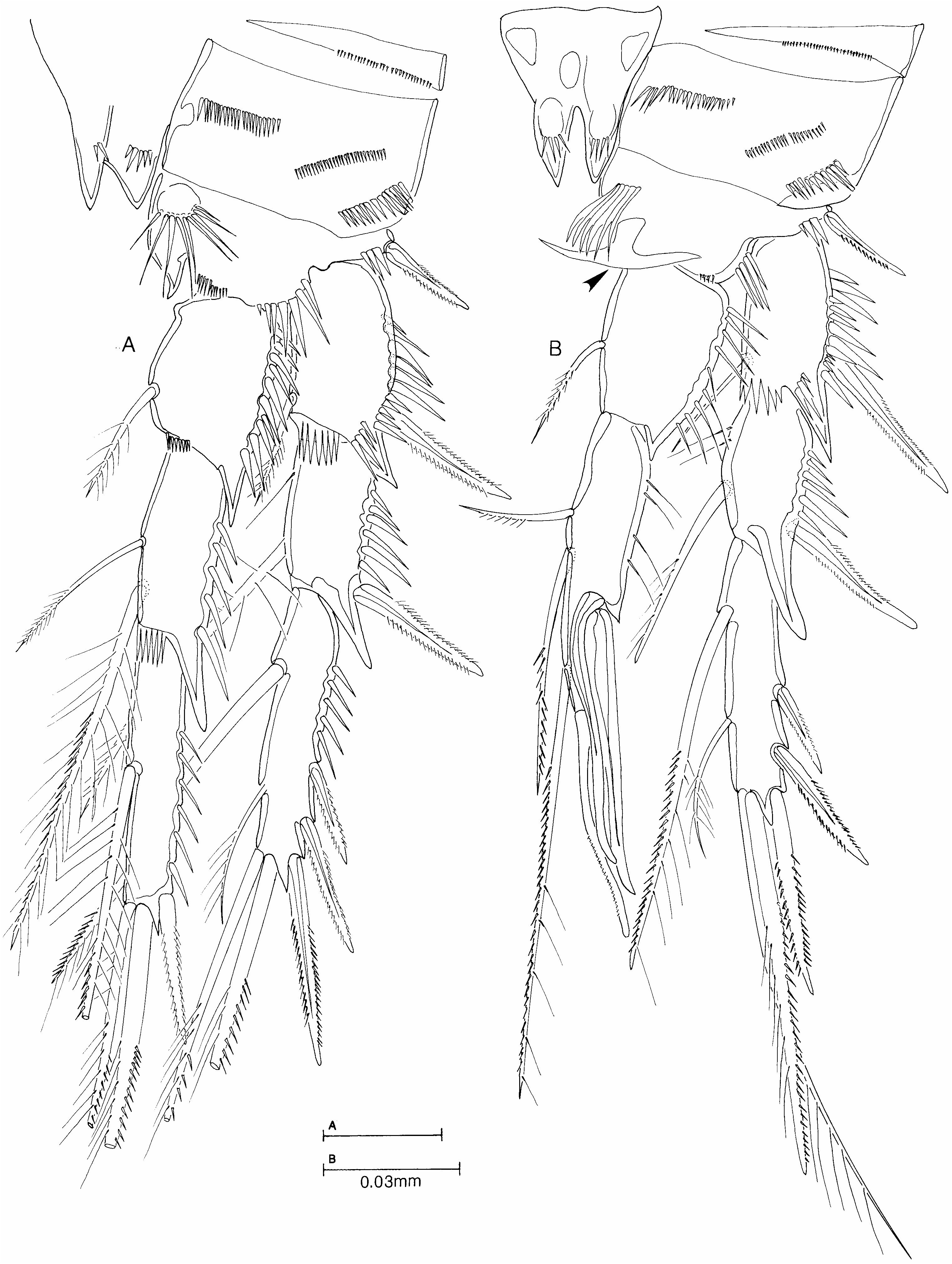
P2 ( Figure 4B View Figure 4 ). Praecoxa and coxa as in female. Basis without chitinous extension of inner margin as in female but with a smooth, anvil-shaped, hyaline structure near inner distal margin (arrowed in Figure 4B View Figure 4 ). Exopod as in female. Endopod modified, two-segmented; enp 1 as in female except inner seta slightly shorter; distal segment with three pinnate setae on inner margin, a seta (with a bluntly rounded tip and small pinnules) on distal margin and a large spine and a sinuous process (with a smooth rounded tip) articulating subdistally on outer margin.
P3 ( Figure 3C View Figure 3 ). As in female except that hyaline tube pore present on anterior face of exp 3.
P5 ( Figure 6B–D View Figure 6 ). Baseoendopods of each side fused medially. Endopodal lobe with two terminal pectinate spines and a few spinules on outer margin; outer peduncle elongate. Exopod about 1.5 times as long as broad with a short row of spinules at base of distal inner seta, a small tube pore on anterior surface and outer distal corner attenuated into a chitinous process; bearing four or five setae, of which inner two setae strongly developed and pinnate, distal and proximal outer setae naked, long and slender, distal outer seta either absent ( Figure 6D View Figure 6 ) or vestigial and only visible under ×40 ( Figure 6C View Figure 6 ) or ×100 ( Figure 6B View Figure 6 ) oil immersion objectives.
Etymology
The specific name is the Latin for anvil, reflecting the shape of the hyaline structure on the male P2 endopod.
Variability
The ornamentation of the urosome was consistent in the presence of fine spinule rows on urosomites 4 and 5 in the female and urosomites 3–5 in the male but the extent of the spinule patches ventro-laterally varied from a few spinules to many spinules.
There was no discernible variation in the structure and setation of the oral and swimming appendages except for the presence and size of the distal outer vestigial seta on the male P5.
Remarks
This material from the two Scottish lochs has been assigned to a new species primarily on the basis of the structure on the basal segment of the male P2 endopod, on the form of the male P5 exopod and on the ornamentation of the urosome in both sexes.
Mu and Gee (2000) first noted and figured (their Figures 13A, 14D) the presence of a peculiar flexible, semi-hyaline, papillate, spine-like structure on the basis of the P2 of the male in their specimens of B. imus which was not present in any of their Chinese material. They concluded that this structure was homologous to the chitinous apophysis present on the basis of the female but otherwise absent in the male of B. imus . The same structures are found on the basis of the P 2 in female and male specimens from Scotland but here the semi-hyaline structure in the male is completely different in shape, being smooth-walled and T-shaped, very much like a blacksmith’s anvil.
Mu and Gee (2000), in their Figure 13B, showed that the exopod of the male P5 of B. imus was only slightly longer than broad and bore six setae, two strongly developed, plumose inner setae, one distal naked seta and three outer setae, the proximal one well developed and pinnate and the two distal setae well developed and distinctly swollen at the base. In the Scottish material the exopod is about 1.5 times longer than broad with the same inner and distal armature but a very different armature of the outer margin. At the outer distal corner is a large chitinous apophysis which has almost certainly been formed by the enlargement and fusion with the segment of the distal outer swollen seta of B. imus . In some specimens there is still an indication of a suture line at the base of the apophysis on the posterior face. Conversely, the middle outer seta has become vestigial (very often its presence can only be discerned under ×100 oil immersion objectives) or has been lost entirely. Drastic modification of the two distal outer setae of the male P5 exopod has also been reported for B. chappuisi by Rouch (1962), who describes and figures an exopod more than twice as long as broad with both the distal outer setae fused to the segment and forming chitinous projections. In this species Rouch (1962) also indicates in his Figure 34 (but does not mention in the text) that the swollen distal outer seta of the female P5 exopod is also fused to the segment.
Whilst these features clearly distinguish the male of B. incus from the male of B. imus , the only distinguishing characteristic in the female is in the abdominal ornamentation. Mu and Gee (2000) showed that in female B. imus there is a group of two strong spinules on the postero-lateral border of urosomite 3 and a median ventral row of strong spinules on the posterior border of urosomite 4 and never any spinules on the posterior border of urosomite 5. In the Scottish material of B. incus there is always a complete row of very fine spinules on the posterior border of urosomite 5; always a ventral row and a small to large ventro-lateral patch of fine spinules on the posterior border and lateral face of urosomite 4 and occasionally a few fine spinules on the posterior border of urosomite 3. Mu and Gee (2000) showed that the urosome ornamentation of B. imus was very consistent, even between two different populations in the North Sea, and it was the difference in somatic ornamentation which first alerted the present author to the possibility that the Scottish material was a different species.
As a result of this discovery it is advisable to treat many of the records of B. imus with caution, particularly those from areas of high organic pollution on the west coast of Scotland, e.g. Moore and Pearson (1986). In this connection, it is interesting to note that although the description and figures of Stenhelia reflexa by T. Scott (1895) is only of the female, this species has definite characteristics of B. incus rather than B. imus , with which species it was synonymized by Lang (1948). In Scott’s drawings he clearly shows a female with a row of spinules on the preanal somite (urosomite 5) which is never found in B. imus . Further, he illustrates seta V of the caudal ramus with a swelling in the fracture zone (not found in specimens of B. imus I have studied), and he illustrates the exopod of the mandible with only one lateral seta on the proximal segment (as in B. incus but B. imus has two).


| V |
Royal British Columbia Museum - Herbarium |
| VI |
Mykotektet, National Veterinary Institute |
No known copyright restrictions apply. See Agosti, D., Egloff, W., 2009. Taxonomic information exchange and copyright: the Plazi approach. BMC Research Notes 2009, 2:53 for further explanation.