
Brachionus calyciflorus, Pallas, 1776
|
publication ID |
https://doi.org/ 10.1080/00222933.2023.2279255 |
|
DOI |
https://doi.org/10.5281/zenodo.10480018 |
|
persistent identifier |
https://treatment.plazi.org/id/03B687E6-220B-FFE7-FF42-FF6FFEF075DE |
|
treatment provided by |
Plazi |
|
scientific name |
Brachionus calyciflorus |
| status |
|
Phylum Rotifera View in CoL View at ENA :
Brachionus calyciflorus View in CoL
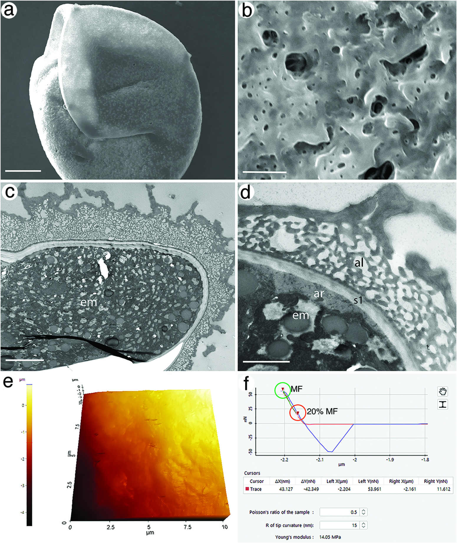
SEM
The ovoid, diapausing embryos measured ca 139 µm in diameter (̄x = 138.4 ± 5.8 μm). Shells appeared largely devoid of ornamentation at lower magnifications (<1000×), but showed a more rugose surface at higher magnifications ( Figure 1a,b View Figure 1 ). Shells often appeared pitted with small pores and craters (0.07–2.96 µm diameter) and occasionally some surface bumps of various textures.
TEM
The shell consisted of two layers – a thick apical layer and a thin basal layer – that together averaged 7.20 ± 1.45 µm (SD) thick ( Figure 1c View Figure 1 ). The surface of the shell had a wavy appearance and consisted of high ridges and numerous small bumps and valleys. Immediately beneath this was a thick zone of empty pockets that were interconnected through a framework of electron-dense bridges; this layer is described by Wurdak et al. (1978) as the alveolar layer (al: Figure 1d View Figure 1 ). Immediately beneath the alveolar layer was a zone [s1: Wurdak et al. (1978)] of less opaque electron density that consisted of two subzones, each containing many fine laminae, and separated by an electron-dense line. An extraembryonic space of variable size separated the physical shell from an amorphous region (ar) that was proximal to the plasma membrane of the embryo.
AFM
Topography of the outer shell layer was similar to that viewed with SEM, but pores were less visible (̄x = 0.23 µm diameter) and surface bumps were less recognisable ( Figure 1e View Figure 1 ). An example force–distance curve is provided ( Figure 1f View Figure 1 ) that displays how Young̾s modulus values were collected for a single spot on one egg. The Young̾s modulus for two diapausing embryos ranged from 9.73 to 17.21 MPa with an average of 13.62 ± 2.43 MPa (SD) ( Table 1 View Table 1 ). Hardness values ranged from 1.62 × 10⁻² to 2.18 × 10⁻² GPa with an average hardness of 1.80 × 10⁻² GPa ± 1.80 × 10⁻³ GPa ( Table 2 View Table 2 ).
No known copyright restrictions apply. See Agosti, D., Egloff, W., 2009. Taxonomic information exchange and copyright: the Plazi approach. BMC Research Notes 2009, 2:53 for further explanation.