
Boophis marojezensis Glaw and Vences, 1994
|
publication ID |
https://doi.org/ 10.1080/00222930600902399 |
|
persistent identifier |
https://treatment.plazi.org/id/19120E4B-B645-0A1C-4571-495EFE27C1B2 |
|
treatment provided by |
Felipe |
|
scientific name |
Boophis marojezensis Glaw and Vences, 1994 |
| status |
|
Boophis marojezensis Glaw and Vences, 1994 View in CoL
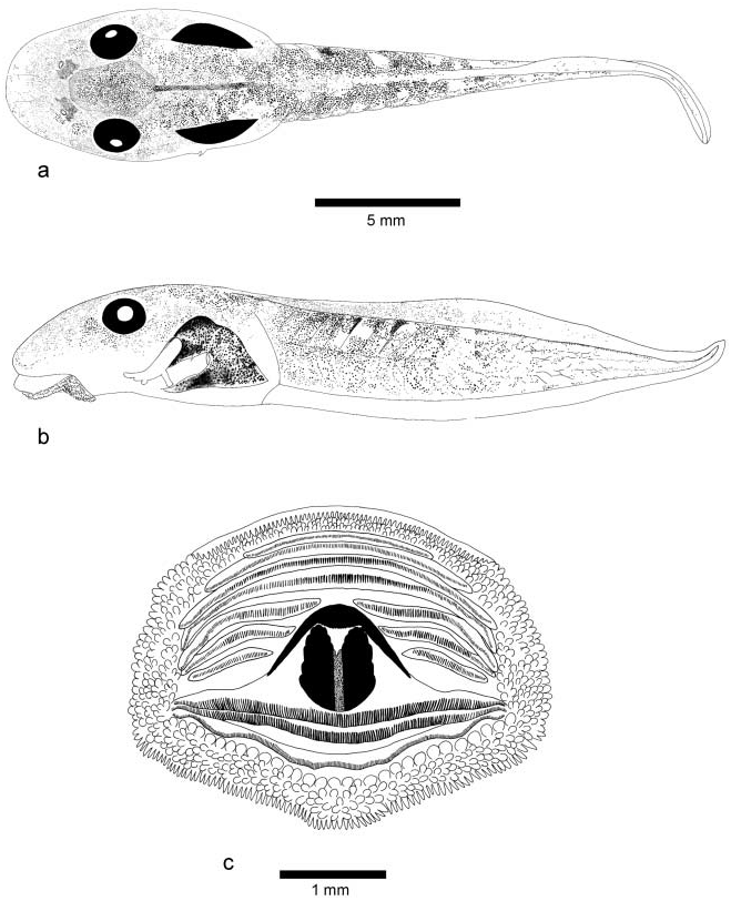
( Figure 6 View Figure 6 )
The following description is based on one tadpole in developmental stage 41 (ZSM 523/2004, field number LR 66, TL 26.5 mm, BL 10.0 mm), from locality 1. Hindlimbs of this specimen were largely used for tissue sampling and are therefore not shown in Figure 6 View Figure 6 .
In dorsal view ( Figure 6a View Figure 6 ), body ovoid elongate, snout rounded. In lateral view ( Figure 6b View Figure 6 ), body greatly depressed, BW 145% of BH, snout almost truncate. Eyes large, ED 16.0% of BL, slightly bulging, not visible in ventral view, positioned dorsolaterally but directed almost laterally, situated at about the anterior third of the body. Nares moderately small, round, rimmed with a very small and flat mediodorsal projection, positioned dorsally, directed anterolaterally and dorsally, much closer to anterior edge of eyes than to snout, RN 169% of NP; NN 44% of PP. Spiracle sinistral, narrow and long, slightly visible from dorsal view, its tip free from body; spiracular opening orientated posterodorsally, much closer to end of body than to snout, SS 76% of BL, situated at the height of the lower part of tail musculature. Vent tube small, short, medial, opening just before the beginning of ventral fin, opening medial, directed more posteriorly than posteroventrally, linked to caudal muscle. Caudal musculature strong, TMH 66% of BH and 59% of MTH, TMW 48% of BW, especially in the anterior half of the tail; height of caudal musculature almost half of total tail height at mid-length of tail, almost reaching tail tip. Caudal fins regular with straight edges, very shallow, MTH 111% of BH; dorsal fin originating next to body–tail junction, convex in its medial part; ventral fin beginning at the level of the ventral terminal of the body, following the caudal muscle; tail tip fine.
Oral disc ( Figure 6c View Figure 6 ) enlarged, ODW 41% of BL and 63% of BW, positioned and directed ventrally, not emarginated. Several uninterrupted rows of marginal papillae around oral disc; no medial gaps in rows of marginal papillae on upper or lower labium. Papillae of internal row round and of moderate size, papillae of external rows small, elongate, and cylindrical. No denticulate papillae. Keratodont row formula 4:3+3/3. About 203 keratodonts on A3 (32–33 per mm). The length of interrupted anterior keratodont rows (A5, A6, and A7) decreases gradually towards the centre of the disc, keratodont rows of lower labium subequal in length. Upper jaw not serrated and weakly developed, median part straight, a black halo on its distal part; lower jaw more developed, narrow V-shaped, ribbed, composed of two parts connected by a less keratinized median area, coarsely serrated.
Coloration in preservative. Dorsally: part anterior to eyes covered with sparse black dots; dark pigmentations concentrated dorsally forming dark patches between eyes and beside nares; black colour on either side of lateral body obscuring intestine area; caudal musculature with scattered dark pigmentation. Laterally: intestinal coils invisible; caudal musculature with sparse black spots, showing some reticulations at its posterior end; dorsal fin with small black spots, ventral fin clear. Ventrally: branchial and cardial region translucent, well visible through ventral body wall; intestinal coils invisible.
Variation. TL and BL of nine tadpoles at stages 29–41 (ZSM 521/2004–524/2004, and LR 61, LR 62, LR 64, LR 65, LR 67), all from locality 1, are 22.1–27.2 and 8.1–10.7 mm, respectively. The ratios vary in the following proportions: BW 118–145% of BH; ED 14.9– 16.8% of BL; RN 143–196% of NP; NN 42–49% of PP; SS 70–81% of BL; TMH 65–76% of BH; TMH 59–84% of MTH; TMW 48–67% of BW; MTH 86–111% of BH; ODW 36– 47% of BL; ODW 63–86% of BW. KRF of the nine tadpoles is 4:3+3/3.
Boophis albipunctatus group Boophis sibilans Glaw and Thiesmeier, 1993
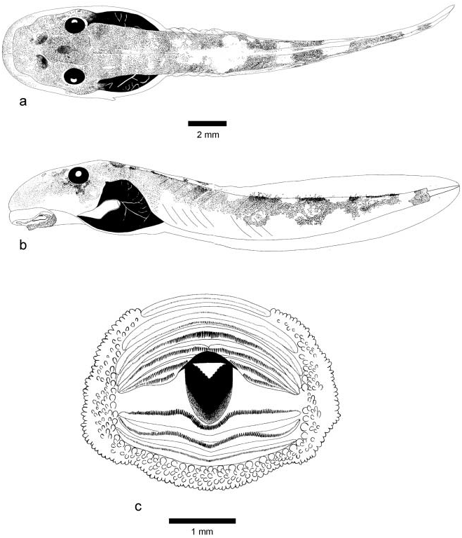
( Figure 7 View Figure 7 )
The following description is based on one tadpole in developmental stage 25 (ZSM 557/ 2004, field number LR 269, TL 25.2 mm, BL 9.0 mm), from locality 4.
In dorsal view ( Figure 7a View Figure 7 ), body elliptical, snout rounded. In lateral view ( Figure 7b View Figure 7 ), body depressed, BW 129% of BH, snout rounded. Eyes moderately large, ED 12.2% of BL, bulging, not visible in ventral view, positioned more dorsally than dorsolaterally but directed dorsolaterally, situated at about the anterior third of the body. Nares moderately sized, elliptical, rimmed with a flat mediodorsal projection, positioned dorsally, directed anterolaterally and opening almost dorsally, much closer to anterior edge of eyes than to snout, RN 148% of NP; NN 50% of PP. Spiracle sinistral, narrow and long, slightly visible from dorsal view, its tip free from body; spiracular opening orientated almost posteriorly, much closer to end of body than to snout, SS 72% of BL, situated below the lower part of tail musculature. Vent tube small, medial, directed posteriorly, linked to caudal muscle, opening posterolateral. Caudal musculature strong, TMH 77% of BH and 75% of MTH, TMW 59% of BW, at mid-length of tail height of caudal musculature almost half of total tail height, parallel in its proximal third then gradually tapering, almost reaching tail tip. Caudal fins moderately shallow, MTH 103% of BH; dorsal fin originating next to body–tail junction, shallow in its anterior part then becoming convex towards mid-tail, ventral fin beginning just behind body and reaching its maximum height more posteriorly than dorsal fin; tail tip rounded.
Oral disc ( Figure 7c View Figure 7 ) enlarged, ODW 44% of BL and 89% of BW, positioned and directed ventrally, not emarginated. Several rows of marginal papillae around oral disc, interrupted by a large median gap on the upper labium (DG 53% of ODW); no gap on lower labium. Papillae small, conical with a more or less pointed tip. No denticulate papillae. Keratodont row formula 4:3+3/3. Keratodonts on continuous rows A1, A2, and A3 very small and difficult to count; estimation of keratodonts done on A4 with a total of about 208 (ca 70 per mm). The length of interrupted upper keratodont rows (A5, A6, and A7) decreases gradually towards the centre of the disc, keratodont rows of lower labium subequal in length. Upper jaw not serrated and weakly developed, black, median part straight; lower jaw more developed, V-shaped, ribbed, coarsely serrated, partially pigmented.
Coloration in preservative. Dorsally: body and tail musculature brownish with scattered dark pigmentation; either side of dorsal body black coloured with white stripes (the blood vessels); some dark pigment concentrated dorsally forming dark patches between eyes and beside nares; tail muscle barred black and white. Laterally: intestinal coils not visible through lateral body wall; spiracle not pigmented; caudal fins clear; lower part of caudal musculature less pigmented anteriorly; musculature junctions very distinct anteriorly. Ventrally: opaque branchial and cardial organs slightly visible through ventral body wall; intestinal coils not visible.
Variation. TL and BL of eight tadpoles at stage 25 (ZSM 556/2004–560/2004, and LR 269a, LR 269c, LR 269f) from localities 2 and 4, are 19.1–25.2 and 6.9–9.0 mm, respectively. The ratios vary in the following proportions: BW 118–132% of BH; ED
11.8–13.0% of BL; RN 127–177% of NP; NN 46–53% of PP; SS 70–78% of BL; TMH 59–77% of BH; TMH 59–75% of MTH; TMW 54–61% of BW; MTH 97–113% of BH; ODW 44–53% of BL; ODW 88–96% of BW. KRF of the eight tadpoles is 4:3+3/3.
Boophis luteus group Boophis luteus (Boulenger, 1882)
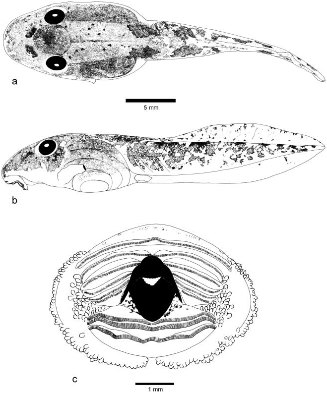
( Figure 8 View Figure 8 )
The following description is based on one tadpole in developmental stage 33 (ZSM uncatalogued, field number LR 218, TL 33.1 mm, BL 13.1 mm), from locality 3.
In dorsal view ( Figure 8a View Figure 8 ), body ovoid, snout rounded. In lateral view ( Figure 8b View Figure 8 ), body depressed, BW 125% of BH, snout rounded. Eyes moderately large, ED 14.5% of BL, bulging, not visible in ventral view, positioned dorsolaterally but directed laterally, situated at about one-third of the body. Nares moderately sized, nearly oval, not rimmed, positioned and directed dorsally, closer to the anterior edge of eyes than to tip of snout, RN 133% of NP; NN 64% of PP. Spiracle sinistral, moderately small, slightly tubular, well visible from dorsal view, its tip free from body; spiracular opening oval, orientated posterodorsally, positioned at about three-quarters of body length, SS 76% of BL, and situated well below the longitudinal axis of tail musculature. Vent tube short, dextral, opening at ventral edge of fin, opening directed posterolaterally, linked to caudal muscle, its right wall displaced anteriorly. Caudal musculature moderately strong, TMH 54% of BH and 65% of MTH, TMW 48% of BW, height of caudal musculature about two-fifths of the total height at mid-tail, reaching tail tip. Caudal fins shallow anteriorly, deepest at about half of their length, their height decreasing progressively up to tail tip, MTH 82% of BH; dorsal fin originating well posterior to the dorsal tail–body junction, ventral fin starting just behind the body; tail tip obtuse.
Oral disc ( Figure 8c View Figure 8 ) large, ODW 37% of BL and 63% of BW, positioned anteroventrally and directed ventrally, not emarginated. Oral disc bordered by one or two rows of marginal papillae interrupted by a large median gap on the upper labium (DG 94% of ODW), and by a small median gap on the lower labium. Submarginal papillae clustered in the corners of labia. Papillae of moderate size, cylindrical elongate with rounded tip. No denticulate papillae. Keratodont row formula 1:5+5/1+1:2. About 238 keratodonts on A1 (ca 63 per mm). The length of the interrupted anterior rows A2, A3, and A4 decreases towards centre of disc; keratodont rows of lower labium subequal in size. Both jaw sheaths serrated and fully black pigmented; upper jaw a wide flattened arch convex medially; lower jaw Vshaped.
Coloration in preservative. Dorsally: body and tail musculature dark pigmented; dark pigmentation concentrated dorsally forming obvious dark patches between eyes and immediately next to nares. Laterally: intestinal coils well visible; dorsal fin pigmented; ventral fin almost clear, musculature mottled. Ventrally: branchial and cardial organs apparent through the transparent surface of body.
Variation. TL and BL of 28 tadpoles at stages 25–41 (LR 160, LR 189, LR 227f, LR 227g, LR 227h, LR 227i, LR 227j, LR 227l, LR 227m, LR 238, LR 238a, LR 267, LR 267a, LR 72, LR 73, LR 227e, LR 227k, LR 227n, LR 227q, LR 218, LR 227b, LR 24, LR 227o, LR 227p, LR 227a, LR 227c, LR 227d, LR 250), from localities 1–5, are 17.7–41.4 and 6.7–16.1 mm, respectively. The ratios vary in the following proportions: BW 121–145% of BH; ED 10.0–16.7% of BL; RN 100–182% of NP; NN 40–64% of PP; SS 58–88% of BL; TMH 54–69% of BH; TMH 58–76% of MTH; TMW 42–65% of BW; MTH 84–113% of BH; ODW 26–44% of BL; ODW 49–75% of BW. KRF of the 28 tadpoles varies from 1:3+3/1+1:2 to 1:5+5/1+1:2.
No known copyright restrictions apply. See Agosti, D., Egloff, W., 2009. Taxonomic information exchange and copyright: the Plazi approach. BMC Research Notes 2009, 2:53 for further explanation.