
Blanus cf. gracilis (Rocek, 1984)
|
publication ID |
https://doi.org/ 10.5281/zenodo.4665621 |
|
persistent identifier |
https://treatment.plazi.org/id/03D08790-FFD2-FFF0-5641-A66AFEEC55EA |
|
treatment provided by |
Felipe |
|
scientific name |
Blanus cf. gracilis |
| status |
|
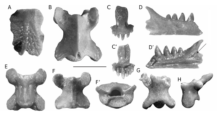
Blanus cf. gracilis ( Fig. 13 View FIG C-F’)
MATERIAL EXAMINED. — One premaxilla ( ISER Tt-0470), four fragmentary dentaries ( ISER Tt-0471/1-4), 60 vertebrae ( ISER Tt-0472/1-60).
DESCRIPTION
Premaxilla
The only specimen belonged to a small sized individual ( Fig.13C View FIG , C’). The anterodorsal surface of the premaxilla is strongly convex and the nasal process is rather high (its distal portion is broken off) with a flat surface. The base of the nasal process displays a weak waisting and laterally it is pierced by two large foramina of oval shape. The inner surface of the nasal process is provided with a prominent medial keel tapering distally. In each side of the keel, near the base of the nasal process, there is a dorsal large foramen and a ventral smaller one. However, on the right side the bony wall between the foramina is not complete. It is worth to mention that a connection exists between the outer and inner foramina. The maxillary process is broken off on both sides, while the lamina horizontalis is rather thin. The dentition is distinctly proterodont with a rather large median tooth and successively decreasing posterolateral teeth. The number of tooth positions in the premaxilla is seven. The teeth are cylindrical and have small circular resorption pits at their base. The tooth crown is monocuspid and bears faintly developed lingual and labial crests which show an anterolingual and posterolabial orientation respectively.
Dentary
The dentaries belonged to individuals of different size. In ISER Tt-0471/1 and 2, which preserve the anterior part of the tooth row, the Meckel’s groove opens at the level of the first tooth position, and widens posterior to the fifth tooth position. The symphyseal process, situated at the level of the second tooth position, is relatively short and flattened dorsoventrally. The crista splenialis is rounded lingually and delimits a weakly defined subdental
shelf. The ventral margin of the posterior third of the crista splenialis bears an elongated imprint left by the splenial. The coronoid process is relatively large and slanting posterodorsally. ISER Tt-0471/1 demonstrates that the dorsoposterior limit of the coronoid process is higher than the largest mandibular tooth ( Fig. 13D View FIG , D’). The surangular process is also well defined but its posterior terminus in all the specimens is broken off. ISER Tt-0471/1 preserves a small remnant of the anterior part of surangular in the original position. The dentition is heterodont with eight teeth in a complete dentary. The first and second teeth are broken off on all the dentaries, but they were presumably smaller than the third tooth. The latter is distinctly larger than the fourth tooth, while beginning from the fifth tooth their size decreases posteriorly. The teeth are conical with smooth apex and they are slightly recurved; some of them preserve a faintly developed anteroposterior crest.
On the labial side of ISER Tt-0471/2 three alveolar foramina are observed. The first foramen opens between the levels of the first and second tooth positions, the second one opens between the third and fourth teeth, while the third one opens at the level of the seventh tooth position.
Vertebrae
All the available specimens belonged to small individuals, the centrum length in 12 measured trunk vertebrae ranging between 1.7 and 2.03 mm. The vertebrae are flattened dorsoventrally. The ventral surface of the centrum is flat or slightly convex and provided with two subcentral foramina. In few specimens, which probably belonged to the anterior region of the vertebral column, a faintly defined subcentral keel is observed ( Fig. 13E View FIG ), while another specimen preserves a well-developed and posteriorly hooked hypapophysis. The lateral margins of the centrum are parallel but the centrum slightly widens near the condyle. The condyle and cotyle are flattened dorsoventrally. The neural ridge is more or less present in all the specimens, finishing in a somewhat widened posterior tuberosity ( Fig. 13F View FIG , F’). In several larger vertebrae there is a tuberosity situated near the anterior margin of the neural arch, while another one is placed near the
posterior margin of the neural arch.The zygosphene- zygantrum complex is lacking and the neural arch exhibits a convex or pointed anterior margin. The synapophyses, situated below the prezygapophyses, are of hemispherical shape. The pre- and postzygapophyses are of oval shape and the prezygapophyseal processes are lacking.
REMARKS
The morphology of the dentary differs from that of Palaeoblanus Schleich, 1988 , the latter being provided with a distinctly larger first tooth ( Schleich 1988; Böhme 1999), but it closely resembles that of the extant genus Blanus . Another fossil blanid species was described under the name Omoiotyphlops gracilis Roček, 1984 from the late early Miocene (MN 4) of Dolnice, Czech Republik ( Roček 1984). The diagnosis given and the morphology of the figured specimens concord with that of the genus Blanus . In fact, the small lingual process near the top of the coronoid process, which has been used by Roček (1984) to diagnose O. gracilis , is variably present in Recent Blanus also. From the same locality, a premaxilla (DP FNSP 317) described by Roček (1984) and assigned to Squamata , family indet. II (see Roček 1984: 61, text-fig. 5) quite probably belonged to Blanus (= Omoiotyphlops ) gracilis too. If the above assignment is correct, then B. gracilis appears as the most widely distributed European blanid. After Böhme (2002) Palaeoblanus and Blanus were contemporaneous at least during MN 3-MN 5, or even over a much longer period (MP 30-MN 5).
| ISER |
Institutul Speologie Emil G. Racovita |
No known copyright restrictions apply. See Agosti, D., Egloff, W., 2009. Taxonomic information exchange and copyright: the Plazi approach. BMC Research Notes 2009, 2:53 for further explanation.