
Aulacophoroides millettiae, Qiao, Gexia, Jiang, Liyun & Martin, Jon H., 2006
|
publication ID |
https://doi.org/ 10.5281/zenodo.173595 |
|
DOI |
https://doi.org/10.5281/zenodo.5669546 |
|
persistent identifier |
https://treatment.plazi.org/id/643E4C68-FFEE-FFF2-FEE2-F91DFA957D4B |
|
treatment provided by |
Plazi |
|
scientific name |
Aulacophoroides millettiae |
| status |
sp. nov. |
Aulacophoroides millettiae sp. nov.
Locus typicus. China ( Hong Kong).
Etymology. The new species is named after its host genus.
Diagnosis. The new species is related to A. formosana , but differs from the latter as follows: ultimate rostral segment 2.44 times as long as its basal width, with 4 accessory hairs, occasionally 2 or 3 (in formosana : 2.13 times, with 2 accessory hairs); length of hairs on antennal segment III shorter than widest diameter of the segment (in formosana : longer than widest diameter of segment); abdominal tergite I with 15–19 hairs (in formosana : 8 hairs); subgenital plate usually with 8–10 anterior hairs, occasionally only 5 hairs (in formosana : 4–6 hairs); cauda shorter, up to 1.25 times as long as its basal width (in formosana : longer, up to 1.80 times basal width); siphunculi over 3 times as long as cauda (in formosana : up to 2.28 times).
Description
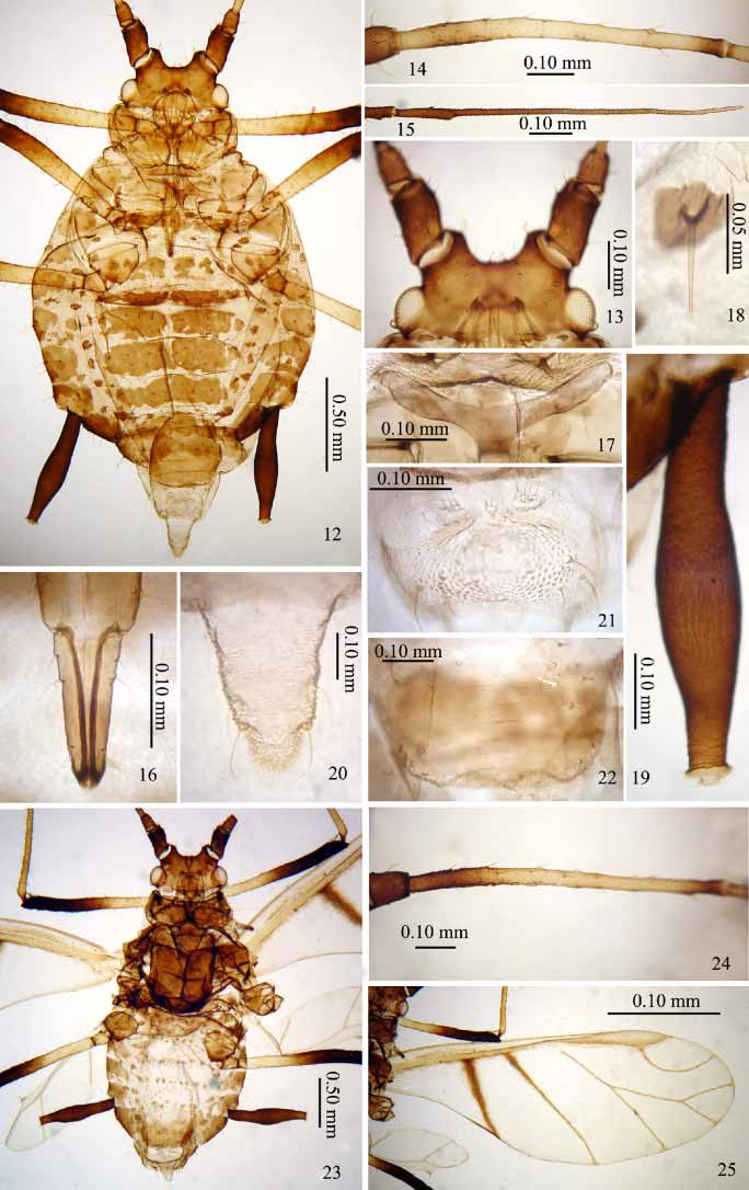
Apterous viviparous female: Body large, rather rotund ( Fig. 12 View FIGURES 12 – 25 ). Dark brown to black in life. General measurements—see Table 1.
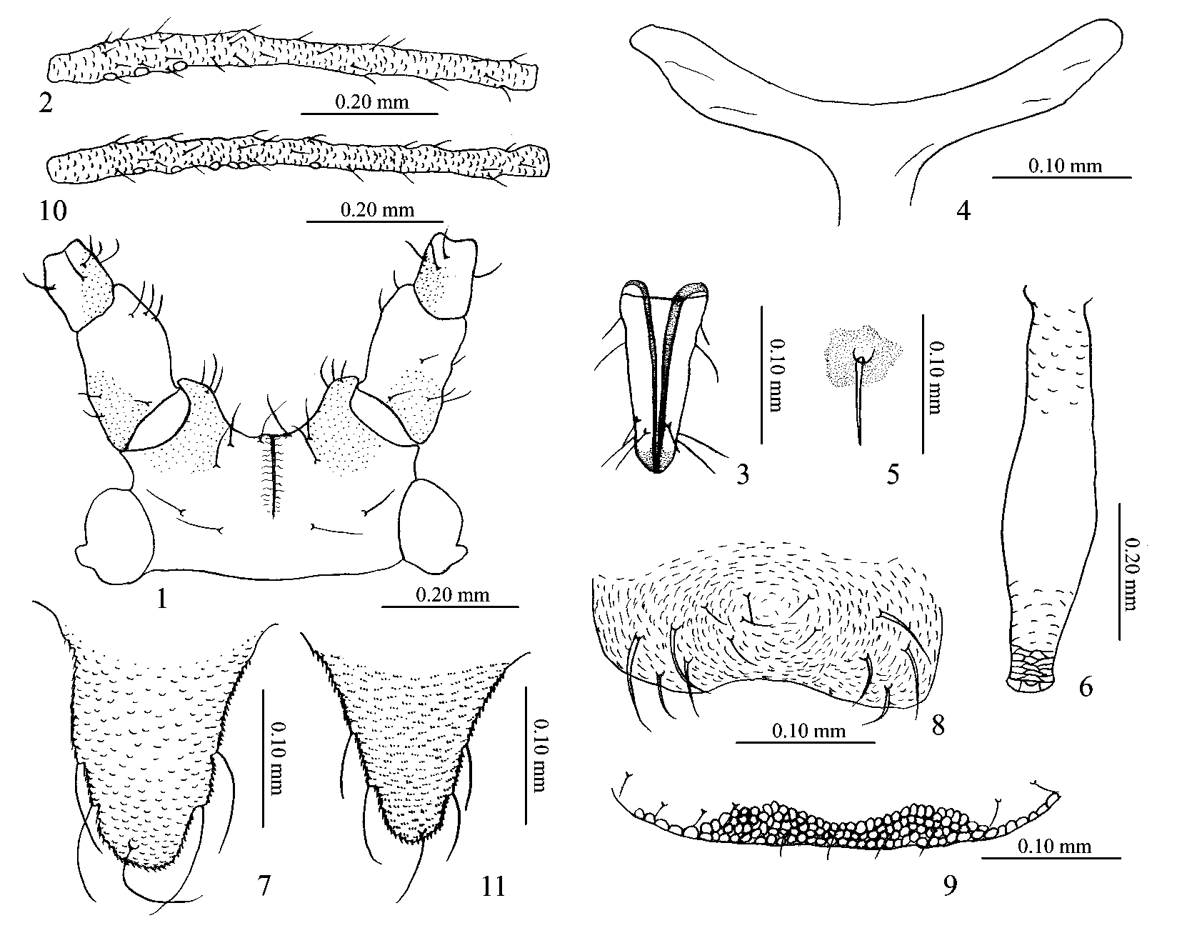
Mounted specimens: Antennal tubercles, antennal segments I–II, distal 1/3 of segment IV, distal half of segment V and base of segment VI, distal 1/3 of fore and midfemora, distal half of hind femora and siphunculi blackish brown; basal and distal parts of antennal segment III, basal half of segment IV, basal half of processus terminalis, ultimate rostral segment, distal of tibia, tarsi and genital plate dark brown; other antennal segments and other appendages brown; cauda and anal plate pale brown. Dorsum of head dark brown ( Figs 1 View FIGURES 1 – 11 , 13 View FIGURES 12 – 25 ). Pronotum and mesonotum brown. Metanotum to abdominal tergites IV each with 1 pair of brown marginal patches, 1 pair of broad spinal bars and 2–3 pairs of small pleural patches, each of which bears a dorsal hair ( Fig. 12 View FIGURES 12 – 25 ). Antesiphuncular and postsiphuncular sclerites well developed, and fused with each other. Abdominal tergites with large sclerites that loosely coalesce to form transverse brown bands, excepting tergite VIII which is much paler.
Ventral surface of head coarsely spinulose, but dorsum of head and antennal tubercles more faintly spinulose and median line of head clear; thorax ventrally coarsely spinulose, but abdominal sternites with much finer transverse spinulose bands; antennal segments I and II ventrally spinulose ( Figs 1 View FIGURES 1 – 11 , 13 View FIGURES 12 – 25 ); antennal segments III–VI imbricate throughout; coxae with spinulose bands; distal halves of femora with fine spinules; second tarsal segments with imbrications but not spinules; siphunculi ( Figs 6 View FIGURES 1 – 11 , 19 View FIGURES 12 – 25 ) distinctly swollen in middle 1/3, with the narrowed apical part bearing a few spinulose imbrications and with 2–3 complete striate bands before the flange, the basal 2/3 smoother but with curious poorlydefined narrow rugae; cauda ( Figs 7 View FIGURES 1 – 11 , 20 View FIGURES 12 – 25 ) and anal plate ( Fig. 8 View FIGURES 1 – 11 , 21 View FIGURES 12 – 25 ) coarsely spinulose; anterior of subgenital plate smooth, posterior margin reticulate ( Figs 9 View FIGURES 1 – 11 , 22 View FIGURES 12 – 25 ); dorsum of abdomen generally smooth. Basal half of antennal segment I with reticulations that may be wax glands. Dorsal body hairs stout but acute ( Figs 5 View FIGURES 1 – 11 , 18 View FIGURES 12 – 25 ). Head with hairs a shown in Fig. 1 View FIGURES 1 – 11 . Pronotum with 9 spinal hairs, 1 pair of pleural and 1 pair of marginal hairs. Abdominal tergite I with 15–21 hairs; tergites II–V each with 6–9 spinal, 3–4 pairs of pleural and 3 pairs of marginal hairs; tergite VI with 3 pairs of spinal, 1 pair of pleural and 3 pairs of marginal hairs; tergite VII with 8–12 hairs; tergite VIII with 4 hairs. Length of cephalic hairs, length of marginal hairs on abdominal tergite I, length of dorsal hairs on tergite VIII 0.80–1.33 times, 1.20–1.67 times and 1.40–1.78 times as long as widest diameter of antennal segment III, respectively. Mesosternal furca with short stem ( Figs 4 View FIGURES 1 – 11 , 17 View FIGURES 12 – 25 ). Spiracles large and kidneyshaped, each on the posterior part of its sclerite. Dorsum of head with a pale spinal suture ( Figs 1 View FIGURES 1 – 11 , 13 View FIGURES 12 – 25 ) which is bordered by a spinulefree zone. Median frontal tubercle littledeveloped; antennal tubercles welldeveloped, their inner margins distinctly divergent. Antennae 6segmented, 1.05–1.27 times as long as body; length in proportion of segments I–VI: 25:14:100:78:62:21+111, respectively; processus terminalis 4.87–5.58 times as long as base of segment VI ( Fig. 15 View FIGURES 12 – 25 ); antennal hairs short, thick, but apically acute; length of hairs on segment III 0.031–0.041 mm, 0.60–0.88 times as long as widest diameter of the segment. Antennal segment III with 2–5 small round secondary rhinaria on basal 1/3–2/5 part ( Figs 2 View FIGURES 1 – 11 , 14 View FIGURES 12 – 25 ). Rostrum reaching abdominal segment I, ultimate rostral segment thick wedgeshaped ( Figs 3 View FIGURES 1 – 11 , 16 View FIGURES 12 – 25 ), 0.14–0.15 mm long, 2.25–2.55 times as long as its basal width, up to 1.5 times as long as second hind tarsal segment; with 1–3 (usually 2) pairs of rather short accessory hairs. Legs normal; hind femur 0.86–1.09 mm long, 1.32–1.43 times as long as antennal segment III; hind tibia 1.52–2.04 mm long, 0.64–0.79 times as long as body; hairs on legs robust but apically acute; length of hairs on hind tibia 0.051–0.062 mm, 1.10–1.38 times as long as midwidth of the segment. First tarsal chaetotaxy 3, 3, 3. Siphunculi with median 1/3 clavate ( Figs 6 View FIGURES 1 – 11 , 19 View FIGURES 12 – 25 ); 0.49–0.64 mm long, 0.20–0.25 times as long as body, 2.94–3.71 times as long as cauda, 5.00–6.11 times as long as its basal width, 4.31–5.00 times as long as diameter of swollen part. Cauda short and roundedtriangular ( Figs 7 View FIGURES 1 – 11 , 20 View FIGURES 12 – 25 ), 0.35–0.42 mm long, 1.06–1.42 times as long as its basal width, usually with 5 hairs. Anal plate as shown ( Figs 8 View FIGURES 1 – 11 , 21 View FIGURES 12 – 25 ), with 12–15 hairs. Subgenital plate ovoid ( Figs 9 View FIGURES 1 – 11 , 22 View FIGURES 12 – 25 ), with 5–10 (usually 8–10) anterior hairs and 10–17 posterior hairs. Gonapophyses three, each with 5–8 short and pointed hairs ( Fig. 21 View FIGURES 12 – 25 ).
Alate viviparous female: Body less rotund than in apterae ( Fig. 23 View FIGURES 12 – 25 ), 3.26 mm long, 1.40 mm wide. Black in life.
Mounted specimen: Antennal tubercles, antennal segments I–II, basal 1/3 and distal of segment III, distal 1/3 of segment IV, segment V and base of segment VI, apex of rostrum, distal 3/5 of femora, distal of tibia and siphunculi blackishbrown; other antennal segments and appendages brown; cauda and anal plate pale brown. Dorsum of head and thorax dark brown. Abdominal tergite I–VII each with 1 pair of marginal patches, tergite I with wider spinopleural transverse band, tergites II–VIII each with 3–11 hairbearing sclerites ( Fig. 23 View FIGURES 12 – 25 ).
Venter of head and dorsum of antennal tubercles with sparse spinules, ventral pleural areas of thorax with spinulose short transverse stripes; abdominal tergites VII–VIII and ventral of abdomen with spinulose transverse bands; inner side of antennal segment I and basal outer part of segment II with spinulose areas, segments III–VI with imbrications; coxae with spinulose bands; distal 2/3 of femora with sparse spinulose imbrications; second tarsal segments imbricate; siphunculi subapically with 3–4 striae before the flange; cauda roundedtriangular ( Fig. 11 View FIGURES 1 – 11 ); anal plate with bands of spinules; anterior of subgenital plate smooth, posteriorly with reticulations. Dorsal hairs of body apically blunt; head with similar pattern of hairs to that seen in apterae. Pronotum with 1 pair of posterior spinal hairs, 1 pair of anterior pleural and 1 pair of marginal hairs. Abdominal tergite I with 14 hairs; tergite VIII with 4 hairs. Length of cephalic hairs 0.051 mm, length of marginal hairs on abdominal tergites I 0.065 mm, length of dorsal hairs on tergite VIII 0.085 mm, 1.00 times, 1.29 times and 1.71 times as long as widest diameter of antennal segment III, respectively. Median frontal tubercle absent but antennal tubercles welldeveloped, their inner margins distinctly diverging. Antennae 6segmented, 4.90 mm long, 1.51 times as long as body; processus terminalis 7.14 times as long as base of segment VI; antennal hairs short and blunt or acute at apices, apex of processus terminalis with 4 hairs; length of hairs on antennal segment III 0.051 mm, about as long as widest diameter of the segment. Antennal segment III with 6 small round secondary rhinaria on its basal half ( Figs 10 View FIGURES 1 – 11 , 24 View FIGURES 12 – 25 ). Rostrum reaching midcoxae, ultimate rostral segment wedgeshaped, 0.19 mm long, 2.36 times as long as its basal width, 1.30 times as long as second hind tarsal segment; with 2 pairs of accessory hairs. Hind femur 1.54 mm long, 1.41 times as long as antennal segment III; hind tibia 2.82 mm long, 0.87 times as long as body; hairs on legs thick, slightly longer than antennal hairs, blunt or acute at apices; length of hairs on hind tibia 0.058 mm, 1.07 times as long as midwidth of the segment. First tarsal chaetotaxy 3, 3, 3. Forewings ( Fig. 25 View FIGURES 12 – 25 ) with media twicebranched, hind wing with two oblique veins, venation and pigmentation as shown. Siphunculus with median 1/3 part clavate; 0.73 mm long, 0.22 times as long as body, 3.85 times as long as cauda, 7.69 times as long as its own basal width, 4.00 times as long as diameter of its swollen part, 9.09 times as long as its own distal width below flange. Cauda ( Fig. 11 View FIGURES 1 – 11 ), 0.19 mm long, 0.68 times as long as its basal width, with 5 hairs. Anal plate with 11 hairs. Subgenital plate ovoid, with 7 anterior hairs and 13 posterior hairs. Gonapophyses three, each with 5 or 6 short and pointed hairs.
Material examined. Holotype apterous viviparous female, 2001XII07, CHINA ( Hong Kong, Pok Fu Lam Country Park, below High West), on Millettia sp., vine (J. H. Martin #7536) ( BMNH). Paratypes, 13 apterous viviparous females, 1 alate viviparous female, 3 nymphs, same data as holotype ( BMNH, ZMCAS); 7 apterous viviparous females, 4 firstinstar nymphs, 2005XII01, same locality, on unidentified vine (J.H.Martin #8236) ( BMNH).
Comments. The new species is most closely allied to A. formosana (Takahashi) , but differs from it as shown in the key. On each occasion on which this aphid has been found, the small colony was feeding on the activelygrowing tip of a woody vine, an exceptionally narrow substrate to support large aphids. Although the host has only been identified once, it is considered likely that the same host was involved on both occasions.
No known copyright restrictions apply. See Agosti, D., Egloff, W., 2009. Taxonomic information exchange and copyright: the Plazi approach. BMC Research Notes 2009, 2:53 for further explanation.