
Archaeohuysia huysi, Gómez, 2021
|
publication ID |
https://doi.org/ 10.11646/zootaxa.5051.1.12 |
|
publication LSID |
lsid:zoobank.org:pub:A99E653A-EBDF-48B1-BF24-0194136E03F9 |
|
DOI |
https://doi.org/10.5281/zenodo.5563563 |
|
persistent identifier |
https://treatment.plazi.org/id/10744E2C-9B90-49E2-8A5D-B15A98ED94C7 |
|
taxon LSID |
lsid:zoobank.org:act:10744E2C-9B90-49E2-8A5D-B15A98ED94C7 |
|
treatment provided by |
Plazi |
|
scientific name |
Archaeohuysia huysi |
| status |
sp. nov. |
Archaeohuysia huysi sp. nov.
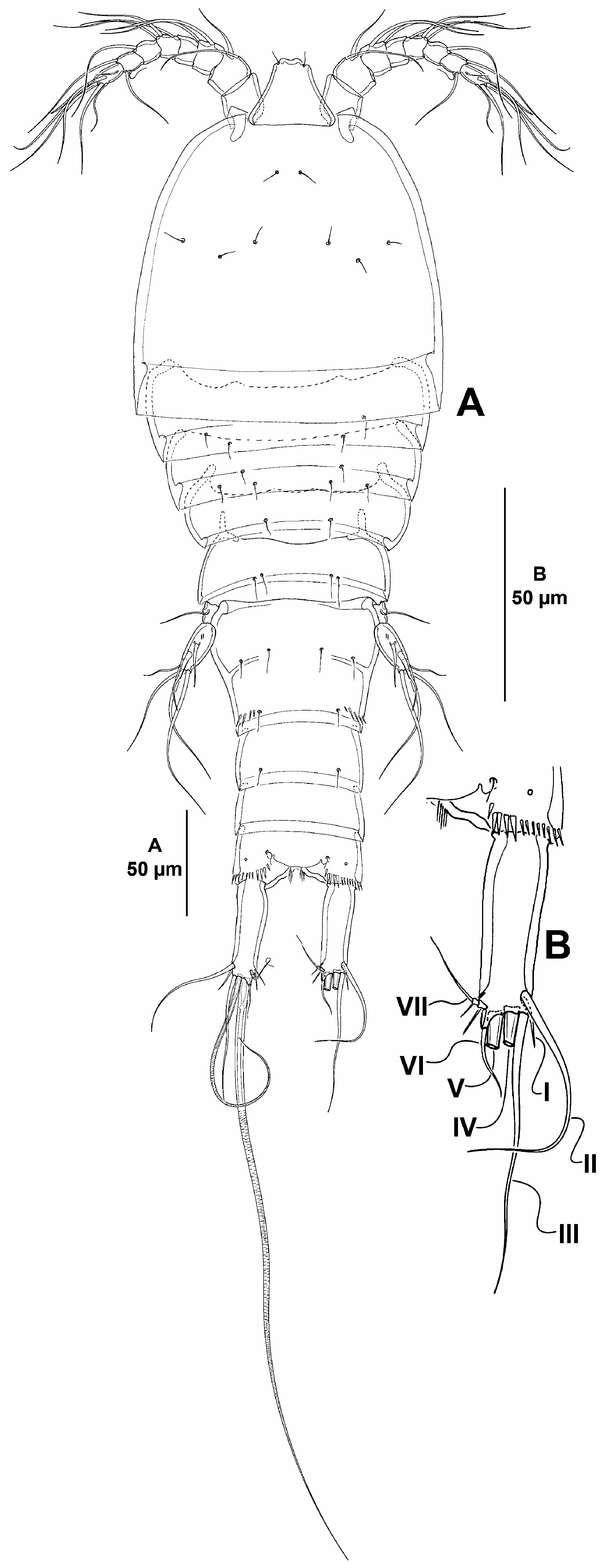
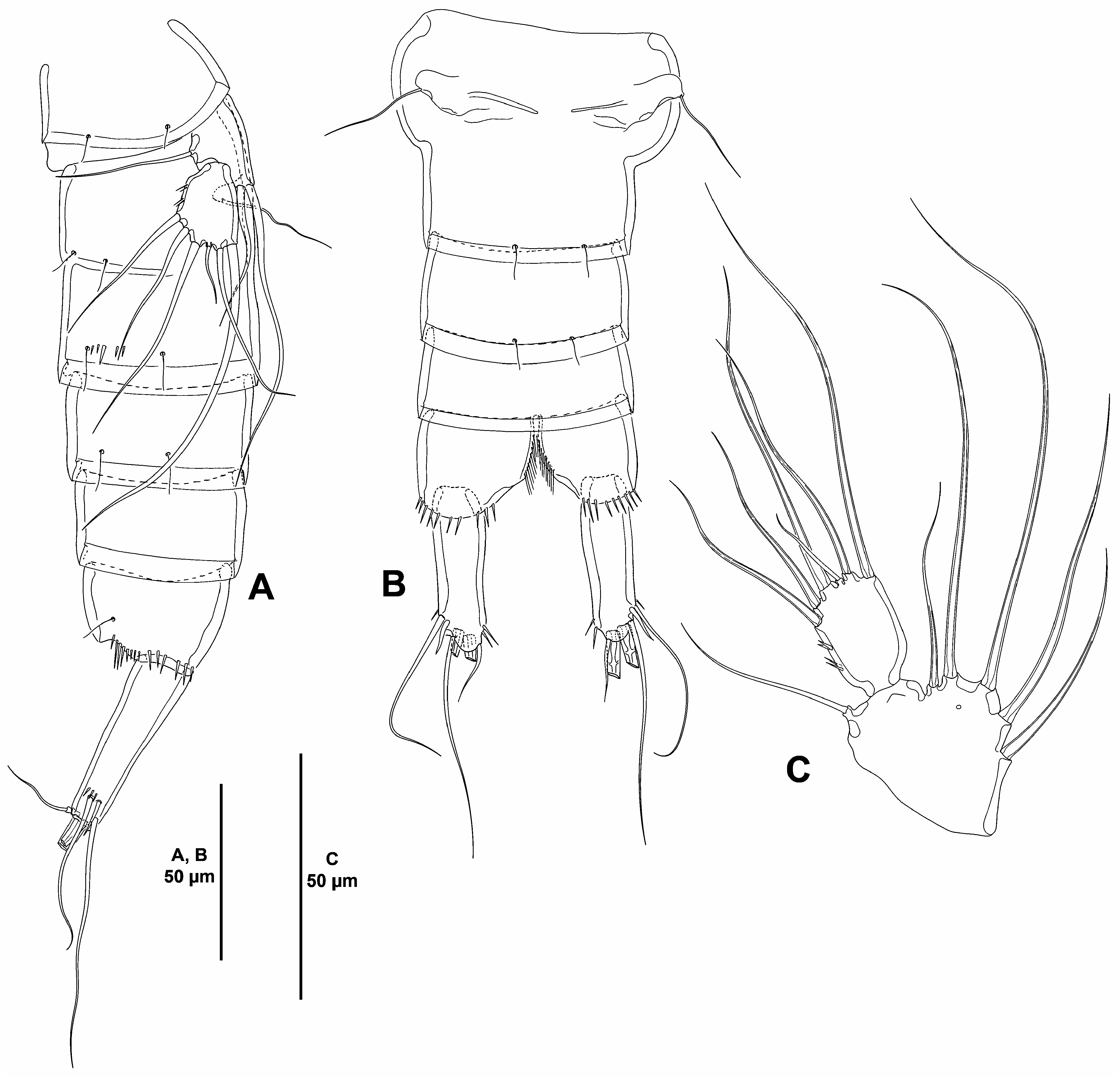
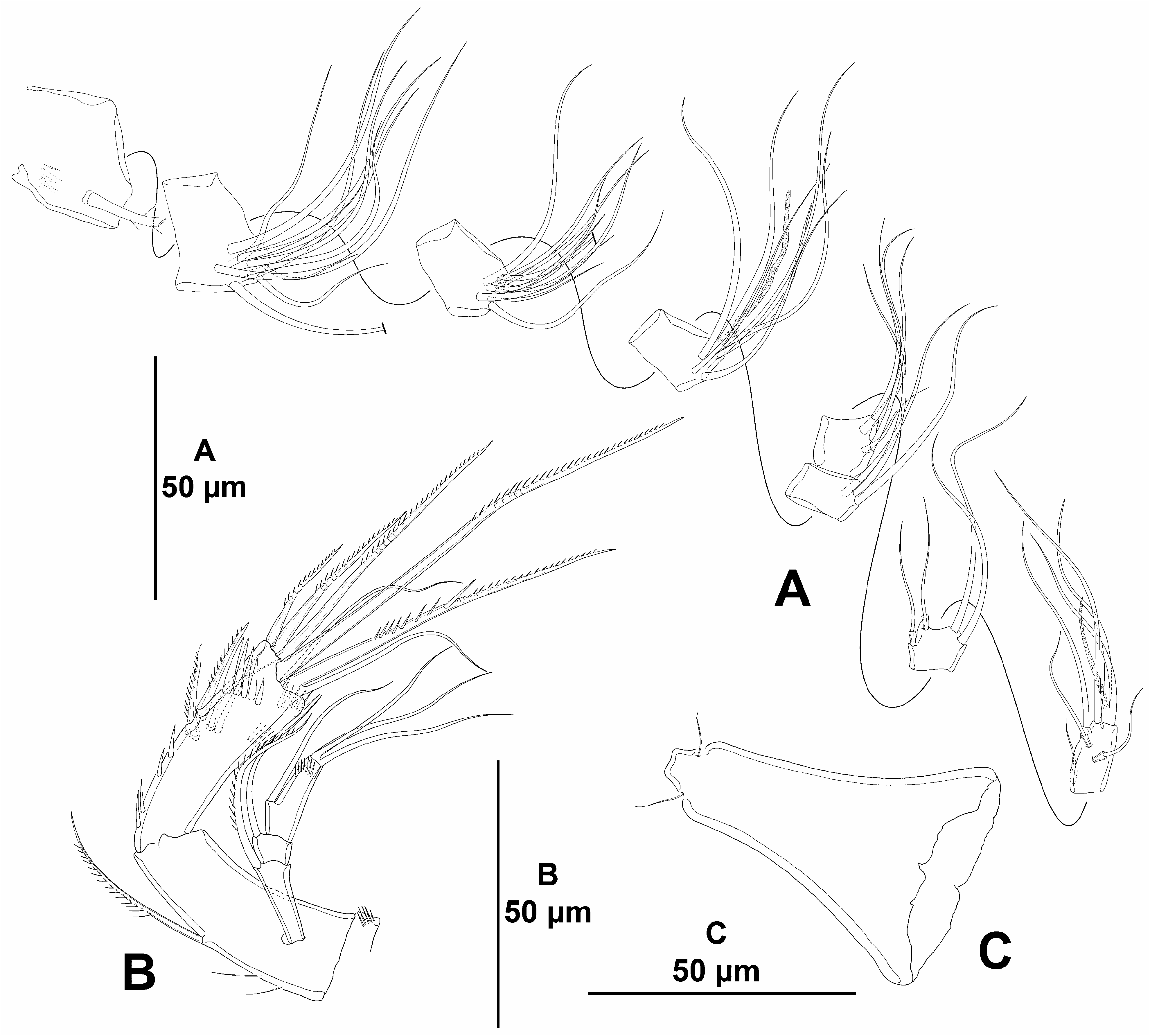
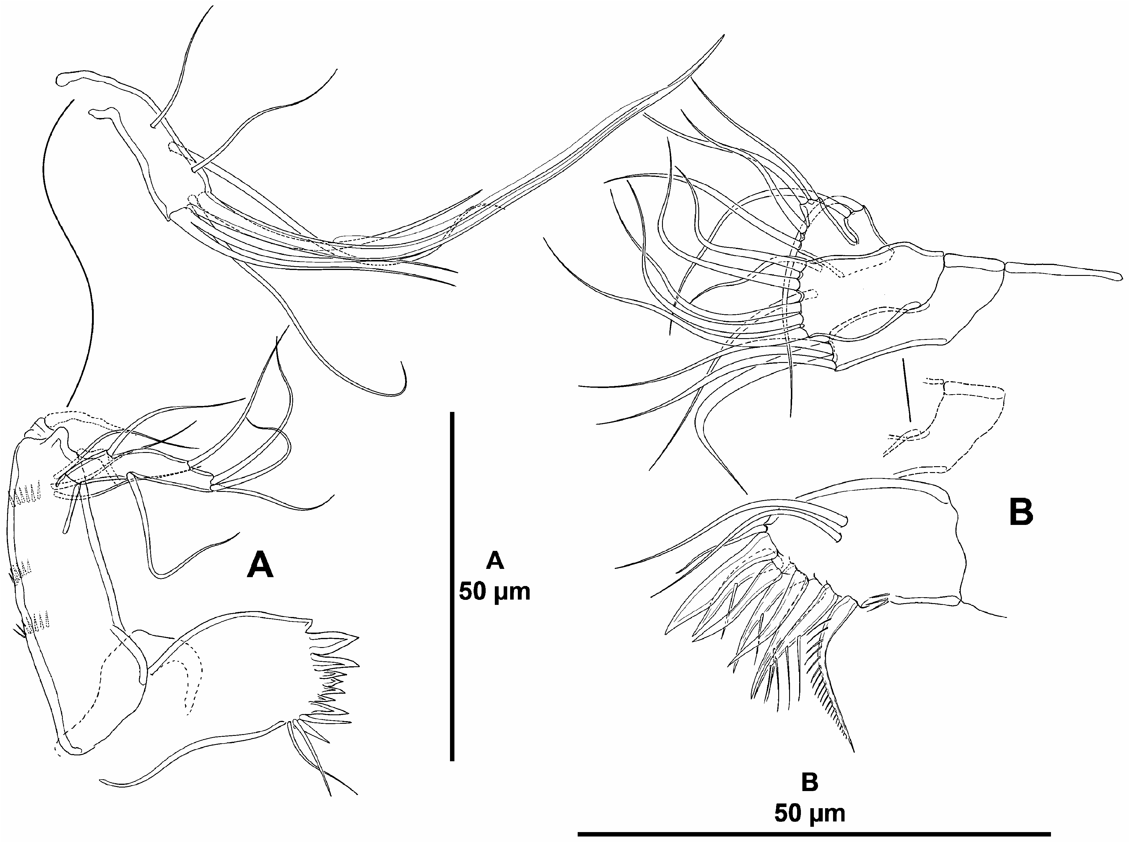
( Figs. 24–30 View FIGURE 24 View FIGURE 25 View FIGURE 26 View FIGURE 27 View FIGURE 28 View FIGURE 29 View FIGURE 30 )
urn:lsid:zoobank.org:act:10744E2C-9B90-49E2-8A5D-B15A98ED94C7
Type locality. San Isidro Basin , off west coast of Baja California (Eastern Tropical Pacific), Mexico; Talud XVIB cruise, sampling station 21 (30.9247°N, 116.8267°W); depth 2,037 m; organic carbon content, 2.21%; organic matter content, 3.81%; sand, 1.08%; clay, 12.54%; silt, 86.38 GoogleMaps %.
Specimens examined. Adult female holotype dissected and mounted onto nine slides (EMUCOP-280514-02); May 28, 2014; coll. S. Gómez.
Etymology. The species is named in honour of Dr. Rony Huys (Natural History Museum, London) for his contribution to the systematics and taxonomy of harpacticoid copepods. It is a noun in the genitive case. Gender masculine.
Description of female. Total body length measured from tip of rostrum to posterior margin of caudal rami, 440 µm; habitus pyriform, widest at posterior end of cephalothorax, tapering posteriad ( Fig. 24A View FIGURE 24 ); cephalothorax/body length ratio, 0.35.
Prosome and pedigerous somites ( Figs. 24A View FIGURE 24 ) largely as in previous species.
Urosome ( Figs. 24A View FIGURE 24 , 25A–B View FIGURE 25 ) consisting of fifth pedigerous somite (first urosomite), genital double-somite (genital—second urosomite—and third urosomites fused), two free urosomites, and anal somite. Urosomites without expansions laterally nor dorsally; integument weakly sclerotized.
Fifth pedigerous somite ( Fig. 24A View FIGURE 24 ) narrower than preceding somites; with some sensilla dorsally ( Fig. 24A View FIGURE 24 ) and laterally ( Fig. 25A View FIGURE 25 ), without spinular ornamentation.
Second and third urosomites completely fused dorsally and ventrally forming genital double-somite ( Fig. 25B View FIGURE 25 ), with dorsolateral trace of division ( Figs. 24A View FIGURE 24 , 25B View FIGURE 25 ); genital double-somite 1.4 times as long as wide, widest part measured close to P6; proximal half with sensilla and without spinules dorsally ( Fig. 24A View FIGURE 24 ), ventrally without sensilla nor spinules ( Fig. 25B View FIGURE 25 ); distal half with dorsolateral sensilla and spinular rows ( Figs. 24A View FIGURE 24 , 25A View FIGURE 25 ), ventrally with few sensilla and without spinules ( Fig. 25B View FIGURE 25 ); posterior hyaline fringe broad and smooth; genital complex hardly distinguishable, copulatory pores not exposed, paired genital apertures located ventrolaterally and covered by P6 ( Fig. 25B View FIGURE 25 ).
Fourth urosomite ( Figs. 24A View FIGURE 24 , 25A–B View FIGURE 25 ) as distal half of genital double-somite but without spinular ornamentation; no pores detected.
Fifth urosomite without sensilla nor spinules ( Figs. 24A View FIGURE 24 , 25A–B View FIGURE 25 ); no pores detected.
Anal somite about 2.5. times as wide as long ( Fig. 24A View FIGURE 24 ); with spinules around joint of caudal rami ( Figs. 24A View FIGURE 24 , 25A–B View FIGURE 25 ); medial cleft with row of setules ventrally ( Fig. 25B View FIGURE 25 ); with one dorsal pore on each side ( Fig. 24A View FIGURE 24 ); anal operculum without spinular ornamentation, semicircular, flanked by one sensilla on each side ( Fig. 24A View FIGURE 24 ).
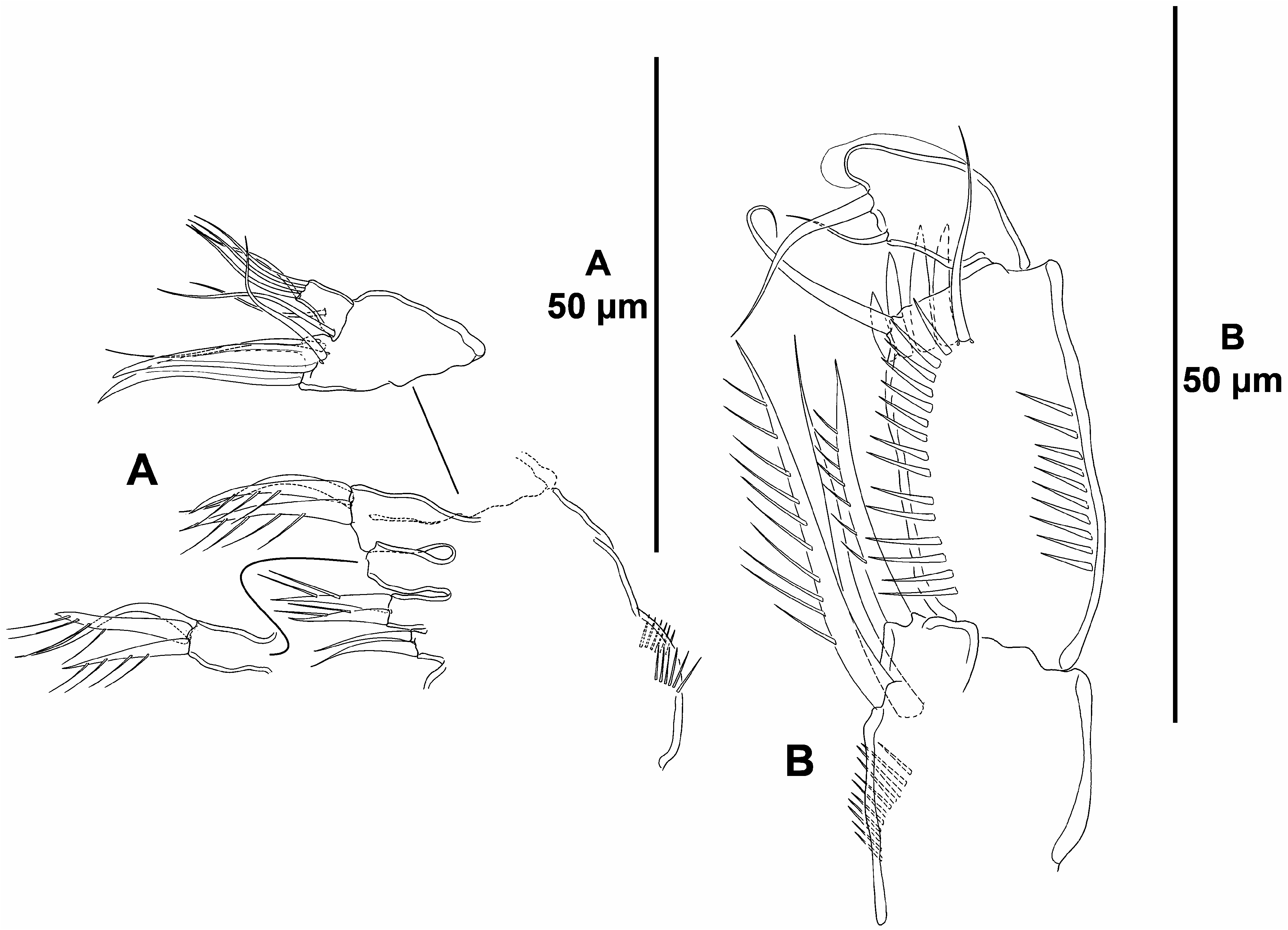
Caudal rami elongate, about 3 times as long as wide ( Figs. 24A–B View FIGURE 24 ) and about as long as fifth and anal somites combined; outer margin nearly straight, inner margin slightly convex proximally; with outer spinules at base of setae I and II ( Fig. 25A View FIGURE 25 ), and with inner spinules subdistally ( Figs. 24A–B View FIGURE 24 , 25B View FIGURE 25 ); with seven elements ( Fig. 24B View FIGURE 24 ); setae I and II subdistal, lateral, seta I spine-like and ventral to seta II, the latter long; seta III subdistal, arising ventrally ( Figs. 25A–B View FIGURE 25 ); setae IV and V distal, rat-tail like in distal half, with fracture plane; seta VI small, issuing at inner distal corner; dorsal seta VII triarticulate at base, situated subdistally close to inner margin.
Rostrum ( Fig. 26C View FIGURE 26 ) trapezoidal, elongate, not fused to cephalothorax, weakly bifid, with two subdistal sensilla, without dorsal pore.
Antennule ( Fig. 26A View FIGURE 26 ) eight-segmented; all segments smooth, except for first segment with proximal spinular row; first segment without pore. All setae smooth, except for one pinnate seta on first segment; no setae with fracture plane detected; seventh segment with two, eighth segment with three articulated setae. Armature formula: 1(1); 2(11); 3(10); 4(6 + (1 + ae)), 5(3); 6(4); 7(4); 8(5 + acro). Acrothek consisting of two setae and one minute aesthetasc fused basally.
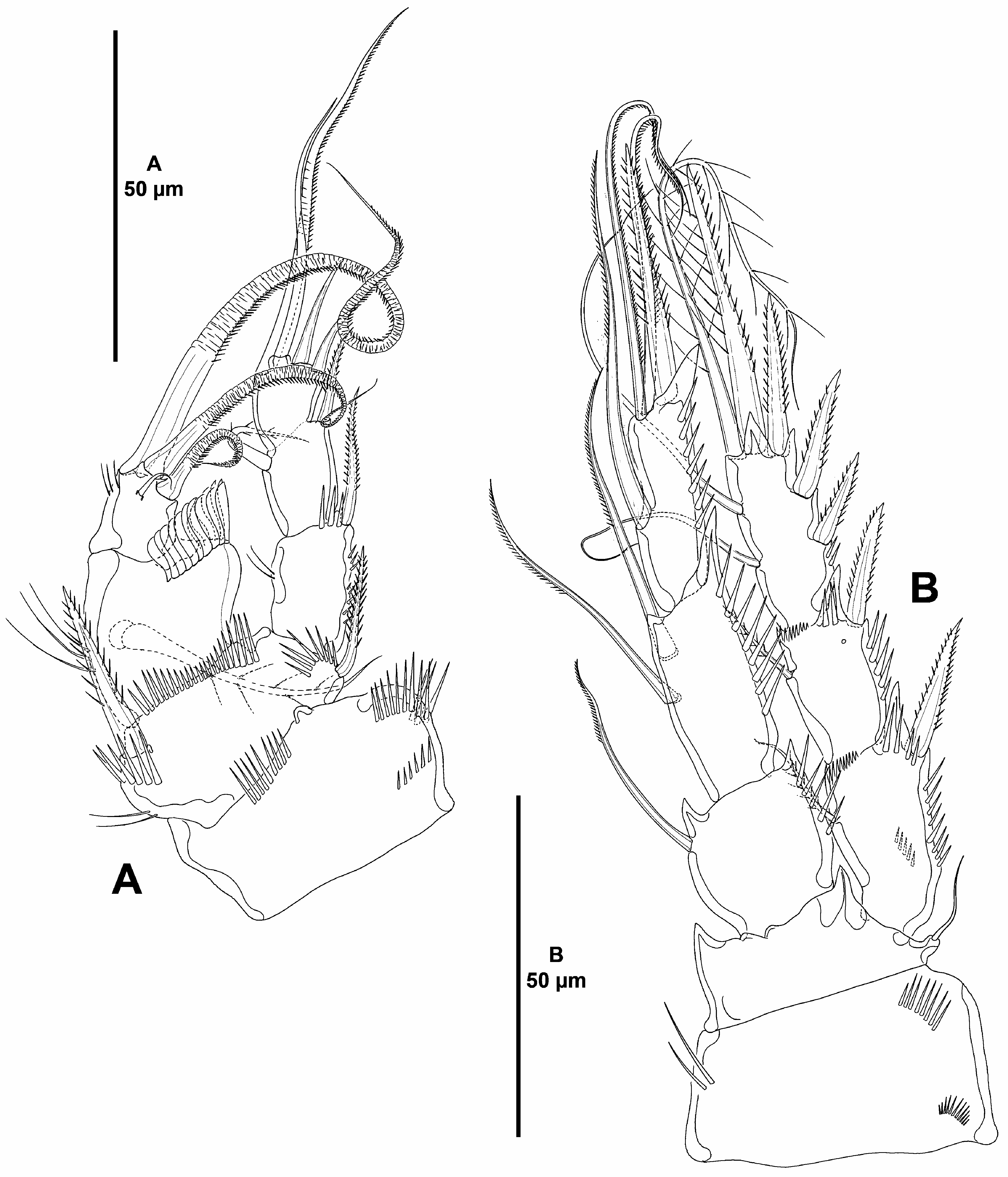
Antenna ( Fig. 26B View FIGURE 26 ). Coxa short, with some outer spinules. Allobasis as long as free endopodal segment; with few slender outer spinules proximally; with one abexopodal seta arising midway inner margin. Free endopodal segment elongate; proximal half with longitudinal row of strong inner spinules, with subdistal outer strong spinules, with two outer subdistal frills; armature composed of two lateral spines and two setae, distally with one inner apical geniculate element, three apical geniculate setae and one slender element, and one outer distal strongly spinulose element fused basally to slender seta. Exopod three-segmented; first and third segments longest; first and middle segment without, third segment with spinules as shown; first and second segments with one distal seta each, third segment with one proximal and three apical setae, two of which seemingly fused basally.
Mandible ( Fig. 27A View FIGURE 27 ). Coxa relatively short. Gnathobase wide; ventral distal corner produced into small sharp semi-hyaline process; with two strong and several smaller teeth, two spines and two setae. Basis elongate, with spinular ornamentation as shown, with three subdistal outer setae. Exopod arising from short pedestal, onesegmented, elongate, about 5 times as long as wide, and 0.5 times as long as basis, with three lateral and three apical setae, none of which fused basally. Endopod recurved, twisted over exopod, with three lateral setae, and five distal elements (three slender setae, two of which fused basally, one strong element, and longest element fused to endopod basally and with hyaline flange in middle part).
Maxillule ( Fig. 27B View FIGURE 27 ). Arthrite of praecoxa with two surface setae and some dorsal spinules; distal armature composed of one ventral seta and seven strong spines as shown, and one lateral pinnate recurved seta. Coxal endite with three setae; spinular ornamentation not detected. Basis with two endites separated by small notch; proximal endite with four, distal endite with three slender setae. Exopod and endopod fused basally, separated from basis, one-segmented; endopod larger than exopod, with four setae; exopod with two setae.
Maxilla ( Fig. 28A View FIGURE 28 ). Large syncoxa with outer spinules as shown; with three endites; proximal endite bilobed, each lobe with two setae; middle and distal endites elongate, the latter slightly longer, with one naked and two spinulose setae each. Basis drawn out into strong claw, with strong spine and two slender setae. Endopod onesegmented, with six slender setae (one arising basally, one medially, and four apically).
Maxilliped ( Fig. 28B View FIGURE 28 ) subchelate, not or weakly prehensile. Syncoxa slightly longer than wide, visibly shorter than basis, with medial inner spinules, with one bare and two spinulose strong elements, of which bare seta and one spinulose element at the same level, the other arising distally from pedestal. Basis longer than syncoxa, rectangular, with some outer spinules, with one anterior and one posterior inner spinular row as figured, with two slender distal setae subequal in length. Endopod one-segmented, with hyaline distal part, with two seta-like elements, of which distalmost homologue to endopodal claw.
P1 ( Fig. 29A View FIGURE 29 ). Coxa massive, 1.5 times as wide as long, with outer and medial spinules as shown. Basis with spinules at base of outer and inner spines and between rami, with long slender inner spinules. Exopod threesegmented, longer than endopod; division between EXP2 and EXP3 observable from posterior view; no pores detected on exopodal segments; EXP1 longest, EXP3 shortest; all segments without outer nor inner acute distal processes; EXP1 with, EXP2 without inner setules and outer spinules; EXP1 without, EXP2 with inner seta; EXP3 without outer spinules, with two outer spines and two apical elements. Endopod characteristic, two-segmented, reaching tip of EXP2, segments without inner nor outer acute distal processes; no pores detected on endopodal segments; ENP1 massive, barely reaching tip of EXP1, nearly as long as wide, visibly longer than ENP2, with few inner long spinules, outer distal corner produced and with semicircular row of modified spinules, with inner seta; ENP2 small, rectangular, about 1.1 times as long as wide, and 0.6 times as long as ENP1, inner distal margin with few slender spinules, without outer spinules, with one inner (medial?) short seta, and two apical and one outer rat-tail like elements.
P2 ( Fig. 29B View FIGURE 29 ). Intercoxal sclerite (not shown) not transversely elongate; trapezoidal; with strong pointed process on distal outer corners; without surface ornamentation. Coxa with outer spinules proximally and subdistally, with few inner long spinules. Basis with strong acute process between rami and at inner distal corner, seemingly without spinular ornamentation; with outer seta. Exopod three-segmented, reaching middle of ENP3; first and third segments longest; EXP1 and EXP2 with outer acute distal process, with outer spinules and with distal inner frill as shown, distal processes of EXP3 as shown; EXP1 with, EXP2 without posterior spinules; EXP1 without, EXP2 with subdistal outer pore; EXP1 and EXP2 with one inner seta; EXP3 with two inner setae, two apical elements and three outer spines. Endopod three-segmented, slightly longer than exopod; ENP1 shortest, about 0.7 times as long as ENP2; ENP2 and ENP3 subequal in length; all endopodal segments with longitudinal row of outer spinules; ENP1 with outer and inner small acute processes subequal in length, outer distal process on ENP2 visibly stronger, ENP3 with one strong outer process only; no pores detected on endopodal segments; ENP1 with one, ENP2 with two stiff inner setae with serrate inner margin; ENP3 with one inner strong element, two apical setae and one outer spine.
P3 ( Fig. 30A View FIGURE 30 ). Intercoxal sclerite (not shown), coxa and basis largely as in P2. Exopod and endopod subequal in length. Exopod as in P2, except for less developed outer acute distal process and for lack of pores on EXP2, and for inner armature of EXP3 (with three inner setae instead of two). Endopod as in P2, except distal processes on ENP3, for inner complement of ENP2 (with one seta instead of two) and ENP3 (with three inner setae instead of two).
P4 ( Fig. 30B View FIGURE 30 ). Intercoxal sclerite (not shown), coxa and basis largely as in P2. Endopod shorter than exopod, reaching proximal third of EXP3. Spinular ornamentation of exo- and endopodal segments as in previous legs; seemingly without pores. Exopod with outer acute distal processes on EXP1 and EXP2 less developed than in P3, EXP3 seemingly without distal process; armature complement as in P3, but inner seta on EXP2 visibly longer. Endopod with outer distal process of ENP1 and ENP2 less developed than in P3, of ENP3 as shown; armature complement as in P3 but inner seta on ENP1 visibly stronger and longer, and ENP3 with two inner setae instead of three.
Setal formula of swimming legs as follows:
P5 ( Fig. 25C View FIGURE 25 ). Baseoendopod pentagonal; endopodal lobe poorly-developed, with subdistal pore, with five setae, of which outermost and adjacent seta set close together; all setae naked. Exopod oval; with two sets of outer spinules, with six setae, of which medial shortest.
P6 ( Fig. 25B View FIGURE 25 ) represented by a minute flap covering ventrolateral genital aperture, fused to somite, without surface ornamentation, with one slender seta.
Male. Unknown.
Variability. No variability was detected in the single female found in the sediment samples.

No known copyright restrictions apply. See Agosti, D., Egloff, W., 2009. Taxonomic information exchange and copyright: the Plazi approach. BMC Research Notes 2009, 2:53 for further explanation.
|
Kingdom |
|
|
Phylum |
|
|
Class |
|
|
Order |
|
|
Family |
|
|
Genus |