
Arachnopsita maya, Junta & Castro-Souza & Ferreira, 2022
|
publication ID |
https://doi.org/ 10.11646/zootaxa.5094.3.3 |
|
publication LSID |
lsid:zoobank.org:pub:50F1BC50-CEAA-491A-8E72-15F3E545BC49 |
|
DOI |
https://doi.org/10.5281/zenodo.6302417 |
|
persistent identifier |
https://treatment.plazi.org/id/0390C840-FFF1-AA7D-CCAD-1AB0FE97FD21 |
|
treatment provided by |
Plazi |
|
scientific name |
Arachnopsita maya |
| status |
sp. nov. |
Arachnopsita maya View in CoL n. sp.
( Figures 2–7 View FIGURES 2–7 , 8–14 View FIGURES 8–14 , 15–18 View FIGURES 15–18 , 19–23 View FIGURES 19–23 , Table 1 View TABLE 1 )
Material examined. Holotype ♂, code ISLA 12418, Guatemala, Alta Verapaz, municipality of Raxruhá, Cúpula de los Murcielagos (15°52’58.71” N; 90°11’20.64” O), 26.vi.2017, Pacheco, G. S. M., leg. GoogleMaps Paratypes, 7 ♂♂ ( ISLA 12410; 12411; 12412; 12413; 12414; 12416; 12419) and 2 ♀ ♀ ( ISLA 12415; 12417), same data of holotype .
Distribution. Cúpula de los Murcielagos, Cueva Blanca, Cueva el Rostro and Cueva del Venado caves, municipality of Raxruhá, Alta Verapaz, Guatemala.
Etymology. Specific epithet “ maya ” refers to the Maya civilization, who inhabited the Mesoamerican region, mainly between A.D. 250 and A.D. 900, known as its Classical Period ( Saunders 2005).
Diagnosis. Combination of the following characters: pseudepiphallic ventral projection acute, triangular shaped, as a spine ( Figs 2, 4–6 View FIGURES 2–7 , Ps.vp); C-sclerite ventral projection globular ( Fig. 4 View FIGURES 2–7 , C-vp); C-sclerite laterobasal spine well developed, thin, hooked shape and projecting towards the Ps.P1 ( Figs 2–6 View FIGURES 2–7 , C-lbs); C-sclerite basal plate broad, concave medially, inclining inward and reaching Ps.P2 ( Figs 3 and 4 View FIGURES 2–7 , C-bp); copulatory papilla with the presence of a convex bulge towards at base, evident laterally ( Fig. 7 View FIGURES 2–7 , b).
Description, male holotype ♂. Body color: dorsal head, pronotum and abdomen uniformly yellowish brown, and whitish ventrally ( Figs 10 and 11 View FIGURES 8–14 ); entire legs brownish, whitish at its proximal portion ( Figs 15–18 View FIGURES 15–18 ); cerci uniformly brown ( Fig. 12 View FIGURES 8–14 ). Head: slightly pubescent and with long bristles at base of vertex (some which were lost probably in fixation), elongated at front view (3.132 and 2.612 mm, length and width respectively), fastigium extending the vertex in an inclined plane; gena with a darkened strip connecting the compound eyes to the mandible insertion, front yellowish brown, clypeus and labrum light greyish, mandibles yellowish brown and sclerotized at apex; all maxillary palpomeres pubescent and whitish brown, first two short and same size, last three are bigger and similar size, fifth palpomere claviform at apex and whitish at the tip ( Figs 8 and 9 View FIGURES 8–14 ), all labial palpomeres pubescent and whitish brown, increasing in size, third palpomere claviform ( Figs 8 and 9 View FIGURES 8–14 ); scape whitish at the base and dark brown next to the pedicel, pedicel dark brown, antennomeres uniformly dark brown ( Figs 8 and 9 View FIGURES 8–14 ); compound eyes black, elongated, border of ommatidia lightly depigmented, ocelli absent ( Figs 8 and 9 View FIGURES 8–14 ). Thorax: pronotum slightly pubescent, anterior, medial and posterior portion with less sclerotized regions (appearance of whitish spots) distributed along the sagittal axis in dorsal view ( Figs 10 and 11 View FIGURES 8–14 ); dorsal disk broader than long, lateral lobes rounded, anterior and posterior margins sub-straight, anterior margin with long bristles, posterior and lateral margins with possibly lost bristles in fixation ( Fig. 10 View FIGURES 8–14 ). Legs: In general, femur, tibia and tarsus pubescent; femur smaller than tibia in length (μ = 9.731 ± 0.790 mm; μ = 11.346 ± 1.127 mm, femur and tibia respectively, Leg III, n = 8) ( Figs 15–18 View FIGURES 15–18 ). Leg I ( Figs 17 and 18 View FIGURES 15–18 ): tibia armed with two same-sized ventral apical spurs, tympanum absent; first tarsomere ventrally serrated and twice longer than second and third together. Leg II ( Figs 17 and 18 View FIGURES 15–18 ): tibia armed with two same-sized ventral apical spurs ( Fig. 17 View FIGURES 15–18 ; ε and Fig. 18 View FIGURES 15–18 ; ζ); first tarsomere ventrally serrated and twice longer than the second and third together. Leg III: femur dilated; tibia serrulated, armed with four subapical spurs on outer side ( Fig. 15 View FIGURES 15–18 ; w, x, y, z), the distal being smaller ( Fig. 15 View FIGURES 15–18 , z), and three on inner side ( Fig. 16 View FIGURES 15–18 ; α, β, γ), three apical spurs on outer ( Fig. 15 View FIGURES 15–18 ; a, b, c) and four on the inner side ( Fig. 16 View FIGURES 15–18 ; d, e, f, g), the inner being the longest ( Fig. 16 View FIGURES 15–18 , e); first tarsomere about twice longer than the second and third together, armed with two apical spurs ( Figs 15 and 16 View FIGURES 15–18 ). Right Tegmen: absent ( Fig. 10 View FIGURES 8–14 ). Abdomen: cerci long and pubescent, mainly in the base ( Fig. 12 View FIGURES 8–14 ): sub-genital plate light yellowish brown, longer than wide, sub-quadrangular, pubescent, with long bristles in distal margin, proximal margin slightly wider ( Figs 12 and 13 View FIGURES 8–14 ); supra-anal plate light yellowish brown, quadrangular, pubescent, proximal margin lightly V-shaped and with two lateral projections, concave distally in side margins, and with two small distal-lateral globular projections with long bristles ( Figs 12 and 14 View FIGURES 8–14 ).
Observations in Paratypes. Male phallic sclerites (paratype ISLA 12414, Figs 2–6 View FIGURES 2–7 ) Pseudepiphallus: arm long and slightly cambered inward ( Fig. 3 View FIGURES 2–7 , Ps.arm); ventral projection acute, triangular shaped, as a spine ( Figs 2, 4–6 View FIGURES 2–7 , Ps.vp); inner bars well sclerotized, curved inward forming a central acuminate projection ( Fig. 3 View FIGURES 2–7 , Ps.ib); membranous shield broad and flat ( Figs 4 and 5 View FIGURES 2–7 , Ps.ms); paramere 1 well developed, cone shaped frontally and globular dorsally, with a band less sclerotized, forming a smaller portion dorsally and a larger portion ventrally ( Figs 3–5 View FIGURES 2–7 , Ps.P1); paramere 2 reduced and undeveloped, connected with Ps.P1, flattened and projecting towards C-sclerite basal plate (C-bp) ( Figs 3–5 View FIGURES 2–7 , Ps.P2); A sclerite well sclerotized, starting from the Ps.arm, thin and involving the paramere 1, almost merging with this paramere ( Figs 3, 4 and 6 View FIGURES 2–7 , A). C-sclerite: in general is the most sclerotized part of the sclerite; ventral projection globular ( Fig. 4 View FIGURES 2–7 , C-vp); laterobasal spine well developed, thin, hooked shape and projecting towards the Ps.P1 ( Figs 2–6 View FIGURES 2–7 , C-lbs); basal plate broad, concave medially, inclining inward and reaching Ps.P2 ( Figs 3 and 4 View FIGURES 2–7 , C-bp). Ectophallic invagination: arc developed, upper and lower central part curved ( Fig. 2 View FIGURES 2–7 , Ect.Arc); lateral bars elongated and projected inwards ( Fig. 2 View FIGURES 2–7 , Ect.lb); apodemes developed, flatted, dilated and projected outwards of the sclerite, at dorsal and ventral view, with its distal portion acuminate at the apex ( Figs 3 and 6 View FIGURES 2–7 , Ect.ap). Endophallus: endophallic fold small and V-shaped ( Figs 2, 5–6 View FIGURES 2–7 , End.F); sclerotized extension of endophallic fold reduced and horizontally projected ( Figs 2 and 6 View FIGURES 2–7 , End.s); apodemes curved dorsally and close to each other, apex dilated and less sclerotized ( Figs 3 and 6 View FIGURES 2–7 , End.Ap).
Female: same appearance in relation to males, body size slightly bigger than male (♀ µ = 20.389 ± 2.044 mm, n = 2); apterous; femur always smaller than tibia; sub-genital plate light yellowish brown and pubescent, short, Vshaped, distal margin forked ( Fig. 19 View FIGURES 19–23 ); supra-anal plate whitish brown and pubescent, distal margin rounded with long bristles, proximal with two small projections ( Fig. 20 View FIGURES 19–23 ); ovipositor yellowish brown and elongated, sword shaped, with a constriction near the apex, pointed apex ( Figs 21–23 View FIGURES 19–23 ). Female genitalia ( ISLA 12415, Fig. 7 View FIGURES 2–7 ). Copulatory papilla elongated and flat ventrally, lateral margins sub-straight ventrally and dorsally, with the apex rounded and sharper than the base ( Fig. 7 View FIGURES 2–7 , a and c); with the presence of a convex bulge towards at base, evident laterally ( Fig. 7 View FIGURES 2–7 , b).
Arachnopsita maya n. sp.
Ecological Remarks: Individuals of Arachnopsita maya n. sp. were found in 4 caves in the Raxruhá municipality (Cúpula de los Murcielagos, Cueva Blanca, Cueva el Rostro and Cueva del Venado caves). Specimens were also preferentially observed in dark zones, as for A. cavicola and A. uncinata , although few specimens were found in twilight zones. The populational densities are very low, and immature specimens are more easily found. Cúpula de los Murcielagos ( Fig. 75 View FIGURES 74–76 ) and Cueva Blanca ( Fig. 76 View FIGURES 74–76 ) caves present some touristic activity, although only guided groups enter such caves. Furthermore, tourist activities are restricted to some areas of those caves, thus keeping other areas preserved. Cueva del Venado and Cueva el Rostro caves do not present touristic use. The higher densities were observed in the later caves, thus indicating that the presence of human visitors (even occasional) may drive specimens off those caves or eventually reduce their densities, although this is speculative, thus deserving further research. The specimens were observed in both speleothems and cave floor. The main organic resources observed in all caves was bat guano, although some vegetal debris (especially brought by wind) were also observed in some caves, mostly in areas closer to entrances. It is likely that A. maya n. sp. also feed in several organic debris, as for other cave crickets, being an opportunistic detritivore. The main predator of this species is also the phrynid amblypygid Paraphrynus williamsii Moreno, 1940 , observed in several caves in the area.
The external forests surrounding the caves are well preserved mainly over the “Mogotes” (steep-sided residual hills of carbonate rock surrounded by alluvial plains) and their immediate surroundings ( Fig. 74 View FIGURES 74–76 ). In most of the areas in between the mogotes, there are crops or pastures. Considering that this species is troglophilic (as A. cavicola and A. uncinata ), external populations certainly do exist, being also advisable to both protect some caves as their surroundings.


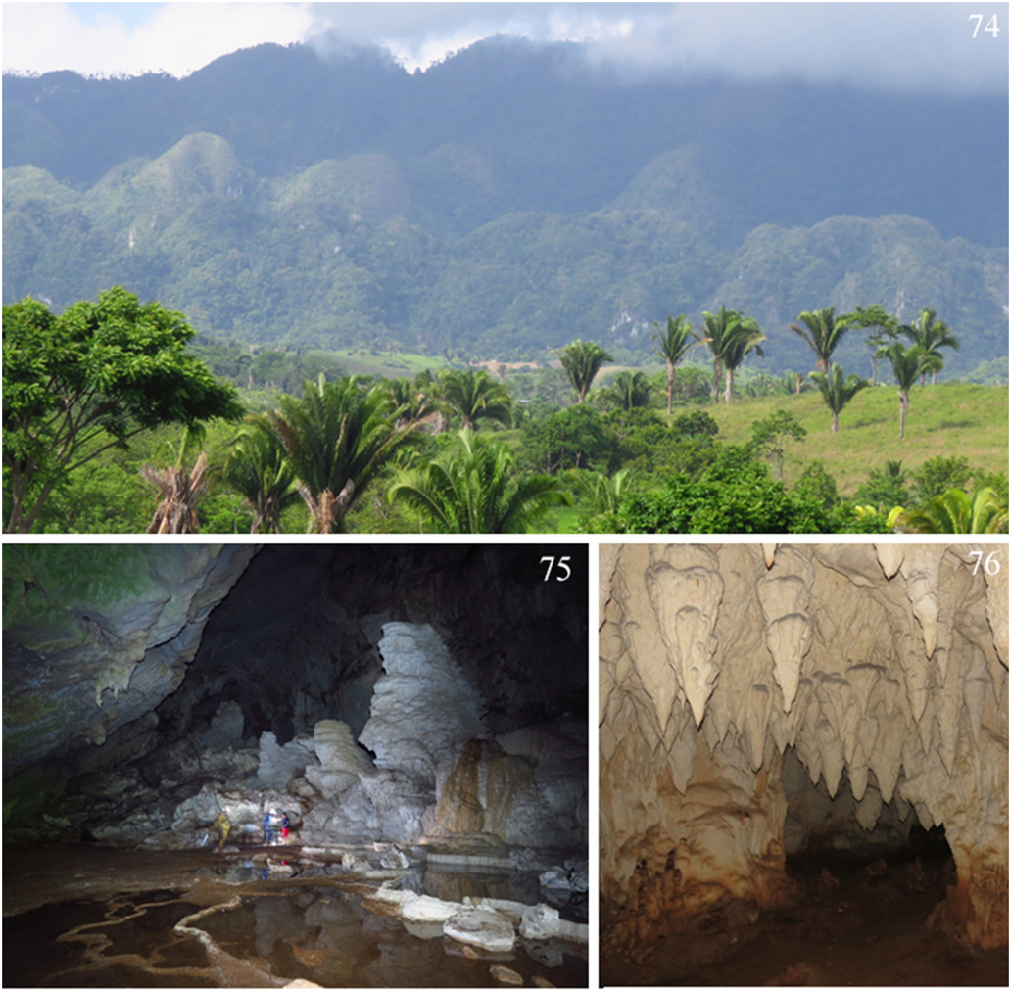
TABLE 1. Arachnopsita maya n. sp., adult male (n = 8) and female (n = 2) morphological measurements (mm), mean (Med.) and stand deviation (D.P.).
| ♂ | 12410 | 12411 | 12412 | 12413 | 12414 | 12416 | 12418 | 12419 | Med. | D.P. |
|---|---|---|---|---|---|---|---|---|---|---|
| Head width | 2.355 | 2.581 | 2.974 | 2.827 | 2.655 | 2.470 | 2.612 | 2.796 | 2.659 | 0.201 |
| Head length | 2.968 | 3.202 | 3.523 | 3.482 | 3.045 | 3.160 | 3.132 | 3.335 | 3.231 | 0.199 |
| Intraocular | 1.662 | 2.041 | 2.407 | 2.247 | 2.161 | 2.019 | 2.040 | 2.148 | 2.091 | 0.216 |
| Femur III | 7.992 | 9.771 | 9.821 | 10.173 | 9.850 | 9.855 | 9.606 | 10.777 | 9.731 | 0.790 |
| Tibia III | 9.023 | 10.864 | 11.387 | 12.817 | 11.532 | 11.145 | 11.723 | 12.280 | 11.346 | 1.127 |
| Body | 11.849 | 13.356 | 15.177 | 17.172 | 12.867 | 14.031 | 13.565 | 11.797 | 13.727 | 1.782 |
| Pronotum width | 2.107 | 2.201 | 2.744 | 2.791 | 2.787 | 2.485 | 2.199 | 2.622 | 2.492 | 0.287 |
| Pronotum length | 1.943 | 2.087 | 2.399 | 2.311 | 2.048 | 1.853 | 1.874 | 1.993 | 2.064 | 0.198 |
| ♀ | 12415 | 12417 | Med. | D.P. | ||||||
| Head width | 3.593 | 3.619 | 3.606 | 0.018 | ||||||
| Head length | 4.494 | 4.662 | 4.578 | 0.119 | ||||||
| Intraocular | 2.784 | 2.851 | 2.818 | 0.047 | ||||||
| Femur III | 13.215 | 12.361 | 12.788 | 0.604 | ||||||
| Tibia III | 15.585 | 15.507 | 15.546 | 0.055 | ||||||
| Body | 18.943 | 21.834 | 20.389 | 2.044 | ||||||
| Pronotum width | 3.240 | 3.316 | 3.278 | 0.054 | ||||||
| Pronotum length | 2.494 | 2.581 | 2.538 | 0.062 | ||||||
| Ovipositor | 9.778 | 9.108 | 9.443 | 0.474 |
No known copyright restrictions apply. See Agosti, D., Egloff, W., 2009. Taxonomic information exchange and copyright: the Plazi approach. BMC Research Notes 2009, 2:53 for further explanation.
|
Kingdom |
|
|
Phylum |
|
|
Class |
|
|
Order |
|
|
Family |
|
|
Genus |