
Antecerococcus keralae Hodgson & Williams
|
publication ID |
https://doi.org/ 10.11646/zootaxa.4091.1.1 |
|
publication LSID |
urn:lsid:zoobank.org:pub:76D13D36-682E-4E91-AC91-693CA9D3D465 |
|
DOI |
https://doi.org/10.5281/zenodo.6081576 |
|
persistent identifier |
https://treatment.plazi.org/id/03F2FF48-816D-0D77-24B6-AC57FD90FE7E |
|
treatment provided by |
Plazi |
|
scientific name |
Antecerococcus keralae Hodgson & Williams |
| status |
sp. nov. |
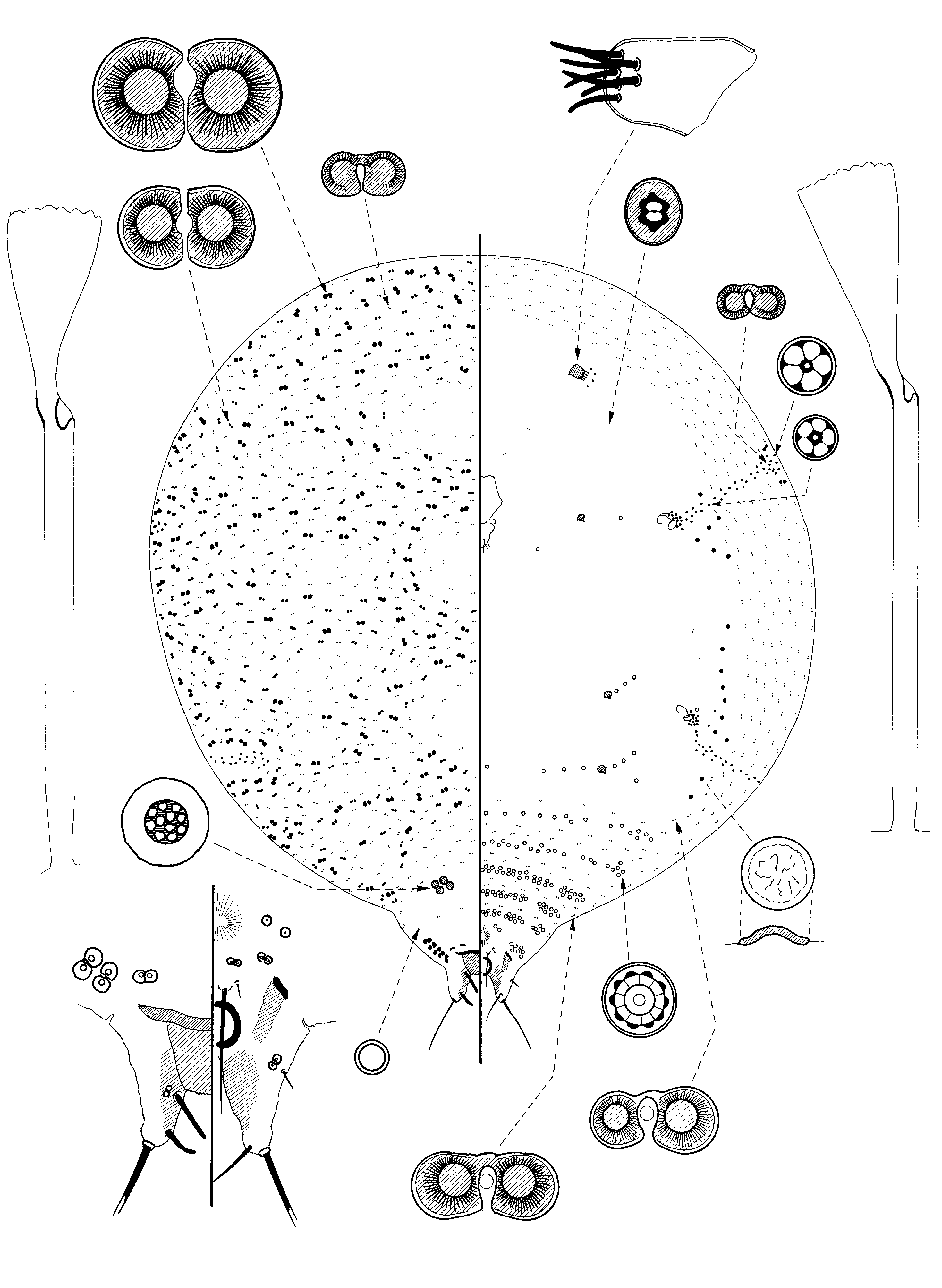
Antecerococcus keralae Hodgson & Williams , sp. nov.
( Fig. 25 View FIGURE 25 )
Material studied. Holotype and paratype ff: INDIA, Kerala, Trivandrum (now known as Thiruvananthapuram), Sreekanyam, on cassava ( Manihot esculenta , Euphorbiaceae ), 1981, no coll. (BMNH): 1/2adff (holotype good but posterior abdominal segments somewhat concertinaed; paratype specimen poor).
Note: description taken almost entirely from holotype specimen.
Mounted material. Body oval to roundly pear-shaped, 1.8 mm long, 1.63 mm wide.
Dorsum. Eight-shaped pores of 4 sizes: (i) largest 18– 22 x 10–13, in sparse bands and swirls throughout most of dorsum anterior to cribriform plates, and with 4 or 5 pores on each lateral margin of posterior abdominal segments; (ii) slightly smaller large pores (each 15–17 x 8–9 µm), usually fairly distinctly smaller than largest pores, but those nearest type (i) pores largest; slightly more abundant than type (i) pores but with similar distribution; (iii) much smaller pores, each 6.5–7.5 x 4.5–5.0 µm, throughout dorsum but smaller (5.0–5.5 x 3.5– 4.0 µm) and very sparse on posterior abdominal segment; and (iv) an even smaller pore, each 4.5–5.0 x 3.0–3.5 µm usually present at apex of each disc-pore band, with 2 in each band. Simple pores very sparse, each about 2 µm wide. Cribriform plates almost round, each 10–14 µm widest, with a broad sclerotized margin and large micropores; in 2 submedial groups of 4 on each side of abdominal segment IV. Dorsal setae few, each setose. Tubular ducts each 25–27 µm long; outer ductule clearly wider than those on venter; abundant. Anal lobes membranous apart from inner margin which is distinctly sclerotized and slightly reticulated; each lobe about 65 µm long with a long apical seta, both broken; dorsal fleshy setae short and stout and straight, each seta near apex 16–23 µm long, more anterior fleshy seta longer and straighter, each 23–28 µm long; ventral setae near apex strong, each 33–35 µm long; medioventral setae only present on one side, 16 µm long; each lobe with 1 intermediate and 1 small 8-shaped pore. Median anal plate 26–36 µm long and 35–40 wide at base; with a slightly serrate apex. Anal ring with 4 pairs of setae, each 80–112 µm long.
Venter. Ventral 8-shaped pores, each 7.5– 10 x 4.5–5.5 µm, present in a fairly narrow marginal band and sparsely across posterior abdominal segments; also with a few anterior to mouthparts. Simple pores not detected. Small bilocular pores, each about 5 x 4 µm, present medially on head and thorax. Spiracular disc-pores small, each 3–5 µm wide (largest near apex of band) with mainly 5 loculi, in rather sparse narrow bands extending onto dorsum; each band with a total of about 50 pores; posterior band appears to bifurcate as usual but more anterior branch quickly becomes obsolete laterally, posterior branch as above; each apex usually with 2 minute 8-shaped pores; also with 4 or 5 disc-pores near each antenna. Sclerotized convex closed pores, each 3–5 µm wide, present as follows: with only 1 anterior to anterior spiracle; with a group of 3–5 just posterior to each anterior spiracle; a line of 7 or 8 anterior to posterior spiracle and a line of 2 posterior to posterior spiracle. Multilocular disc-pores, each about 7 µm wide with mainly 10 loculi, distributed as follows: VIII possibly 0; VII 6 or 7 on each side near margin and 2 on each side more medially; and then in lines across segments: VI 5 or 6 submarginally + 23 medially; V 4–8 submarginally + 25–32 medially; IV 5–12 submarginally + 24–43 medially; III 3–7 submarginally + 21–27 medially, II 5 or 6 submarginally and a sparse line of 11 medially; metathorax with 2–6 laterad to each leg stub + 7 medially, and with 2–4 near each mesothoracic leg, and 1 near each anterior spiracle. Tubular ducts similar to those on dorsum but narrower, present throughout. Ventral setae sparse; preanal setae each 63–70 µm long; companion seta short. Leg stubs fairly small. Antennae unsegmented, each 36–40 µm long, without either an apical cone-like point or a setal cavity. Clypeolabral shield 125 µm long. Spiracular peritremes each 26–28 µm wide.
Comment. These specimens were originally identified as A. ornatus but they are clearly very different. Adult females of A. keralae are extremely similar to those of A. gabonensis , differing mainly in the presence of two sizes of larger 8-shaped pores throughout the dorsum in A. keralae (largest pores restricted to posterior abdominal segments on A. gabonensis ). Females of A. keralae differ also in other more subtle differences (character-states for A. gabonensis in brackets): (i) many fewer sclerotized convex closed pores ventrally, with none between each antenna and anterior spiracles (present); (ii) only five large 8-shaped pores on each side of posterior abdominal segments (up to 20 on each side, in two groups); and (iii) each cribriform plate with a broad margin and small area of micropores (narrower margin and with a large area of micropores). Cassava is grown widely in Africa and it is likely that many “sticks” used for planting cassava in India were originally imported from there. Based on this assumption, it could be argued that A. gabonensis could have been imported with it and therefore that the name A. keralae should be considered a synonym of A. gabonensis . However, A. gabonensis has not been collected from cassava and the above morphological differences are considered sufficiently distinct to justify their separation.
The adult female of A. keralae can be separated from those of all other Antecerococcus species (except perhaps A. gabonensis ) in having the following combination of character-states: (i) dorsum with four sizes of 8-shaped pores, largest and intermediate-sized pores sparse throughout; (ii) apices of each stigmatic band with two smallest 8-shaped pores; (iii) lateral margins of posterior abdominal segments each with four or five large 8-shaped pores; (iv) cribriform plates in a submedial group of four on each side of abdominal segment IV; (v) leg stubs present; (vi) posterior stigmatic bands non-bifurcated; (vii) small convex closed pores present in a sparse line between anterior spiracles and metathorax; (viii) multilocular disc-pores present across all abdominal segments and metathorax, and also mesad to each spiracle, and (ix) antennae without either a cone-like apex or a setal cavity.
In having small convex closed pores near the spiracles, it is also similar to A. theydoni Hall but on the latter species (i) the convex closed pores tend to be smaller than the quinquelocular disc-pores (about the same size), (ii) the posterior stigmatic pore band is bifurcated, and (iii) the distribution of the 8-shaped pores on the dorsum is different (see figures).
The adult female of A. keralae falls within Group A in the key to species of Antecerococcus , keying out close to A. gabonensis and A. ovoides .
Name derivation: keralae —Latin genetive of the region Kerala, in southern India, of which Thiruvananthapuram is the capital.
No known copyright restrictions apply. See Agosti, D., Egloff, W., 2009. Taxonomic information exchange and copyright: the Plazi approach. BMC Research Notes 2009, 2:53 for further explanation.