
Anoplodelphys incerta Lafargue and Laubier, 1978
|
publication ID |
https://doi.org/ 10.5281/zenodo.176361 |
|
DOI |
https://doi.org/10.5281/zenodo.5661723 |
|
persistent identifier |
https://treatment.plazi.org/id/C03D8785-045C-FFBA-FF2D-FF49FCFAFA32 |
|
treatment provided by |
Plazi |
|
scientific name |
Anoplodelphys incerta Lafargue and Laubier, 1978 |
| status |
|
Anoplodelphys incerta Lafargue and Laubier, 1978
Material examined: Holotype female, reg. no. ZMA CO.102.602.
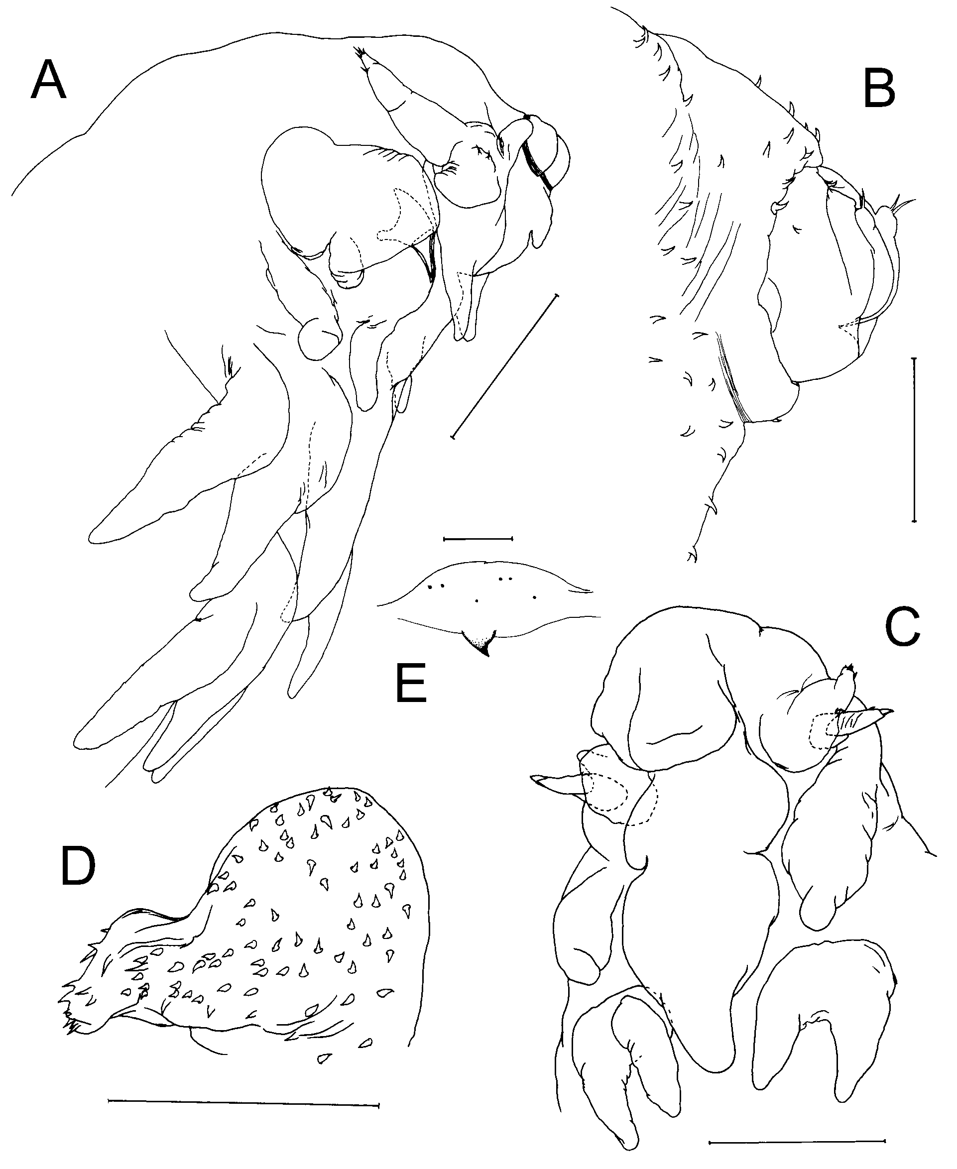
Differential Diagnosis: Body elongate, with indistinct segmentation. Cephalosome with narrow frontal margin bearing rostrum. Rostrum inflated, forming large ventrally-directed lobe ( Fig. 4 View FIGURE 4 C). Post-rostral median lobe present. Labrum strongly inflated at base, tending distally into tapering lobe ( Fig. 4 View FIGURE 4 C). Lateral margin of cephalosome expanded ventrally to form ridge-like swelling produced into process. Antennomedial lobe lacking. Metasome elongate with about 25% of length extending posterior to level of origin of leg 4: metasomal somites bearing small legs ventrally near lateral margins. No mid-ventral processes present between legs. Urosome small, unsegmented; located terminally; with median anal incision, bearing minute paired caudal rami armed with setae distally. Surface of body, rostrum, labrum, cephalosomic processes, and legs densely ornamented with surface setules.
Antennule unsegmented, tapering lobe ( Fig. 4 View FIGURE 4 D). Antenna 2-segmented, with triangular basal segment and distal subchela incorporating sclerotised distal claw, armed with 3 vestigial setal elements. Mandible to maxilliped all lacking. Legs 1–4 biramous; each with protopodal part almost completely incorporated into somite so small lobate rami appearing to originate from somite surface. Ratios of gaps between legs along metasome as in Table 2 View TABLE 2 . Leg 5 represented by tiny sclerotised points ( Fig. 4 View FIGURE 4 E).
Body length of female 2.40–3.65 mm. Male unknown.
Remarks: In their paper on Anoplodelphys, Lafargue and Laubier (1978a) comment upon the very wide intraspecific variability between specimens of Anoplodelphys incerta collected from different host species. They recognised a so-called “typical” form of A. incerta from Didemnum commune (Della Valle, 1877) and an “atypical” form from Didemnum fulgens (Milne Edwards, 1841) , D. protectum (Daumézon, 1908) , D. peyrefittense (Brément, 1913) and D. maculosum (Milne Edwards, 1841) . According to Lafargue and Laubier (1978a), the main distinguishing characters between “typical” and “atypical” forms were: the form and organisation of the elements of surface ornamentation, the general body shape, and the degree of development and position along the body of legs 1–4. Lafargue and Laubier (1978a) selected a specimen of the typical form collected from D. commune as the holotype (ZMA Co. 102.602) thereby fixing the name. Re-examination of this holotype of A. incerta leads us to conclude that the typical and atypical forms are not conspecific.
We propose to restrict the concept of A. incerta to the typical form (found on D. commune ) characterised by the elongate body, the reduced size of legs 1 to 4, and the relatively wide spacing of legs 1 and 2 (18%) along the longitudinal axis of the body ( Table 2 View TABLE 2 ). We here treat the atypical form as Anoplodelphys cf. galli , since the only differences from A. galli relate to the arrangement of the spinular ornamentation, a character that is difficult to assess with our current state of knowledge.
| ZMA |
Universiteit van Amsterdam, Zoologisch Museum |
No known copyright restrictions apply. See Agosti, D., Egloff, W., 2009. Taxonomic information exchange and copyright: the Plazi approach. BMC Research Notes 2009, 2:53 for further explanation.