
Aletheiana tenella, Ng, Peter K. L. & Lukhaup, Christian, 2015
|
publication ID |
https://doi.org/ 10.11646/zootaxa.4039.1.4 |
|
publication LSID |
lsid:zoobank.org:pub:A1F46EFE-0639-4754-9F45-69915F2599D9 |
|
DOI |
https://doi.org/10.5281/zenodo.6106633 |
|
persistent identifier |
https://treatment.plazi.org/id/08492243-FFA3-FF84-6D87-EF2132C29D4D |
|
treatment provided by |
Plazi |
|
scientific name |
Aletheiana tenella |
| status |
sp. nov. |
Aletheiana tenella View in CoL n. sp.
( Figs. 1–4 View FIGURE 1 View FIGURE 2 View FIGURE 3 View FIGURE 4 )
Material examined. Holotype: male (3.6 × 3.3 mm) ( MZB Cru 4386), among roots along banks of cascades at Beteleme-Tomata road, west of Wawopada, Puawu River, Tomori area, 02°1.314´S 121°12.042´E, Central Sulawesi, Indonesia, coll. C. Lukhaup 5 October 2011. Paratypes: 2 females (4.1 × 3.8 mm, 3.5 × 3.2 mm) ( MZB Cru 4387), 1 male (2.8 × 2.6 mm), 2 females (4.8 × 4.3 mm, 3.0 × 2.7 mm) ( MNHN), 1 male (3.2 × 2.9 mm), 1 juvenile male (2.4 × 2.2 mm), 3 females (5.3 × 4.8 mm, 4.2 × 3.8 mm, 3.7 × 3.4 mm) ( ZRC 2015.278), same data as holotype.
Comparative material. See Ng & Chuang (1996), Naruse & Ng (2007), Naruse et al. (2008a, b), Husana et al. (2011).
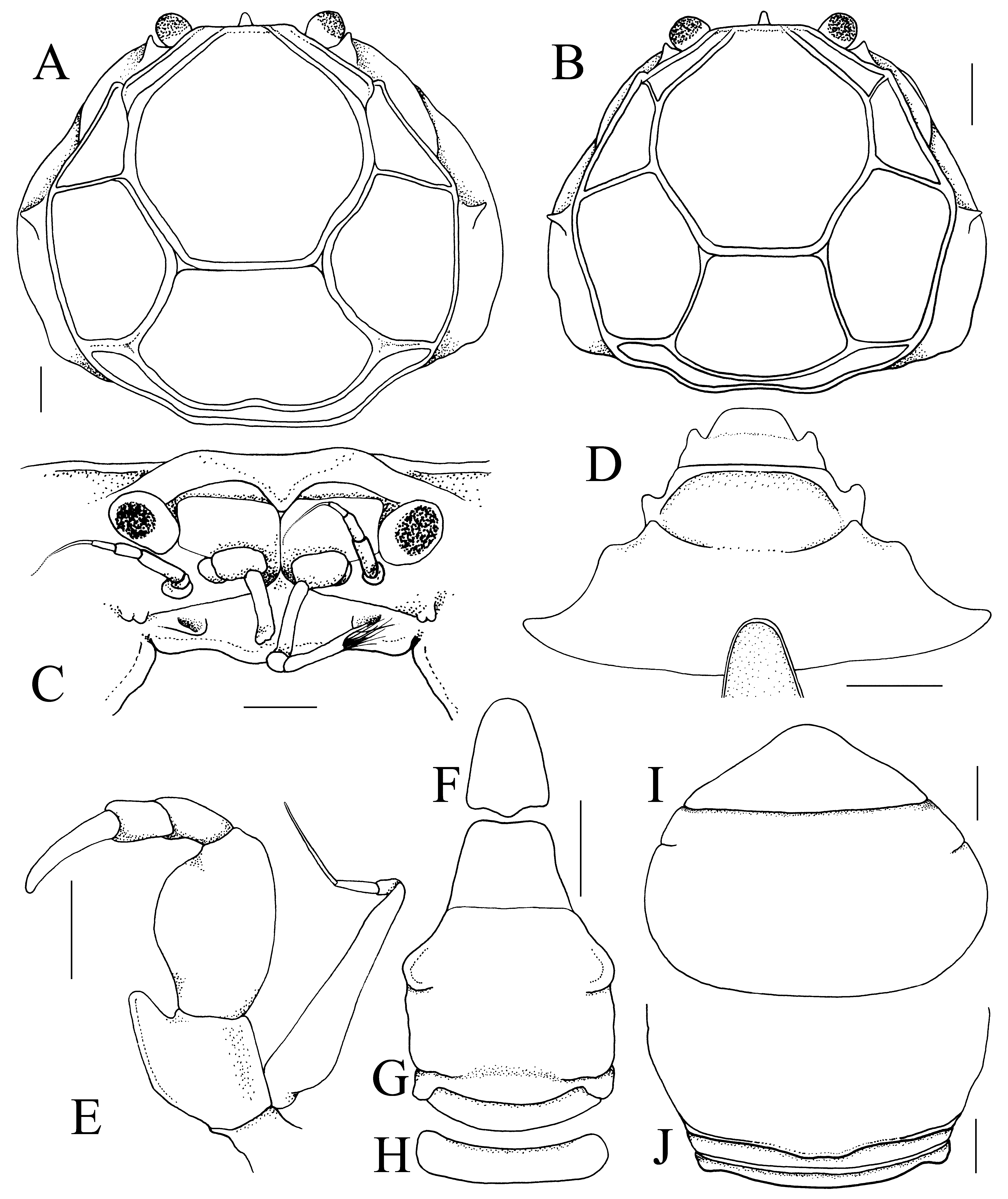
Diagnosis. Carapace slightly wider than long, width to length ratio 1.08–1.11; surrounded by rim, regions demarcated by prominent deep grooves ( Figs. 2 View FIGURE 2 A, 3A, B); rostrum unilobate, no lateral lobes present, median lobe small, triangular, tip rounded, submarginal in position ( Figs. 2 View FIGURE 2 A, 3A–C); lateral margin of carapace with anterodorsally curved spiniform tooth at junction of antero- and posterolateral margins; anterolateral margin with 1 low tooth on anterior half ( Figs. 2 View FIGURE 2 A, 3A, B); eyes well developed, mobile, fully pigmented ( Figs. 1 View FIGURE 1 D–F, 2A, 3A–C); epistome distinct, broad, not projecting anteriorly, almost vertical from lateral view; posterior margin with with 2 low, rounded median lobes ( Fig. 3 View FIGURE 3 C); third maxilliped merus longitudinally ovate, longer than ischium, without anteroexternal angle; ischium with inner distal angle strongly produced to form acute lobe, dactylus elongated, twice length of propodus ( Figs. 3 View FIGURE 3 E); ambulatory legs slender, long, dactylus falciform, with prominent subdistal curved spine and often with additional 1–3 short spines or sharp tubercles ( Figs. 4 View FIGURE 4 B–I); male abdomen-pleotelson triangular, relatively elongate; somite 5 separated from somite 4 by shallow suture, not mobile; male pleotelson elongated, linguiform, base slightly wider than distal margin of somite 6 ( Fig. 3 View FIGURE 3 F–H); G1 twisted medially, tip tapering, inner distal margin lined with long, stiff setae ( Fig. 4 View FIGURE 4 J–L); G2 simple, short, with subspatuliform tip ( Fig. 4 View FIGURE 4 N, M); female abdomen-pleotelson round, somites 3–5 completely fused, somites 1 and 2 free ( Fig. 3 View FIGURE 3 I, J).
Description of male. Carapace subovate, slightly wider than long, width to length ratio 1.08–1.11; dorsal surface gently convex at gastric regions, other regions almost flat, surrounded by continuous rim, regions clearly demarcated by prominent deep grooves, H-shaped gastric groove continuous with cervical groove, cervical groove branching on anterior half of carapace, confluent with anterolateral rim ( Figs. 2 View FIGURE 2 A, 3A, B). Front straight from dorsal view, margin appears unarmed; rostrum unilobate, no lateral lobes present, median lobe small, triangular, tip rounded, just below frontal margin, above septum between antennular fossae, just visible from dorsal view ( Figs. 2 View FIGURE 2 A, 3A–C). Lateral margin of carapace (on branchiostegal surface) with prominent antero-dorsally curved spiniform tooth at junction of antero- and posterolateral margins; anterolateral margin with 1 low tooth on anterior half, behind suborbital tooth, directed obliquely dorsally ( Figs. 2 View FIGURE 2 A, 3A, B). Posterior carapace margin almost straight to gently sinuous ( Figs. 2 View FIGURE 2 A, 3A, B).
Orbits not visible; eyes well developed, mobile, visible dorsally, cornea rounded, fully pigmented ( Figs. 1 View FIGURE 1 D–F, 2A, 3A–C). Antennular fossa shallow, surface gently concave, separated by narrow septum; basal antennular article short, quadrate, not swollen, free, positioned at base of fossa, second and third articles elongated, unable to fold into fossa ( Fig. 3 View FIGURE 3 C). Antenna with long filiform flagellum (sometimes broken off), almost reaching curved spiniform tooth on lateral carapace margin when folded posteriorly; positioned between base of ocular peducle and outer margin of antennular fossa; first article (osmoregulatory opening) present as knob with concave dorsal surface on each side of epistome ( Fig. 3 View FIGURE 3 C). Epistome distinct, broad, not projecting anteriorly, almost vertical from lateral view; posterior margin with gently sinuous lateral margins, with 2 low, rounded median lobes separated by shallow depression; not projecting anteriorly, not visible from dorsal view ( Fig. 3 View FIGURE 3 C). Suborbital tooth distinct, clearly visible from dorsal view ( Figs. 2 View FIGURE 2 A, 3A, B).
Third maxilliped relatively narrow, median part of buccal cavity not covered, exposing mandibles; merus longitudinally ovate, longer than ischium, no anteroexternal angle visible; ischium with inner distal angle strongly produced to form acute lobe, without visible sulcus; palp longer than merus, carpus and propodus subequal in length, dactylus elongated, about twice length of propodus; exopod slender, reaching to just before distal margin of merus, with distinct flagellum ( Figs. 3 View FIGURE 3 E).
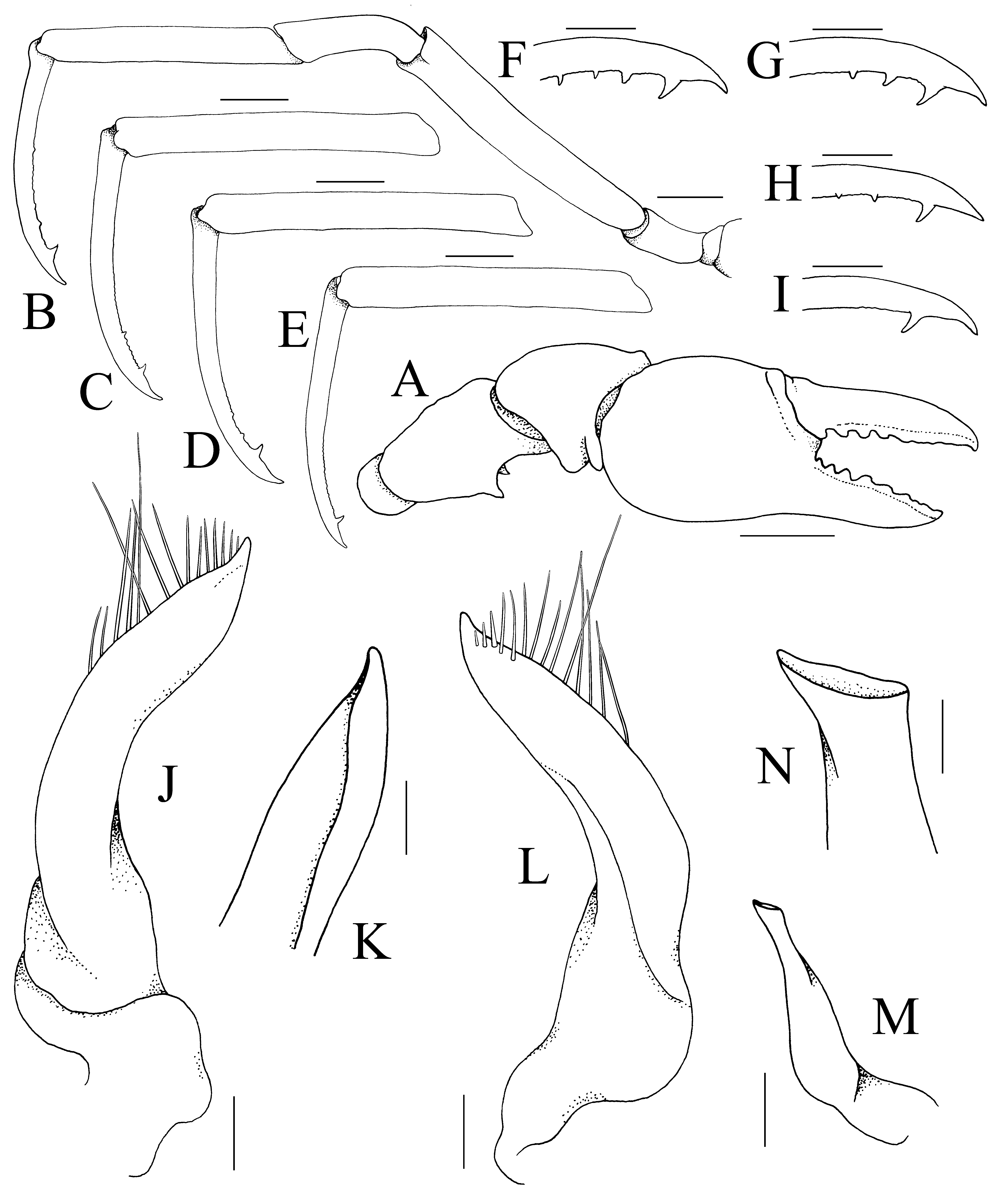
Chelipeds symmetrical, relatively stout; basis and ischium separate, short, unarmed; with short merus, ventral margin with strong subdistal tooth medially and tubercle below it, dorsal margin uneven but not armed; carpus cupshaped, inner angle with low rounded tooth; chela with smooth rounded palm, outer surface gently convex, covered with long dense plumose setae which partially obscures surface and margins; fingers as long as palm, setose on outer surfaces; cutting edges of fingers with 3–5 teeth on proximal half, distal half almost entire, forming blade-like structure, tips curved ( Figs. 1 View FIGURE 1 D–F, 2A, 4A).
Ambulatory legs slender, long, second longest; margins lined with scattered short setae that do not obscure surface; all meri with unarmed but dorso-distal angle discernible, appears dentiform; carpus curved, unarmed; propodus subrectangular, elongated, unarmed; dactylus gently falciform, all with short but prominent subdistal curved spine, margin just posterior to spine crenulated or uneven, may be armed with additional 1–3 short spines or sharp tubercles which are decreasing in size ( Figs. 4 View FIGURE 4 B–I).
Thoracic sternum relatively wide ( Figs. 2 View FIGURE 2 C, 3D); sternites 1 and 2 fused, no suture visible but demarcated by a fold, with sternite 1 bent inwards towards mandibles, sternite 1 semicircular, sternite 2 wide; sternite 2 separated from sternite 3 by distinct groove; sternite 3 trapezoidal, with median part gently depressed, fused with sternite 4 but lateral part of suture visible, disappearing medially ( Fig. 3 View FIGURE 3 D); pterygostome gently pronounced, joining anterolateral extension of sternite 4, separating Milne Edwards openings from base of chelipeds. Milne Edwards openings relatively broad, margins lined with dense setae. Sterno-abdominal cavity deep, reaching to middle of sternite 4; margins with prominent rim; no trace of locking button ( Figs. 2 View FIGURE 2 C, 3D).
Male abdomen-pleotelson triangular, relatively elongate; somite 1 broad, subrectangular, reaching to base of P5, just visible from dorsal view, lateral margins evenly convex; somite 2 as wide as somite 1, separated from somite 3 by suture but not mobile; somites 3 and 4 fused to form subpentagonal plate, posterolateral part forming short subauriculiform projection, somite 4 separated from somite 3 only by shallow lateral fissures; lateral part of somite 4 dilated to form rounded projection with median part gently swollen; somite 5 trapezoidal, separated from somite 4 by shallow suture, not mobile; pleotelson elongated, linguiform, triangular with rounded tip, lateral margins gently convex, base slightly wider than distal margin of somite 6 ( Fig. 3 View FIGURE 3 F–H).
G1 clearly twisted medially, tip tapering, directed towards buccal cavity in situ; inner distal margin lined with long, stiff setae ( Fig. 4 View FIGURE 4 J–L). G2 short, with subspatuliform tip; base not dilated ( Fig. 4 View FIGURE 4 N, M).
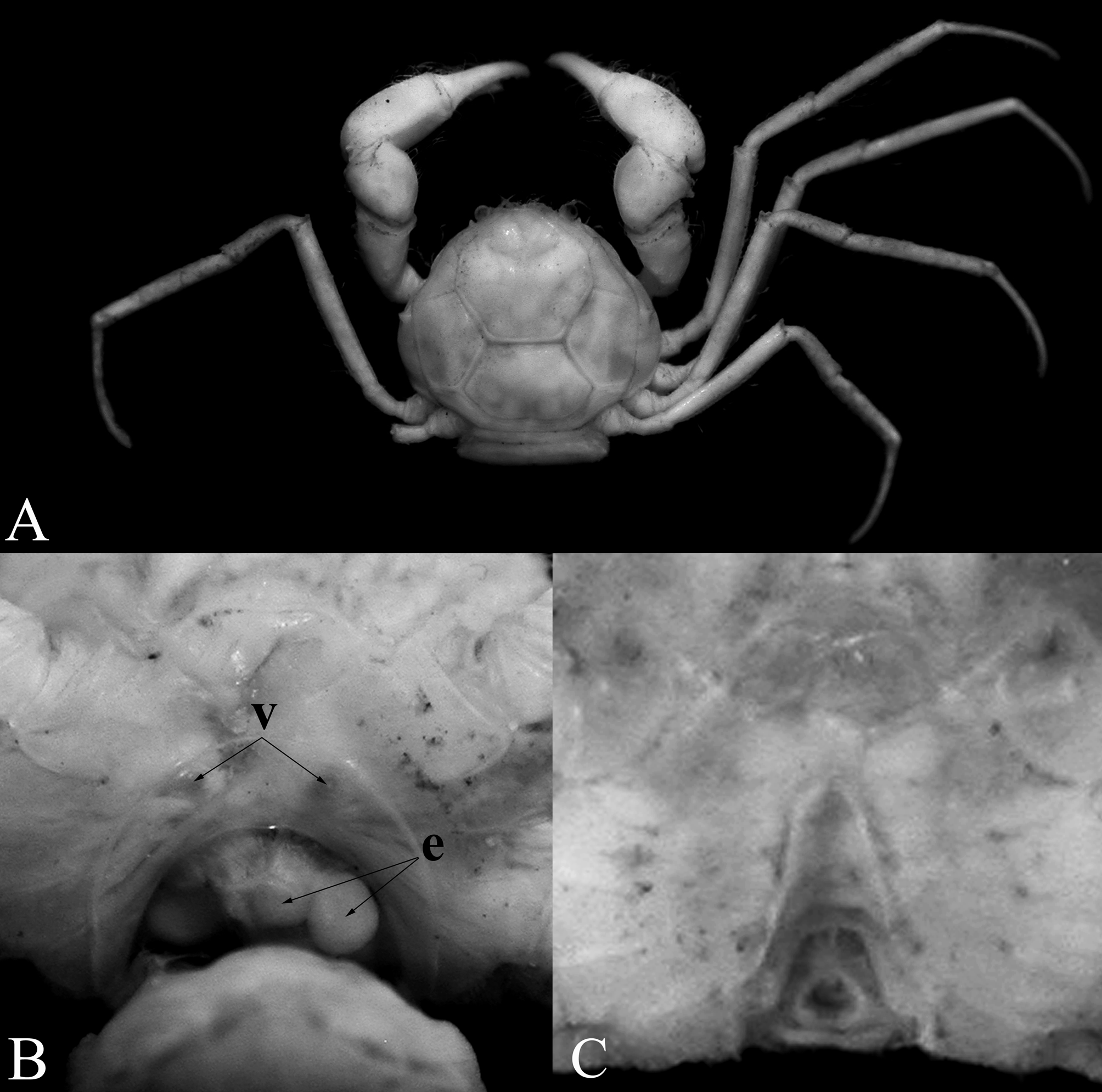
Females. The largest adult females are larger than the adult males and agree in most non-sexual characters, except that the carapace is more rounded and the chelipeds are relatively more slender. The two largest females are ovigerous, with the abdomen rounded and swollen. The pleotelson is a wide triangle with gently sinuous lateral margins and somites 3–5 are completely fused ( Fig. 3 View FIGURE 3 I); with somites 1 and 2 free ( Fig. 3 View FIGURE 3 J). The swollen abdomen is filled with large eggs in the female, with the eggs also seen in the body cavity ( Fig. 2 View FIGURE 2 B). The vulvae are positioned submedially on thoracic sternite 6 and are relatively small, with the diameter less than that of each egg ( Fig. 2 View FIGURE 2 B). This is identical to what has been reported for N. mangalis by Ng & Chuang (1996), and the species almost certainly practices ovovivipary, as is believed to occur in members of the genus. Female specimens below 4 mm in carapace width are still immature, with the abdomen subquadrate, not swollen and not covering most of the thoracic sternum (e.g., 3.5 × 3.2 mm, MZB Cru. 4387). Larger females (e.g., 4.1 × 3.8 mm, MZB Cru 4387) have a more rounded abdomen that covers more of the sternum but it is still not swollen. Only the two largest females (4.8 × 4.3 mm, MNHN; 5.3 × 4.8 mm, ZRC 2015.278) have large swollen abdomens that are filled with eggs. Interestingly, the fusion between somites 3–5 is complete with the sutures not visible, even when they are small.
Etymology. The name is derived from the Latin “tenellus” for delicate; alluding to the small size and slender appendages of the species.
Remarks. See discussion for genus.
Biology. Aletheiana tenella gen. et sp. nov. was found among the submerged fibrous roots of trees at the slow flowing parts of the riverbank near cascades along the Puawu River, in Central Sulawesi ( Fig. 1 View FIGURE 1 A, B). The crabs were observed at the slow flowing parts of the riverbank among roots near cascades of the Puawu River. The water at the time of collection was muddy and had a temperature of 22°C. In the faster flowing parts of the river, no crabs were found. This is a typical habitat for many free-living hymenosomatids (e.g., see Naruse & Ng 2007). A new species of Caridina , C. boehmei (Atyidae) was also recently described from the type locality of Aletheiana tenella gen. et sp. nov. by Klotz & von Rintelen (2013).

No known copyright restrictions apply. See Agosti, D., Egloff, W., 2009. Taxonomic information exchange and copyright: the Plazi approach. BMC Research Notes 2009, 2:53 for further explanation.