
Agalliopsis dutrai Gonçalves, Mejdalani & Coelho, 2009
|
publication ID |
https://doi.org/ 10.5281/zenodo.185901 |
|
DOI |
https://doi.org/10.5281/zenodo.6217216 |
|
persistent identifier |
https://treatment.plazi.org/id/03B387EC-FFF3-497B-3EC9-F93A85E88872 |
|
treatment provided by |
Plazi |
|
scientific name |
Agalliopsis dutrai Gonçalves, Mejdalani & Coelho |
| status |
sp. nov. |
Agalliopsis dutrai Gonçalves, Mejdalani & Coelho View in CoL , sp. nov.
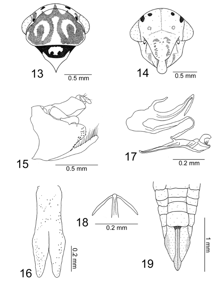
( Figs 13–19 View FIGURES 13 – 19 , 65, 66 View FIGURES 65 – 74 )
Length. Male holotype 3.9 mm; male paratypes 3.8–4.0 mm; female paratypes 3.9–4.4 mm.
Description (holotype). Head and thorax (color). Ground color yellowish-brown. Crown ( Figs 13, 14 View FIGURES 13 – 19 ) with pair of dark brown maculae approximately equidistant between eyes and median line; pair of brown to dark brown maculae near inner eye margins; with three triangular brown maculae contiguous to pronotal maculae. Eyes with red tonality. Face ( Fig. 14 View FIGURES 13 – 19 ) yellow with slender, inverted Y-shaped brown macula between ocelli; tiny brown maculae around ocelli. Frons ( Fig. 14 View FIGURES 13 – 19 ) with small brown maculae forming pair of lateral rows. Pronotum ( Fig. 13 View FIGURES 13 – 19 ) with two large, circular brown stripes, each one forming oval figure including brown macula; stripes fused to each other anteriorly, extending to crown and delimiting pair of anterior yellow areas. Mesoscutum ( Fig. 13 View FIGURES 13 – 19 ) broadly covered by brown marks; mesoscutellum with pair of dark brown maculae at laterobasal portions.
Male genitalia. Pygofer ( Fig. 15 View FIGURES 13 – 19 ), in lateral view, with posterior margin subtruncate. Subgenital plates ( Fig. 16 View FIGURES 13 – 19 ), in ventral view, elongate with basal half narrower than apical half; plates fused basally with valve and fused with each other along basal half; in lateral view, extending beyond pygofer apex, dorsal surface with longer scattered setae. Styles ( Fig. 17 View FIGURES 13 – 19 ), in lateral view, elongate, with few setae at apical portion, outer fork smaller and rounded, inner fork larger with its basal portion strongly curved, subapical portion with small tooth-like process directed downward. Connective ( Fig. 17 View FIGURES 13 – 19 ), in lateral view, linear, fused to aedeagus; subtriangular in dorsal view. Aedeagus ( Fig. 17 View FIGURES 13 – 19 ), in lateral view, strongly curved, basal half directed anteriorly and apical half directed posteriorly, curved region enlarged; apical portion of shaft with pair of long spiniform processes directed ventrally; shaft apex, in dorsal view, rounded, gonopore apical ( Fig. 18 View FIGURES 13 – 19 ). Anal tube ( Fig. 15 View FIGURES 13 – 19 ) with segment X well developed, divided into two parts, with pair of processes expanded towards apex and bearing two tooth-like ventral projections.
Female (color). Abdominal sternite VII ( Fig. 19 View FIGURES 13 – 19 ) mostly yellow; laterotergites from segment VIII yellow; pygofer ( Fig. 19 View FIGURES 13 – 19 ), in ventral view, yellow, with pair of brown maculae laterally; gonoplacs brown. Other features similar to those of the male holotype.
Female genitalia. Abdominal sternite VII ( Fig. 19 View FIGURES 13 – 19 ), in ventral view, transverse, rectangular, posterior margin with lateral portions oblique and central area slightly concave. First valvulae, in lateral view, dorsally curved from base; dorsolateral surface with oblique rows of scale-like processes on distal 1/2 of shaft; ventroapical region with scale-like processes; apex acute. Second valvulae ( Fig. 65 View FIGURES 65 – 74 ), in lateral view, dorsally curved from base, with broadest region at apical 1/4; dorsal hyaline area present; dorsal prominence slightly pronounced; teeth on apical half of dorsal margin, basally small and uniform, increasing in size towards apex ( Fig. 66 View FIGURES 65 – 74 ); ventroapical region with tiny rounded teeth; shaft apex rounded. Gonoplacs, in lateral view, expanded along apical half; apex rounded; surface with tiny spiniform processes and setae at apical portion and extending anteriorly along ventral margin.
Etymology. The new species is described in honor of the late Prof. José Alfredo Pinheiro Dutra (Instituto de Biologia, Universidade Federal do Rio de Janeiro), who has contributed to our knowledge of Brazilian Agalliinae .
Type material. Holotype: male, Brazil, “ Viçosa , MG [Estado de Minas Gerais], Brasil \ Data 22/XII/1992 \ P. S. Fiuza F. [Ferreira]” ( DZRJ). Twenty-three paratypes with same data as holotype, except: one female “ 02/ XII/1981 ” ( DZRJ), one female “ 01/IX/1982 ”, two females “ 13/X/1982 ”, one female “ 24/XI/1982 ”, one female “ 09/II/1983 ”, one female “ 01/VI/1983 ”, one male “ 11/VIII/1986 ”, one female “ 11/III/1987 ”, one female “ 14/X/1987 ”, one female “ 10/XI/1987 ”, one male and two females “ 08/I/1988 ”, one female “ 20/V/ 1988 ”, one female “ 08/X/1992 ”, one female “ 20/X/1992 ”, two females “ 29/XII/1992 ” (UFV), one female “ 10/XI/1982 ”, one female “ 02/II/1983 ”, two males “ 14/X/1987 ” ( MNRJ).

| MNRJ |
Museu Nacional/Universidade Federal de Rio de Janeiro |
No known copyright restrictions apply. See Agosti, D., Egloff, W., 2009. Taxonomic information exchange and copyright: the Plazi approach. BMC Research Notes 2009, 2:53 for further explanation.