
Arostrilepis gulyaevi, Makarikov, Arseny A., Galbreath, Kurt E. & Hoberg, Eric P., 2013
|
publication ID |
https://doi.org/10.11646/zootaxa.3608.6.1 |
|
publication LSID |
lsid:zoobank.org:pub:32AAC94B-5793-4D51-8DCC-AF2D8AD5BCBD |
|
DOI |
https://doi.org/10.5281/zenodo.6147202 |
|
persistent identifier |
https://treatment.plazi.org/id/E07D87D9-FFEA-6507-68BA-FEAFB8C783D3 |
|
treatment provided by |
Plazi |
|
scientific name |
Arostrilepis gulyaevi |
| status |
sp. nov. |
Arostrilepis gulyaevi sp. n.
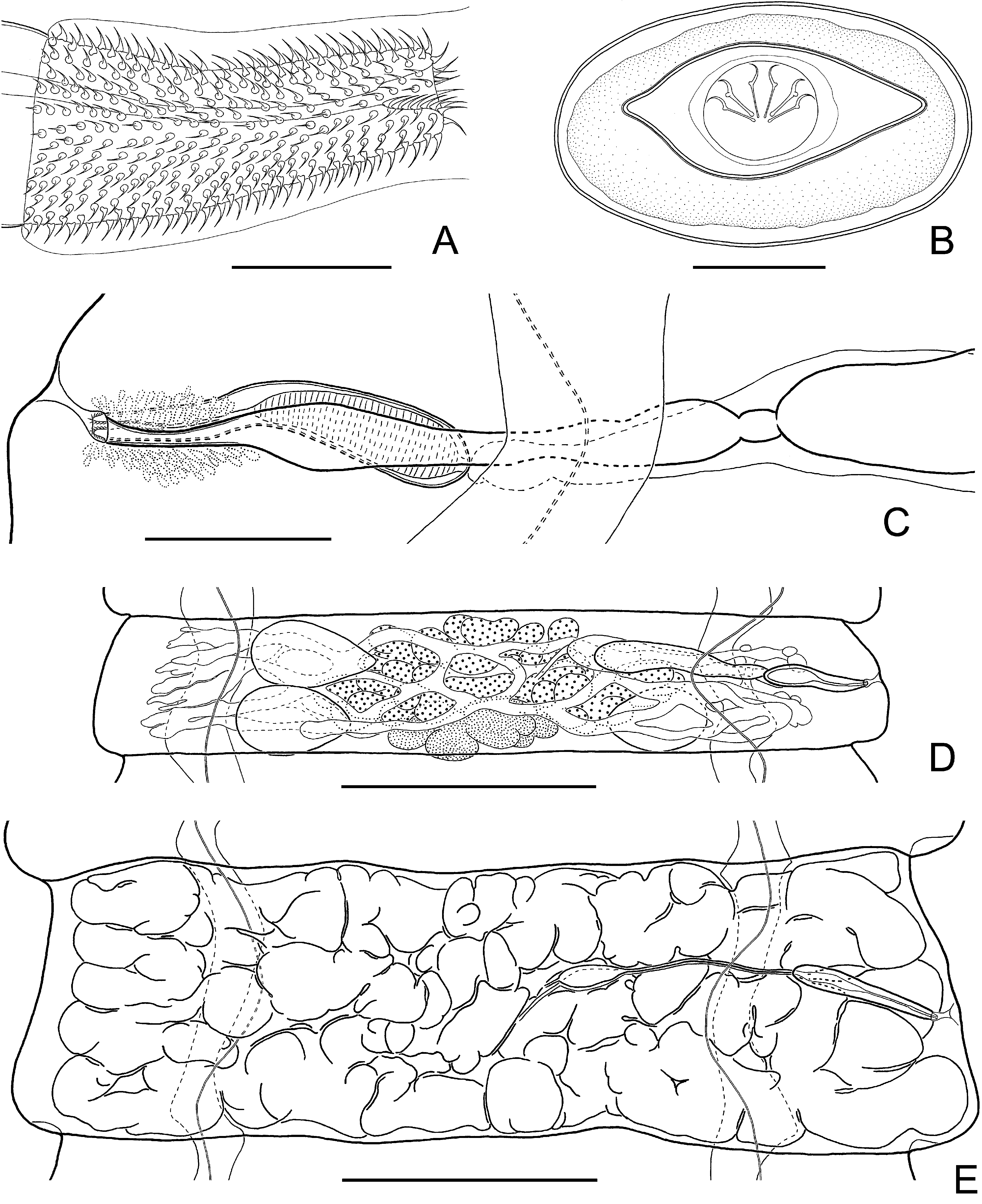
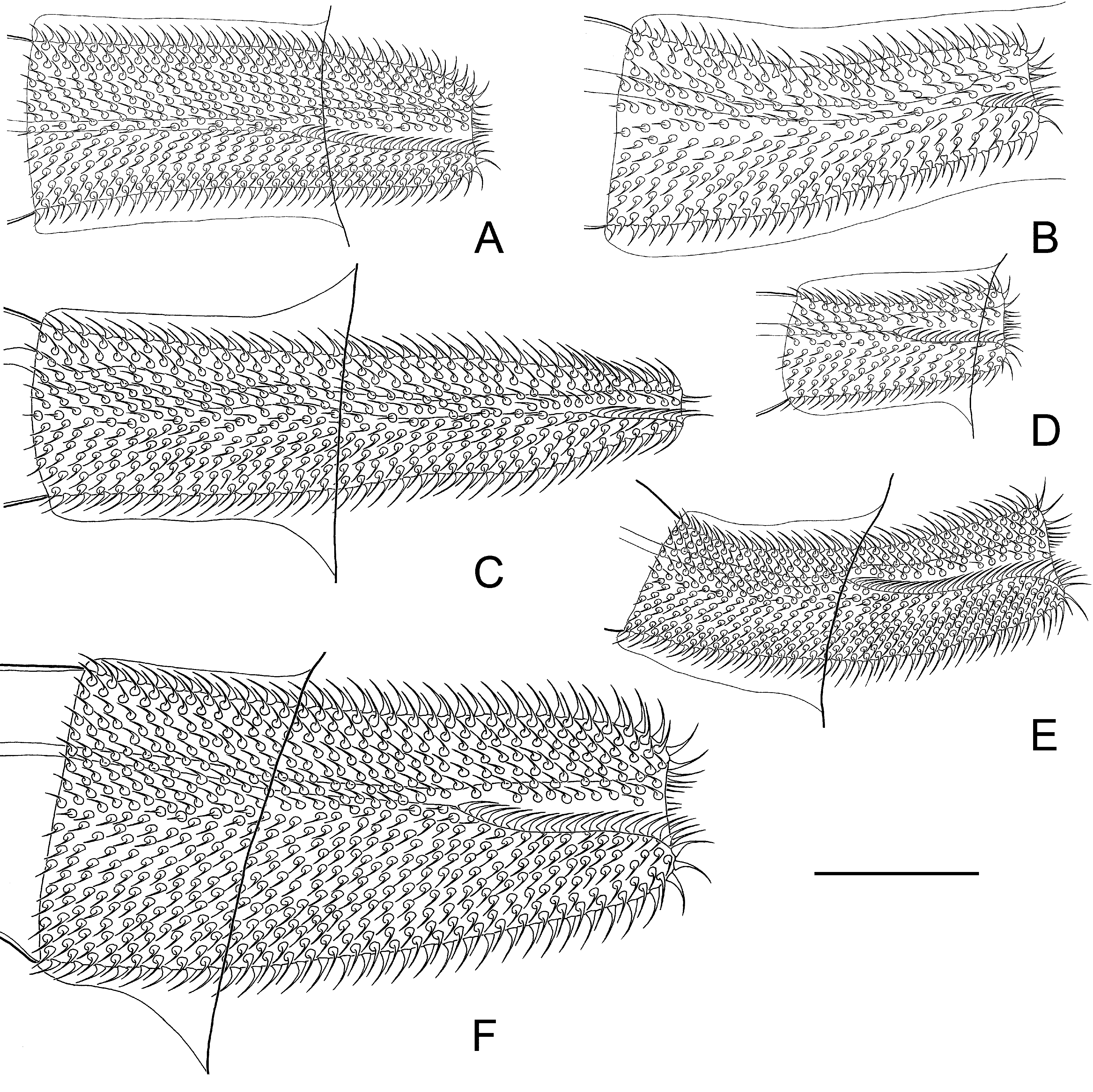
( Figs. 2 View FIGURE 2 , 3 View FIGURE 3 )
Type host: Myodes rufocanus (Sundevall) ( Rodentia : Cricetidae : Arvicolinae ).
Other hosts: Rarely Microtus oeconomus Pallas.
Type locality: Bol'shekhekhtsirskiy Nature Reserve, Khabarovskiy Kray, Russia (ca., 48°16’N, 134°45’E).
Other localities: Baikal Nature Reserve, Republic of Buryatia, Russia (ca., 51o21’N, 105o17’E); Anadyr, Chukotka Autonomous Okrug, Russia, 1 km SE of confluence of Markova and Anadyr Rivers ( 64o40’57”N, 170o26’26”E); Buyunda River, Magadanskaya Oblast’, Russia ( 62o20’N, 153o21’E); Chaun Biological Field Station, Chaun River, Chukotka Autonomous Okrug, Russia ( 69o00’N, 170o50’E).
Type material: Holotype ISEA No. 18.28.8.1 from type host and locality by Y. Melnikova, 18 July 2003. Paratypes from type locality and type host species: MSB Para 1262 (ISEA No.18.28.8.4) by Y. Melnikova, 17 July 2003; ISEA No. 18.28.8.7 by Y. Melnikova, 18 July 2003; No. 18.28.8.8 by Y. Melnikova, 21 July 2003; and No. 18.28.8.10 by Y. Melnikova, 21 July 2003. Paratypes from other localities: MSB Para 1263 (field number RLR 43095) in type host species by L. Smirnova at Chaun Biological Field Station, 16 July 1975; MSB 1259, 1261 (IF 5657/ cyt- b sequence; 5657 C1) in type host species by A. Lahzuhtkin and K.E. Galbreath at Buyunda River, Russia, 4 July 2002; MSB 1253, 1254 (IF 5079 C1/ cyt- b sequence; 5079 C2) in type host species, by N.E. Dokuchaev at Anadyr, Russia, 23 July 2002; and MSB 1256 (IF 5094/ cyt- b sequence) in type host species by N.E. Dokuchaev at Anadyr, Russia, 24 July 2002. See Appendix 1 for additional identified vouchers in M. rufocanus and Microtus oeconomus from the Baikal Reserve and Magadanskaya Oblast’.
Symbiotype: Type host specimen not deposited in a museum archive.
Description: Based on 10 specimens. Fully developed strobila 180–300 mm long, with maximum width at postmature or pregravid gravid proglottides, 1.9–3.4 mm. Strobila flat, consisting of 900–1500 craspedote proglottides. Scolex slightly compressed dorso-ventrally, 250–350 (292, n = 6) wide, clearly wider than neck. Suckers unarmed, ovoid in surface view, 177–253 × 135–185 (221 × 153, n = 12), with thin walls, prominent ( Fig. 2 View FIGURE 2 A, B). Rhynchus and rostellar apparatus absent. Neck relatively long and narrow, 137–185 (154, n = 8) wide.
Two pairs of osmoregulatory canals, without transverse anastomoses. Dorsal osmoregulatory canals thin, 0.5–1.5 (0.9, n = 15) wide, situated predominantly in same sagittal plane as ventral canals. Ventral osmoregulatory canals 60–170 (117, n = 15) wide. Position of dorsal osmoregulatory canals not always constant; loops may be situated laterally to ventral canals. Genital pores unilateral, dextral. Genital ducts may pass dorsally or between longitudinal osmoregulatory canals within single strobila; intersegmental variation not showing regularity ( Fig. 2 View FIGURE 2 C, D). Development of proglottides gradual, protandrous. Strobilar part containing juvenile proglottides without external segmentation; proglottides become externally distinct at level of premature part of strobila.
Mature proglottides 215–300 × 1350–2030 (248 × 1619, n = 10), transversely elongate, trapeziform ( Fig. 2 View FIGURE 2 C, D). Testes relatively large, usually three in number, almost of equal size, 173–375 × 95–216 (238 × 144, n = 20), oval or pear-shaped, commonly situated in triangle; poral testis separated from two antiporal testes by female gonads. Arrangement of testes may vary (from triangle with flat angle to linear). Cirrus-sac relatively short, 192–235 × 32–45 (206 × 39, n = 20), with well-developed external muscular layers ( Figs. 2 View FIGURE 2 D, 3C). Proximal part of cirrus-sac commonly not reaching or rarely overlapping ventral longitudinal canal or rarely overlapping. Genital atrium simple, infundibular, deep, opens laterally about middle or slightly anterior of lateral proglottis margin. Cirrus 53–72 (64, n = 28) long, conical, with relatively wide basal region, 16–24 (19, n = 28) in diameter, and narrow distal region, 9–15 (12, n = 28) wide; armed along entire length with relatively large (up to 4–4.5 long) rosethorn-shaped spines ( Fig. 3 View FIGURE 3 A). Internal seminal vesicle with circular musculature, ovoid, 85–120 × 27–35 (103 × 31, n = 18), shorter than half of cirrus-sac length ( Figs. 2 View FIGURE 2 D, 3C). External seminal vesicle transversely elongate, 195–390 × 55–84 (266 × 69, n = 16), clearly outlined from vas deferens, slightly larger than seminal receptacle or with size approximately equal to that of seminal receptacle.
Ovary 575–715 (633, n = 17) wide, median, fan-shaped, irregularly lobed, ventral to male genital organs, occupying a substantial part of median field, slightly overlapping testes ( Fig. 2 View FIGURE 2 D). Vitellarium 100–142 × 215–386 (111 × 283, n = 17), postovarian, median, scarcely lobed. Vagina tubular, not clearly distinct from seminal receptacle; ventral to cirrus-sac. Distal part of vagina 62–92 × 7–16 (73 × 10, n = 15), thick-walled, covered externally by dense layer of intensely stained cells; proximal part of vagina infundibular ( Fig. 3 View FIGURE 3 C). Conductive part of vagina 255–320 × 9–35 (290 × 19, n = 10), thin-walled, vastly varying in diameter depending on degree of distention with sperm. Seminal receptacle relatively small, transversely elongate, 150–340 × 30–80 (198 × 60, n = 14).
Uterus appears as complex of fine-walled anastomosing tubes of varying length and diameter, positioned ventrally to other organs ( Fig. 3 View FIGURE 3 D). With development of proglottides, tubular structures increase in width and uterus becomes labyrinthine. Uterus may pass dorsally or between longitudinal osmoregulatory canals within same strobila; intersegmental variation does not show any regularity. Testes remain in postmature and pregravid proglottides; cirrus-sac and vagina persist in the gravid proglottides. Gravid proglottides transversely elongate, 340–540 × 1700–3410 (466 × 2343, n = 10). Fully developed uterus labyrinthine, occupying entire median field, extending bilaterally beyond longitudinal osmoregulatory canals ( Fig. 3 View FIGURE 3 E). Uterus contains numerous (up to 3000) eggs. Eggs 30–37 × 58–68, elliptic, with thin outer coat ( Fig. 3 View FIGURE 3 B); oncosphere 13–15 × 16–19. Embryophore fusiform, 16–20 × 40–49, with straight polar processes. Embryonic hooks small, 7.2–8 long.
Etymology: The name of this species has been dedicated to the memory of Vladimir D. Gulyaev, in recognition of his critical studies on hymenolepidid cestodes of rodents and faunistic explorations in central Siberia.
Remarks: Arostrilepis gulyaevi sp. n. is distinguished from 9 recognized congeners by the length and shape of the cirrus (Table 2). In specimens of A. gulyaevi the cirrus is shorter relative to those in A. horrida , A. macrocirrosa , A. microtis and A. intermedia , but longer in comparison to A. beringiensis and A. mariettavogeae . In A. gulyaevi the length and form of the cirrus is similar to those in A. tenuicirrosa and A. microtis : the cirrus with conical basal region and cylindrical distal region, this species can be distinguished from A. tenuicirrosa as its cirrus is twice as wide as that of A. tenuicirrosa and armed with relatively large rosethorn-shaped spines; in the latter species, the cirrus is armed with relatively small needle-shaped spines ( Figs. 11 View FIGURE 11 , 12 View FIGURE 12 ). Arostrilepis gulyaevi is characterized by a relatively wide strobila and ovary (Table 2). The proximal end of the cirrus-sac in hermaphroditic mature proglottides commonly does not reach the ventral longitudinal canal or rarely overlaps it. This species can additionally be distinguished from A. beringiensis as its testes are arranged in a triangle; in the latter species, the testes form a flat angle or are situated in one row. Similarly, the testes form one row in A. microtis and A. mariettavogeae . Furthermore, the gravid proglottides are transversely elongate and the polar processes of the embryophore are straight in A. gulyaevi . This species is a specific parasite of red-backed voles ( Myodes ) from the eastern Palearctic region.


No known copyright restrictions apply. See Agosti, D., Egloff, W., 2009. Taxonomic information exchange and copyright: the Plazi approach. BMC Research Notes 2009, 2:53 for further explanation.
|
Kingdom |
|
|
Phylum |
|
|
Class |
|
|
SubClass |
Eucestoda |
|
Order |
|
|
Family |
|
|
Genus |