
Notanisus oulmesiensis (Delucchi)
|
publication ID |
https://doi.org/ 10.11646/zootaxa.3948.3.4 |
|
publication LSID |
lsid:zoobank.org:pub:E349818A-165B-4CA8-BA29-0E345AFDF6C6 |
|
DOI |
https://doi.org/10.5281/zenodo.5275701 |
|
persistent identifier |
https://treatment.plazi.org/id/D4478723-FF98-D179-299D-AD8AFB18FF6B |
|
treatment provided by |
Plazi |
|
scientific name |
Notanisus oulmesiensis (Delucchi) |
| status |
|
Notanisus oulmesiensis (Delucchi) View in CoL
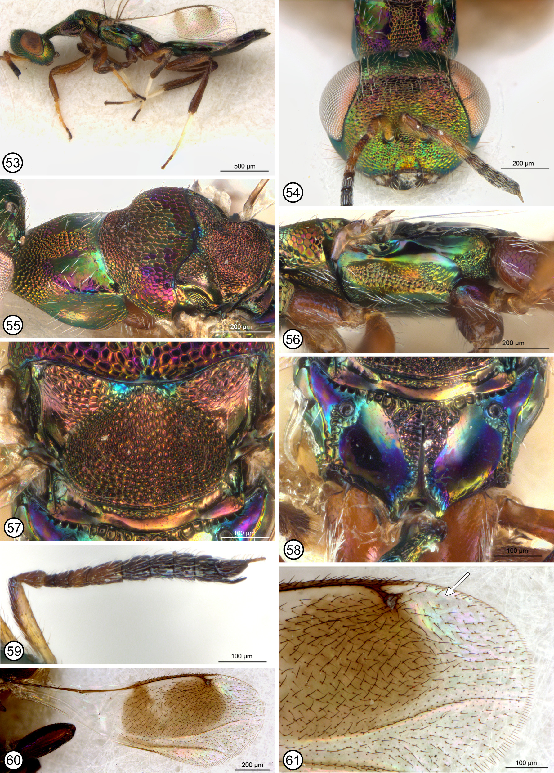
Figs 53–61 View FIGURES 53 – 61
Amarisca oulmesiensis Delucchi, 1962: 12 View in CoL –13, 21 (figs 2, 3). Holotype ♀ (ETHZ; paratypes examined); Baur, 2001: 66 View Cited Treatment (type data). Baur (2001) stated that the holotype is labelled “ex 1. 28.VIII.51 Maroc Oulmès 3703; Regragui; s/xylophages s/ prunus View in CoL ”; AMARISCA oulmesiensis n. V. Delucchi det.; HOLOTYPUS [red]”. The original description stated the following for the type series: “Moyen Atlas, 1250 m ... laboratory reared from cherry wood infested with Scolytus mediterraneus Eggers (Col., Scolytidae View in CoL ) and Anthaxia View in CoL sp. (Col., Buprestidae View in CoL ) between June 13 and August 28, 1951, B. Regragui leg.”.
Notanisus oulmesiensis View in CoL ; Bouček, 1991: 204; Mitroiu and Andriescu, 2008: 312 (♀ keyed), 313 (distribution), 315 (fig. 2, ♀ dorsal habitus), 316 (fig. 6, ♀ dorsal head and mesosoma).
Distribution. Western Palaearctic (see Noyes 2014).
Material examined. CYPRUS. Kalopanayiotis, 2500 ft., 23.IX.1963 / Mavromoustakis (1♀ BMNH). GREECE. Pelopone, Gythion, 13–16.V.1979, Hladilcvi lgt. (1♀ NMPC). Pelop., Petalidion, 27.VIII.1979, Bouček (1♀ NMPC). Samos, Oros Thios, 15.X.1989, M. Koponen leg. (1♀ NMPC). Thessalia, Kalambaka, hillside meadow, 14–20.VII.1979 / BM 1979-312, M.C. Day, G.R. Else, D. Morgan (1♀ BMNH). MOROCCO. 4 ♀ paratypes (3703): 2 ex. 21.VII.51 and 2 ex. 23.VII.51 ( ETHZ). ROMANIA. Dobr. Reserv. Agigea [Agigea Nature Reserve], 29–30.VI.1973, C.G. Nagy legit (1♀ MICO). TURKEY *. Asia Minor, Tr. Çesme, V.1979, T.E. Leiler (1♀ BMNH).? UZBEKISTAN *. Šsafrikan, 28.IV.1980, Kyzyl-kum des., J. Hladil lgt. (1♀ NMPC). YUGOSLAVIA. Mljet, Nacional Park, 19.VIII.1980, A. Hoffer (1♀ NMPC). Ulcinj, Crna Gora, 22.VI.1969, Hoffer (1♀ NMPC).
Description. FEMALE ( Fig. 53 View FIGURES 53 – 61 ). Length 1.9–3.5 mm. Head in frontal view green ( Fig. 54 View FIGURES 53 – 61 ) or with variably extensive coppery to reddish-violaceous lusters, most commonly on frontovertex, face above toruli and on parascrobal region, but sometimes with limited luster also on lower face and gena; frontovertex distinctly differentiated by difference in sculpture at level about two-thirds distance between toruli and anterior ocellus, with larger, more isodiametric and much finer reticulations (shallow but noticeably delineated by raised ridge) dorsad level compared to distinctly reticulate sculpture ventrad level, the sculpture between frontovertex and torulus in particular more transverse reticulate-imbricate; in lateral view lower face and gena posterior to malar sulcus similarly strongly sculptured and colored; in dorsal view OOL 1.1–1.7× maximum diameter of posterior ocellus. Antenna ( Fig. 59 View FIGURES 53 – 61 ) variable in color, rarely entirely yellowish, but at least scape largely yellow, the pedicel and flagellum sometimes entirely dark (smaller females) but usually at least fl2–fl4 yellowish and clava and apical three or four funiculars usually dark; fl1 at least quadrate and usually slightly longer than wide, fl4 slightly shorter than combined length of fl2 and fl3, and funiculars increasing in width and beyond fl4 decreasing in length such that apical funiculars slightly transverse in dorsal view; apical funicular ventrally extending under clava as apically tapered, ventrally bare and shiny, finger-like projection to level equal with apex of clava excluding slender, terminal, setose, spiniform process. Mandible bidentate with two similar teeth ventrally and obliquely angled margin dorsally.
Pronotal collar in lateral view shallowly concave to flat ( Fig. 53 View FIGURES 53 – 61 ) and in dorsal view not distinctly “shoulderlike” posterolaterally; mostly reticulate and green to bright coppery ( Figs 54, 55 View FIGURES 53 – 61 ) except for much more finely sculptured, meshlike coriaceous to smooth, elongate-triangular region posterolaterally, the posterolateral regions usually mostly reddish-violaceous, though rarely green or often with some coppery or bluish luster, but with scattered distinct setae and with longitudinal inner margins differentiating quadrangular median sculptured region over length of collar, the median region mostly punctulate-reticulate to reticulate except extreme posterior margin often more finely sculptured. Mesoscutum ( Fig. 55 View FIGURES 53 – 61 ) anteriorly between incomplete notauli similarly sculptured and colored as pronotum dorsomedially, but with much larger reticulations posteriorly and with broad reddishviolaceous band posteriorly along transscutal articulation extending onto lateral lobes; scutellar-axillar complex green with variably distinct and bright coppery luster to mostly coppery or reddish-violaceous except at least for green to bluish median crenulate region between axillae ( Fig. 57 View FIGURES 53 – 61 ), axilla with reticulate dorsal surface longer than median crenulate region between axillae, and with obliquely angled posterior surface very finely meshlike coriaceous to smooth, and scutellum low convex to distinctively flat ( Fig. 53 View FIGURES 53 – 61 ) and more or less uniformly punctatereticulate to reticulate ( Fig. 57 View FIGURES 53 – 61 ), though reticulations sometimes smaller mesally or more elongate mediolongitudinally and more reticulate-imbricate laterally. Tegula variably extensively yellowish to brown. Macropterous; fore wing ( Fig. 60 View FIGURES 53 – 61 ) marginal vein about 6.0–7.2× length of stigmal vein; apex of postmarginal vein extending to or slightly beyond apex of uncus; uncus ( Fig. 61 View FIGURES 53 – 61 ) diverging from stigmal vein at or near apex so as to at most differentiate small, rounded stigma posterior to uncus, and apex separated from posterior margin of postmarginal vein by distance greater than width of postmarginal vein but less than maximum height of stigma plus uncus; costal cell ventrally with 2–9 setae within about basal third and rarely with 1 or 2 setae within about apical third; disc ( Fig. 60 View FIGURES 53 – 61 ) with distinct brownish region behind venation except narrowly hyaline behind marginal vein mesally, with hyaline band within basal half of marginal vein extended posteriorly for at least half distance to medial fold as variably conspicuous tapered hyaline region, and with cubital fold and often membrane behind cubital fold variably extensively similarly or lighter brown, and with distinct brown setae, the setae at most only slightly shorter in hyaline regions, except for narrow bare band behind parastigma and about basal half of marginal vein, but dorsally without distinct bare band beyond stigmal vein ( Fig. 61 View FIGURES 53 – 61 ); marginal fringe present except anteroapically beyond postmarginal vein ( Fig. 61 View FIGURES 53 – 61 ). Prepectus extensively setose with short white setae. Mesepimeron ( Fig. 56 View FIGURES 53 – 61 ) bare along posterior margin. Metapleuron ( Fig. 56 View FIGURES 53 – 61 ) bare, with about ventral half reticulate and dorsal half smooth and shiny. Metasternum comparatively long, with base of mesocoxa distinctly anterior to, and apex of mesocoxa about level with base of metacoxa. Legs ( Fig. 53 View FIGURES 53 – 61 ), including coxae, yellowish- to orangeybrown or with meta- and/or mesofemora and tibiae darker brown, but basal three tarsomeres of meso- and metatarsi white and apical two tarsomeres of all legs darker brown; metacoxa with at least 2 setae dorsobasally and usually with distinct patch of several white setae in larger specimens ( Figs 56, 58 View FIGURES 53 – 61 ). Propodeum ( Fig. 58 View FIGURES 53 – 61 ) with crenulate band along anterior margin recurved into posteriorly tapered, V-shaped crenulate-rugulose region on either side of complete median carina; panels otherwise smooth and shiny, and callus smooth and shiny; callus anteriorly and propodeal plical region laterally between foramen and spiracle variably dark reddish-violaceous to purple, though callus posterior to spiracle often variably extensively green, median sculptured region often coppery to green, and smooth region adjacent to medial sculptured region usually variably green or greenish-coppery to blue or purple depending on angle of light.
Petiole dark with at least slight green to bluish-green lusters, dorsally sculptured, and about 1.4–2.0× as long as median width. Gaster uniformly dark brown or basally with green to reddish-violaceous lusters; presyntergal tergites very finely meshlike coriaceous except posterior margins smooth and basal tergite usually with more obscure, effaced sculpture (base of Gt2 often also variably extensively smooth depending on extent covered by Gt1), with Gt2 at least similar in length and often longer than Gt1 or Gt3 depending on extent covered by Gt1.
MALE. Unknown.
Hosts. Type material was reared originally from the wood of cherry, likely Prunus avium (L.) ( Rosaceae ), infested with Scolytus rugulosus (Mueller) (= Scolytus mediterraneus (Eggers)) ( Coleoptera : Curculionidae : Scolytinae ) and Anthaxia sp. ( Coleoptera : Buprestidae ).
Remarks. The original description includes line drawings of the female antenna and fore wing (Deluchhi 1962, figs 2, 3), and states that the species was described from the holotype and eight paratypes, of which Baur (2001) located all but one paratype. I examined four paratypes, all of which are covered with particulate matter and incomplete to varying extents. The gaster was missing from three and only two had one complete antenna each.
Although not given as part of the description, females of N. oulmesiensis vary in how compressed the head and mesosoma appear, some female having these quite conspicuously compressed such that the dorsal surface of the mesosoma is almost flat ( Figs 53, 56 View FIGURES 53 – 61 ). Specific status and putative features differentiating females from those of N. kansensis are discussed under the latter species. Mitroiu and Andriescu (2008) noted that of the species with associated males, N. versicolor and N. sexramosus , males were much more common than females. It therefore seems unusual that males are unrecognized for such a relatively commonly collected species as N. oulmesiensis . Although at least some of the shared features listed in couplet two of the key likely represent symplesiomorphies, several suggest N. oulmesiensis (and N. kansensis ) is more closely related to N. yemenensis and N. vanharteni than to N. brevipetiolus and N. longipetiolus . Based on this assumed relationship, the unknown males of N. oulmesiensis therefore more likely have a ramose than a pedicellate flagellum. However, if with another type of flagellar structure they may not be readily associated with females or recognized as Notanisus males.
No known copyright restrictions apply. See Agosti, D., Egloff, W., 2009. Taxonomic information exchange and copyright: the Plazi approach. BMC Research Notes 2009, 2:53 for further explanation.
|
Kingdom |
|
|
Phylum |
|
|
Class |
|
|
Order |
|
|
Family |
|
|
Genus |
Notanisus oulmesiensis (Delucchi)
| Gibson, Gary A. P. 2015 |