
Notanisus brevipetiolus, Gibson, Gary A. P., 2015
|
publication ID |
https://doi.org/ 10.11646/zootaxa.3948.3.4 |
|
publication LSID |
lsid:zoobank.org:pub:E349818A-165B-4CA8-BA29-0E345AFDF6C6 |
|
DOI |
https://doi.org/10.5281/zenodo.5275693 |
|
persistent identifier |
https://treatment.plazi.org/id/D4478723-FF8D-D167-299D-A91CFA07FCCE |
|
treatment provided by |
Plazi |
|
scientific name |
Notanisus brevipetiolus |
| status |
sp. nov. |
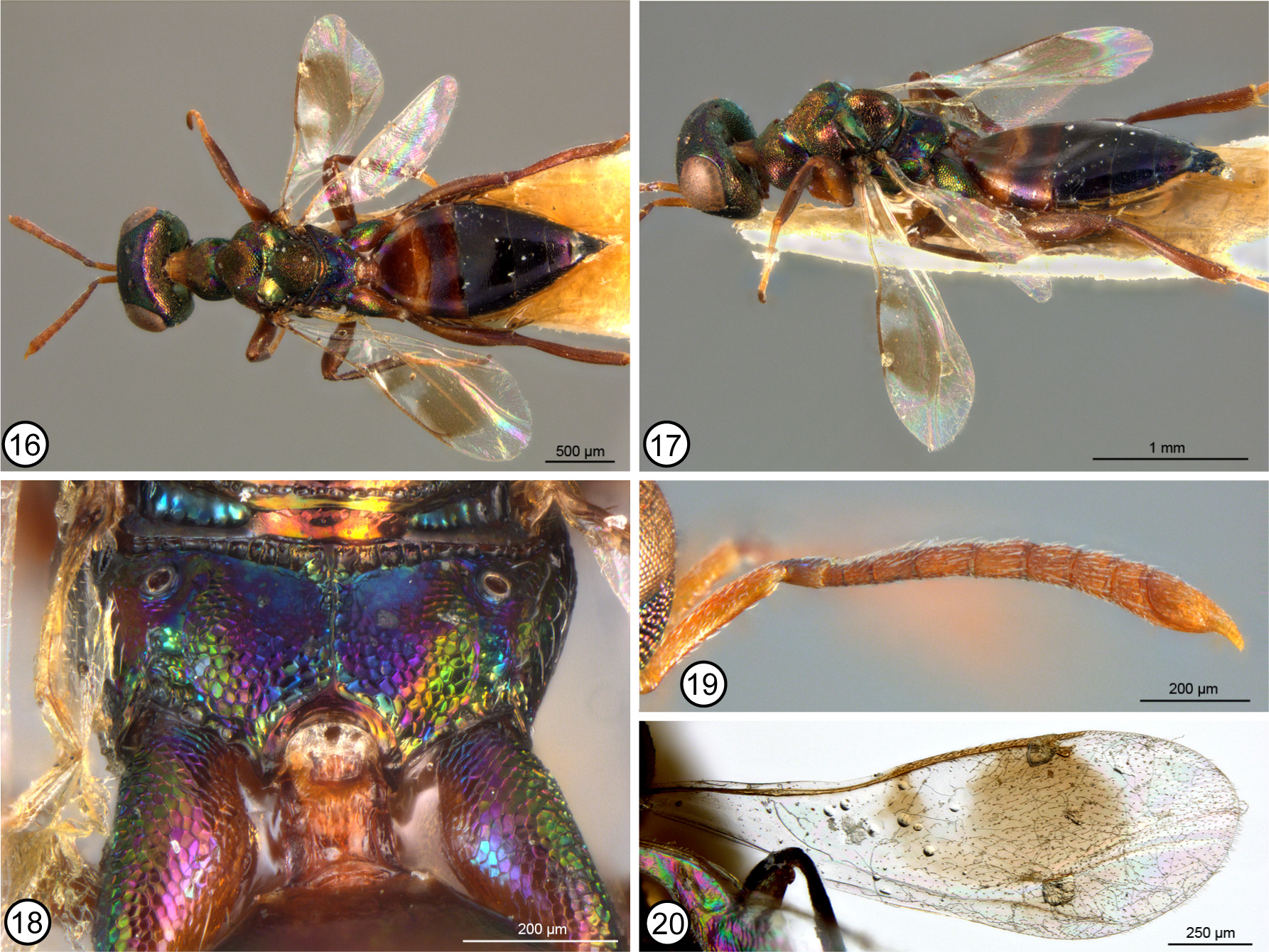
Notanisus brevipetiolus n. sp.
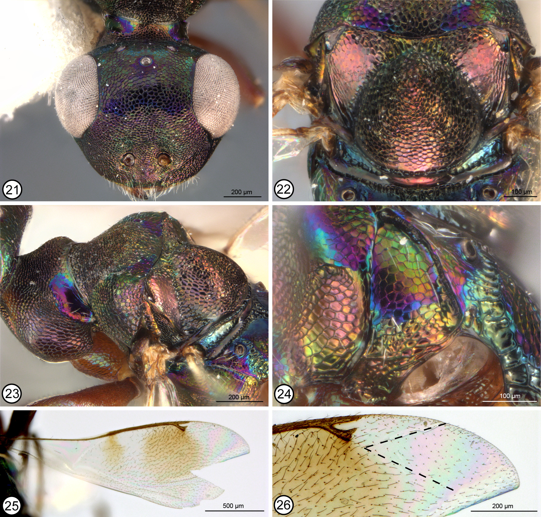
Figs 16–26 View FIGURES 16 – 20 View FIGURES 21 – 26
Type material. Holotype ♀ ( NMPC). N. Rhodesia [ Zambia]: Congo Border, Niankasa / 126.29 / 22.XI.1929, H. Silvester Evans / Pres. by Imp. Inst. Ent., Brit. Mus. 1932.1545 [entire but glued ventrally along length to point, with mouthparts and some other ventral features obscured by glue].
Paratype. UGANDA. Lake Victoria, XI.1971, H. Falke (1♀ CNC).
Etymology. Formed from the Latin words brevis (short) and petiolus (stalk) in reference to its comparatively short petiole.
Description. FEMALE ( Figs 16, 17 View FIGURES 16 – 20 ). Length about 1.9 mm. Head in frontal view ( Fig. 21 View FIGURES 21 – 26 ) with face and frontovertex mostly dark or with slight violaceous luster under some angles of light, except face above scrobes and sometimes scrobes somewhat more distinctly green, and frontovertex sometimes more extensive green to partly reddish-violaceous; frontovertex distinctly differentiated by difference in sculpture at level about midway between toruli and anterior ocellus, with larger, shallower, more isodiametric meshlike reticulation dorsad level compared to much smaller, more transversely punctate-reticulate sculpture ventrad level; in lateral view lower face and gena posterior to malar sulcus similarly strongly sculptured and colored; in dorsal view OOL 2.1–2.2× maximum diameter of posterior ocellus. Antenna ( Fig. 19 View FIGURES 16 – 20 ) yellow; fl1slightly transverse, fl4 almost 0.8× length of fl2 and fl3, and funiculars increasing in width and beyond fl4 decreasing in length such that apical funicular slightly transverse in dorsal view; apical funicular ventrally extending under clava as apically tapered, ventrally sparsely setose, finger-like projection to level where clava tapers into terminal, setose, ventrally curved finger-like process. Mandibular dentition not clearly visible.
Pronotal collar ( Figs 16 View FIGURES 16 – 20 , 23 View FIGURES 21 – 26 ) in lateral view flat but in dorsal view with abruptly declivitous concave regions posterolaterally such that pronotum appears more or less “shoulder-like” on either side; dorsally punctulatereticulate and mostly dark or green with coppery luster except neck yellowish and much more finely sculptured and shinier posterolateral declivitous regions violaceous to partly blue or green laterally where more distinctly meshlike coriaceous, with each region having line of obvious white setae along lateral margin and transversely across region behind transverse anterior margins that together delineate posterior margin of broadly V-like convergent dorsal sculptured region. Mesoscutum ( Figs 16 View FIGURES 16 – 20 , 23 View FIGURES 21 – 26 ) anteromesally between incomplete notauli dark and similarly punctulate-reticulate as pronotum dorsally, but over about posterior half and on lateral lobes posteriorly with larger, more mesh-like reticulations and mostly dull to bright greenish-coppery, though with some reddishviolaceous anterior to axilla; scutellar-axillar complex ( Fig. 22 View FIGURES 21 – 26 ) dark with coppery to green or reddish-violaceous lusters depending on angle of light, particularly axillae, axilla with slender dorsal reticulate surface at most about as long as length of median crenulate region between axillae and with obliquely angled posterior surface much more finely meshlike coriaceous, and scutellum convex ( Fig. 23 View FIGURES 21 – 26 ), reticulate-punctate dorsally but more reticulate to reticulate-imbricate laterally and posteriorly ( Fig. 22 View FIGURES 21 – 26 ), the dorsal sculpture intermediate in size between anteriorly and posteriorly on mesoscutum. Tegula brown. Macropterous; fore wing ( Figs 20 View FIGURES 16 – 20 , 25, 26 View FIGURES 21 – 26 ) marginal vein about 6.2– 7.5× length of stigmal vein; postmarginal vein extending slightly but distinctly beyond level of uncus; uncus ( Fig. 26 View FIGURES 21 – 26 ) diverging from stigmal vein apically so distinct stigma not differentiated and apex separated from posterior margin of postmarginal vein by distance slightly greater than width of postmarginal vein or uncus and subequal to or less than maximum height of stigma plus uncus; costal cell bare; disc ( Figs 20 View FIGURES 16 – 20 , 25 View FIGURES 21 – 26 ) with variably large yellowish-brown region behind parastigma and larger, similarly colored region behind about apical half of venation anterior to medial fold, the two infuscate regions sometimes continuous posteriorly to form a U-shaped region ( Fig. 20 View FIGURES 16 – 20 ), with longer brownish setae in infuscate regions and shorter, less conspicuous setae apically, except bare behind parastigma and marginal vein basally, the bare region conspicuously expanded posteriorly within hyaline area between infuscate regions, and dorsally with comparatively large, elongate bare region beyond stigmal vein ( Fig. 26 View FIGURES 21 – 26 ); marginal fringe present except anteroapically beyond postmarginal vein. Prepectus bare or with a few short, inconspicuous setae posteroapically. Mesepimeron in dorsal half (upper mesepimeron) with short white setae along extreme posterior margin ( Fig. 24 View FIGURES 21 – 26 ). Metapleuron ( Fig. 24 View FIGURES 21 – 26 ) reticulate ventrally but more finely sculptured, almost isodiametric meshlike coriaceous over at least dorsal two thirds and with sparse white setae anteroventrally and dorsally and posteriorly in more finely sculptured region. Metasternum comparatively short, with bases of meso- and metacoxae about on same level and apex of mesocoxa projecting conspicuously beyond base of metacoxa. Legs ( Figs 16, 17 View FIGURES 16 – 20 ) with procoxa orange to orangey-brown, mesocoxa darker brown with slight metallic luster, and metacoxa dorsally dark brown with variably distinct reddish-violaceous or green lusters depending on angle of light but yellowish- to orangey-brown ventrally; femora, tibiae and tarsi orangey-brown to dark brown except basal three or four tarsomeres of meso- and metatarsi more yellowish compared to somewhat more darker brown apical tarsomeres; metacoxa bare dorsally ( Fig. 18 View FIGURES 16 – 20 ). Propodeum ( Fig. 18 View FIGURES 16 – 20 ) with crenulate band along anterior margin recurved posteromedially into narrowly V-shaped rugose to crenulate region on either side of partial to complete median carina; panels otherwise reticulate except for comparatively small smooth and shiny region anteromesally on either side of median sculptured region, and multicolored, variably blue to purple anteriorly in smoother regions to partly green or reddish-violaceous over sculptured part of panels; callus finely though quite distinctly coriaceous and multicolored similar to panels.
Petiole ( Fig. 18 View FIGURES 16 – 20 ) yellowish-brown, subquadrate with parallel sides, only slightly longer than wide. Gaster ( Figs 16, 17 View FIGURES 16 – 20 ) dark brown except for two lighter, more yellowish bands basally, one across apical smoother part of Gt1 and Gt2, and one across apical smoother part of Gt3; presyntergal tergites isodiametric mesh-like coriaceous except smoother along posterior margins and basal two tergites smooth and shiny or Gt1 with only subeffaced mesh-like sculpture basally, with Gt2 strongly transverse, much shorter than other tergites, and Gt4 the largest tergite.
MALE. Unknown.
Host. Unknown.
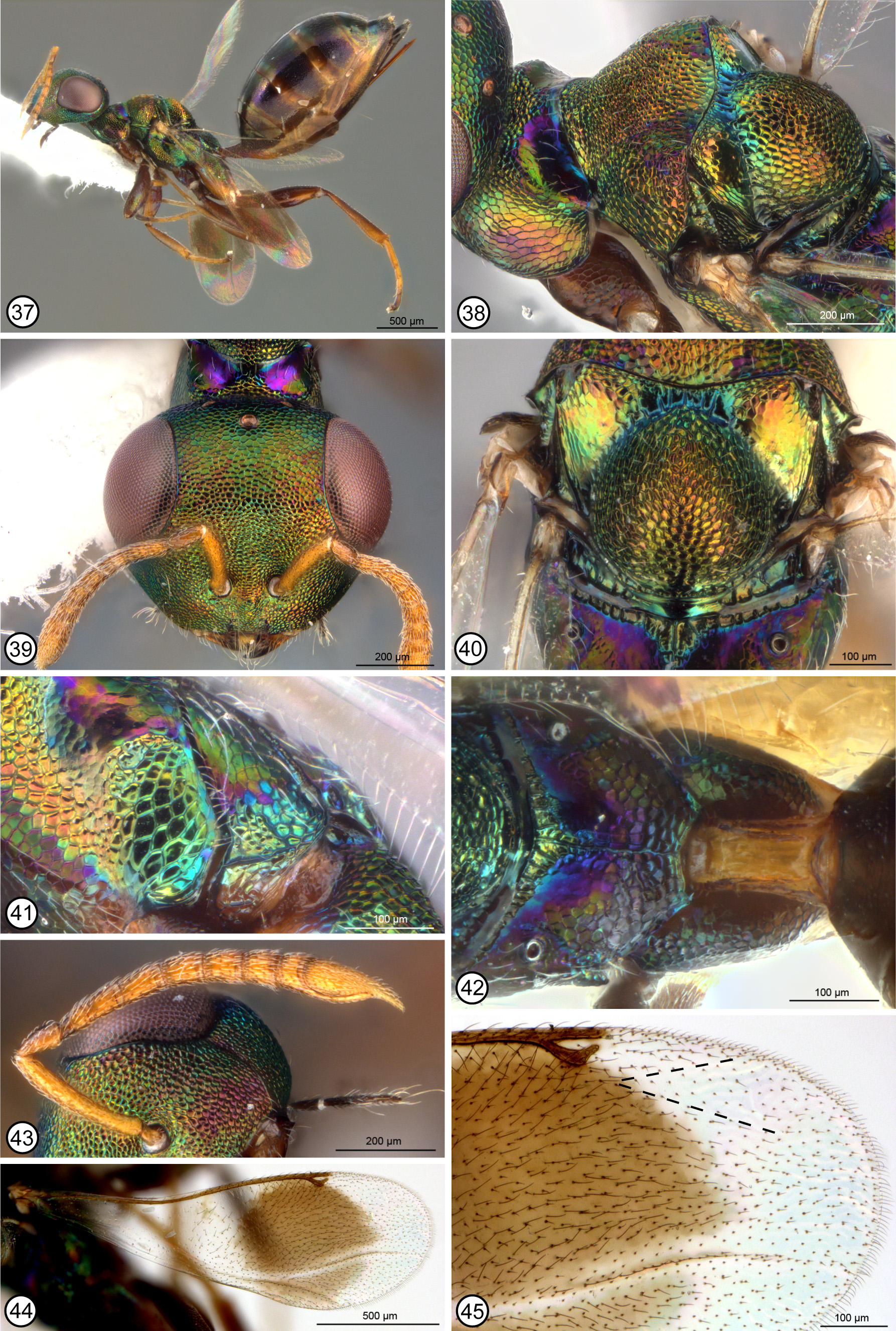
Remarks. Minimally, colour of the petiole, structure of the antennal clava (Fig. 28), and a distinctively short, transverse-bandlike Gt2 ( Yang 1996, fig. 144) are shared between N. brevipetiolus , N. longipetiolus and N. gracilis . However, the latter feature is also shared with N. kansensis and N. oulmesiensis within the oulmesiensis -group, and the total number of features that females of the three species share is uncertain because the holotype of N. gracilis was not studied for comparison. Females of N. brevipetiolus and N. longipetiolus are most easily separated from each other by relative length of the petiole (cf. Figs 18 View FIGURES 16 – 20 , 42 View FIGURES 37 – 45 ) and gastral color pattern (cf. Figs 16, 17 View FIGURES 16 – 20 , 37 View FIGURES 37 – 45 ). Additional females are needed to determine whether the apparently less extensively setose posterior margin of the mesopleuron (cf. Figs 24 View FIGURES 21 – 26 , 41 View FIGURES 37 – 45 ) is an additional differential feature for N. brevipetiolus . Among observed Notanisus , setae along the posterior margin of the mesopleuron is unique to females of N. brevipetiolus and N. longipetiolus . Such setae are also possessed by many species of Cleonymus , but not observed Callocleonymus . Males of N. brevipetiolus are unknown, but based on presumed relationships with N. longipetiolus they likely have a pedicellate flagellum, the metapleuron partly setose (similar to females) but the posterior margin of the mesopleuron bare, and the pronotum entirely reticulate, though possibly with a difference in color differentiating posterolateral regions.
No known copyright restrictions apply. See Agosti, D., Egloff, W., 2009. Taxonomic information exchange and copyright: the Plazi approach. BMC Research Notes 2009, 2:53 for further explanation.