
Petrobia (Tetranychina) hispaniola, Martínez, Leocadia Sánchez, Flechtmann, Carlos H. W. & De Moraes, Gilberto J., 2014
|
publication ID |
https://dx.doi.org/10.11646/zootaxa.3846.4.3 |
|
publication LSID |
lsid:zoobank.org:pub:12F1BF5E-F6C0-44CB-8712-7ED7677A4DC9 |
|
persistent identifier |
https://treatment.plazi.org/id/7B512D59-1F3E-FFCE-54AF-FDD21D22F8CA |
|
treatment provided by |
Plazi (2016-04-12 13:46:59, last updated 2022-01-30 07:48:14) |
|
scientific name |
Petrobia (Tetranychina) hispaniola |
| status |
n. sp. |
Petrobia (Tetranychina) hispaniola n. sp. Sánchez & Flechtmann (Figs 1–8)
DIAGNOSIS. A Petrobia ( Tetranychina ) with long body setae; legs I twice as long as idiosomal length (not including gnathosoma); peritreme ending in anastomosing chambers forming a globular structure; tibia of leg I in males with seta b trichobothrium-like and with 41 solenidia.
FEMALE (holotype + 8 paratypes). Idiosoma (Fig. 1 A) broadly oval 638 (638–820); 545 (358–600) wide. All dorsal setae robust, finely pilose and set on strong tubercles (Fig. 1 B). Dorsopropodosomal setae: v 2 100 (85–110), 85 (78–85) apart; sc 1 145 (120–153), 125 (118–130) apart; sc 2 88 (73–88). Hysterosomal setae: c 1 190 (188–200), 163 (175–188) apart; c 2 183 (163–188); c 3 88 (65–83); d 1 208 (190–213), 83 (50–75) apart; d 2 193 (165–190); e 1 195 (173–200), 58 (50–88) apart; e 2 185 (163–188); f 1 195 (175–203), 30 (25–50) apart; f 2 153 (125–163); h 1 115 (85–113), 90 (75–112) apart. Integumental striae irregularly transverse, tending to longitudinal between bases of setae e 1; striae granulated (Fig. 1 C). Prodorsum punctated centrally and outlined by striae. Gnathosoma: stylophore rounded anteriorly (Fig. 1 D). Peritreme ending in anastomosing chambers forming a globular structure (Fig. 1 E). Palp robust ( Fig. 4 View FIGURE 4 A); trochanter and femur indistinctly separated; genu with one posterolateral seta, tibia with a dorsal “claw” and three setae; tarsus with seven setiform structures.
Venter: integument transversally striated to setae 3 a (intercoxal 3); posterior striae irregularly longitudinal with a small transversely striate rhomboid area posterior to setae 4 a (intercoxal 4) ( Fig. 4 View FIGURE 4 B). Pseudanal setae: h 2 11 (9–14), h 3 10 (11–13).
Legs: legs I approximately twice as long as idiosoma not including gnathosoma. Legs I 1327 (1042–1340), II 570 (518–595), III 645 (577–677), IV 1017 (767–1055).
Leg chaetotaxy, from coxae to tarsi (solenidia shown in parentheses, eupathidia shown in square brackets):
I: 2 – 1 – 9 – 4 – 15 (+ 11) – 16 (+ 7)[+ 3] + 2 duplexes ( Figs 2 View FIGURE 2 B, C)
II: 2 – 1 – 7 – 5 – 11 – 11 (+ 1)[+ 3] + 2 duplexes ( Figs 2 View FIGURE 2 D, E)
III: 1 – 1 – 5 – 5 – 10 – 14 (+ 1) ( Figs 3 View FIGURE 3 A, B)
IV: 1 – 1 – 5 – 5 – 10 – 14 (+ 1) ( Figs 3 View FIGURE 3 C, D)
Variation in leg I chaetotaxy: tibia I: three specimens with 15 (9); one specimen with 15 (13) and one specimen with 14 (12); tarsus I: four specimens with 18 (8) + 2 duplexes.
Empodia I–IV clawlike, distally recurved, with two rows of 12 tenent hairs directed ventrally; claws (true claws) padlike, about one third length of uncinate empodium, with tenet hairs ( Fig. 2 View FIGURE 2 A).
MALE (n = 1 paratype) Much smaller than female, 325 long not including gnathosoma, 365 long including gnathosoma, 175 wide ( Fig. 5 View FIGURE 5 ).
Most dorsal idiosomal setae much shorter and broader than those of female ( Fig. 5 View FIGURE 5 ): v 2 31, 35 apart; sc 1 61, 77 apart; sc 2 40; c 1 38, 58 apart; c 2 37; c 3 27; d 1 25, 36 apart; d 2 33; e 1 20, 28 apart; e 2 20; f 1 22, 10 apart; f 2 25; h 1 39, 30 apart. Palp robust ( Fig. 8 View FIGURE 8 A); trochanter and femur indistinctly separated; genu and tibia also indistinctly separated, with four setae; tarsus with seven setiform structures.
FIGURE 1. Petrobia (Tetranychina) hispaniola Sánchez & Flechtmann n. sp., female. A. Dorsal idiosoma dorsal; B. Seta sc 1; C. Detail of integument; D. Dorsal outline of stylophore; E. Peritreme.
Legs: relative lengths similar to female. Leg I 1235, II 532, III 585, IV 863. Leg chaetotaxy, from coxae to tarsi, solenidia in parentheses:
I: 2 – 1 – 9 – 4 – 3 (+ 41) + 5 duplexes + b (a trichobothrium-like seta) – 10 (+ 15)[+ 2] + 5 duplexes ( Figs 6 View FIGURE 6 A, B, C)
II: 2 – 1 – 8 – 5 – 11 – 13 (+ 2)[+ 2] + 1 duplex ( Figs 6 View FIGURE 6 D, E)
III: 1 – 1 – 5 – 5 – 10 (+ 1) – 14 (+ 1) ( Figs 7 View FIGURE 7 A, B)
IV: 1 – 1 – 5 – 5 – 11 – 14 (+ 1) ( Figs 7 View FIGURE 7 C, D)
On tibia of leg I seta d has the typical aspect of a trichobothrium. Tarsus I with four duplex setae.
Venter: setae h 2 13, h 3 14 ( Fig. 8 View FIGURE 8 B).
Aedeagus similar to those of other species for which the male is known: lanceolate, narrowing gradually and apically rounded; with two lateral acicular appendices.
ETYMOLOGY: The name hispaniola refers to the island shared by Haiti and Dominican Republic.
TYPE MATERIAL: Female holotype and five female paratypes from orange ( Citrus sinensis Osbeck ; Rutaceae ) leaves, and three female and one male paratypes from rose ( Rosa sp.; Rosaceae ) leaves, all from Río Seco, La Vega, Dominican Republic (Caribbean), collected by a group of undergraduate students in Nov. 2010; all deposited at Departamento de Entomologia e Acarologia (ESALQ-USP), Piracicaba-SP, Brasil.
REMARKS: Petrobia (T.) hispaniola n.sp. resembles P. (T.) harti Ewing, 1909 sensu Pritchard & Baker (1955) and Baker & Tuttle (1994), in the aspect of the dorsal body setae and length of female leg I, but it is distinguished by the different peritreme (straight, ending in a simple chamber in P. (T.) harti ). It also resembles P. (T.) kleptes Kamran & Afzal, 2004 by having dorsal setae long and inserted on tubercles; however, it is distinguished by the different peritreme (distally hooked and ending in a simple chamber in P. (T.) kleptes ).
In this new species, tibial seta d has a trichobothridial aspect, which is found only in some Bryobiinae ; in those cases, that seta is denoted by db ( Grandjean, 1943; Lindquist, 1985). In the new species here described, that type of seta is only found in the male. This is the first species of Petrobia ( Tetranychina ) reported from roses and the third from Citrus [together with P. (T.) harti and P. (T.) kleptes ]. Although Lindquist (1985) had mentioned the number of solenidia on tibia I of male to be up to 30, in the species here described the number is much higher (41).
Baker, E. W. & Tuttle, D. M. (1994) A guide to the spider mites (Tetranychidae) of the United States. Indira, Michigan, 347 pp.
Ewing, H. E. (1909) New species of Acarina. Transactions of the American Entomological Society, 35, 401 - 418 + 4 plates.
Grandjean, F. (1943) Les trichobothries pedieuses des Acariens er leur priorite chez les Bdelles. Comptes Rendus de la Societe de physique et d'histoire naturelle de Geneve, 60 (3), 241 - 246.
Kamran, M. & Afazal, M. (2004) A new species of the genus Petrobia (Acarina: Tetranychidae) on citrus from District of Layyah, Punjab. Pakistan. Pakistan Entomologist, 26 (1), 121 - 123.
Lindquist, E. E. (1985) 1.1. Anatomy, Phylogeny and Systematics. 1.1. 1. External anatomy. In: Helle, W. & Sabelis, M. W. (Eds.), Spider Mites. Their Biology, Natural Enemies and Control. Elsevier, Amsterdam, Vol. 1 A, pp. 3 - 28.
Pritchard, A. E. & Baker, E. W. (1955) A revision of the spider mite family Tetranychidae. Memoirs of the Pacific Coast Entomological Society, 2, 1 - 472.
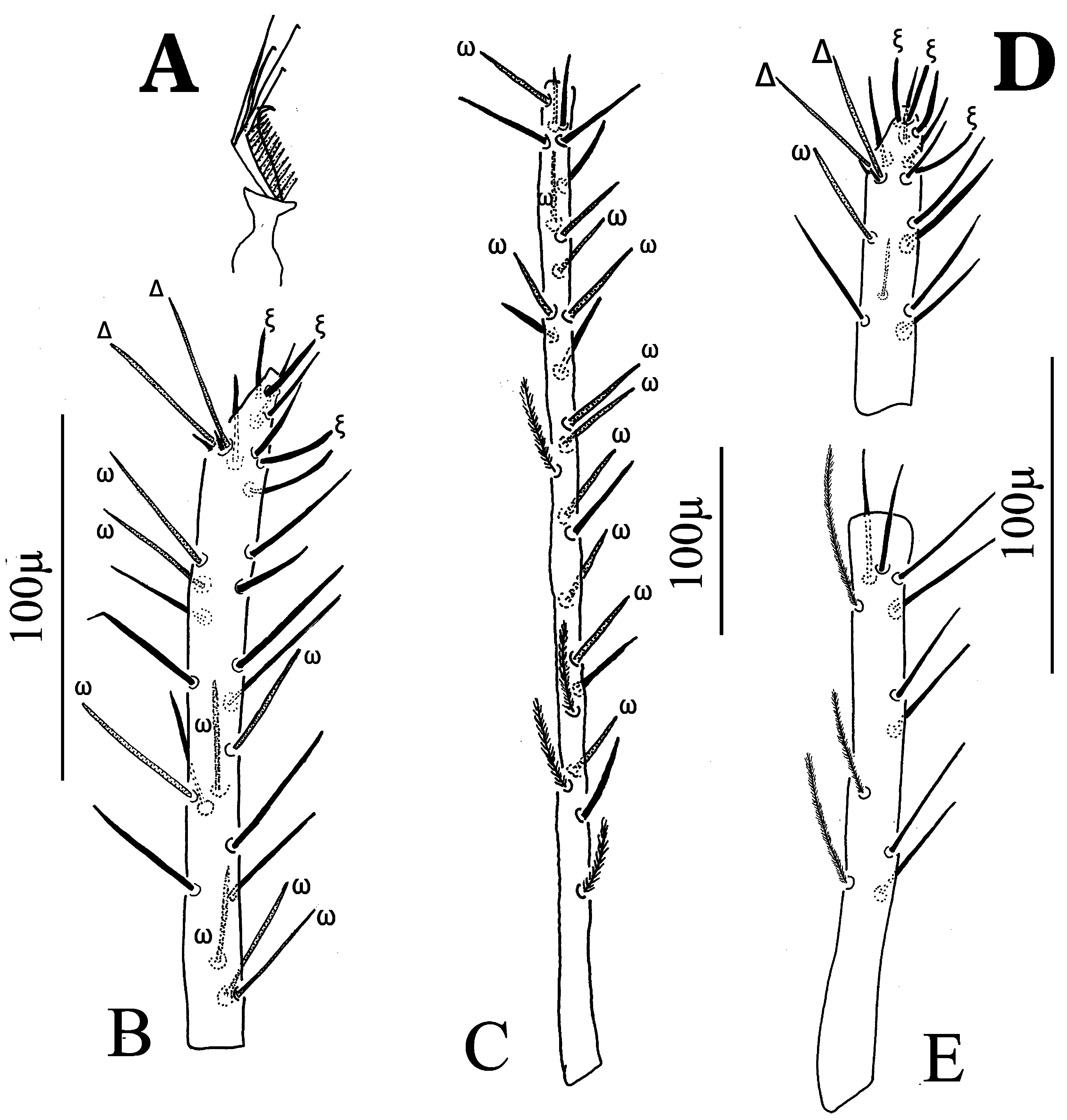
FIGURE 2. Petrobia (Tetranychina) hispaniola Sánchez & Flechtmann n. sp., female, legs. A. Empodium; B. Tarsus I; C. Tibia I; D. Tarsus II; E. Tibia II. Tactile setae unmarked; solenidia, eupathidia and duplex setae indicated by ω, ξ and Δ.

FIGURE 3. Petrobia (Tetranychina) hispaniola Sánchez & Flechtmann n. sp., female, legs. A. Tarsus III; B. Tibia III; C. Tarsus IV; D. Tibia IV. Tactile setae unmarked; solenidia indicated by ω.
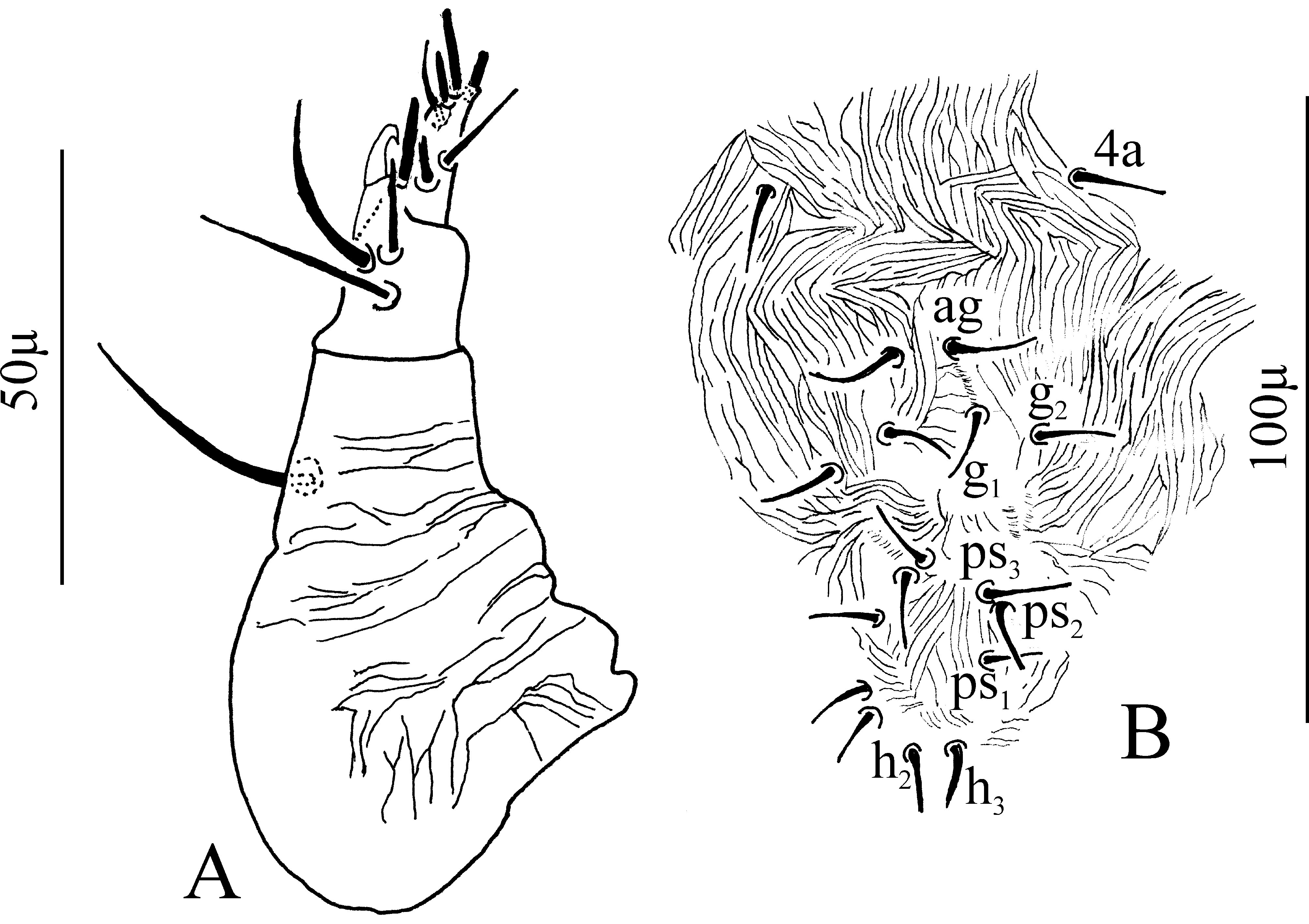
FIGURE 4. Petrobia (Tetranychina) hispaniola Sánchez & Flechtmann n. sp., female. A. Palpus; B. Genito-anal region.

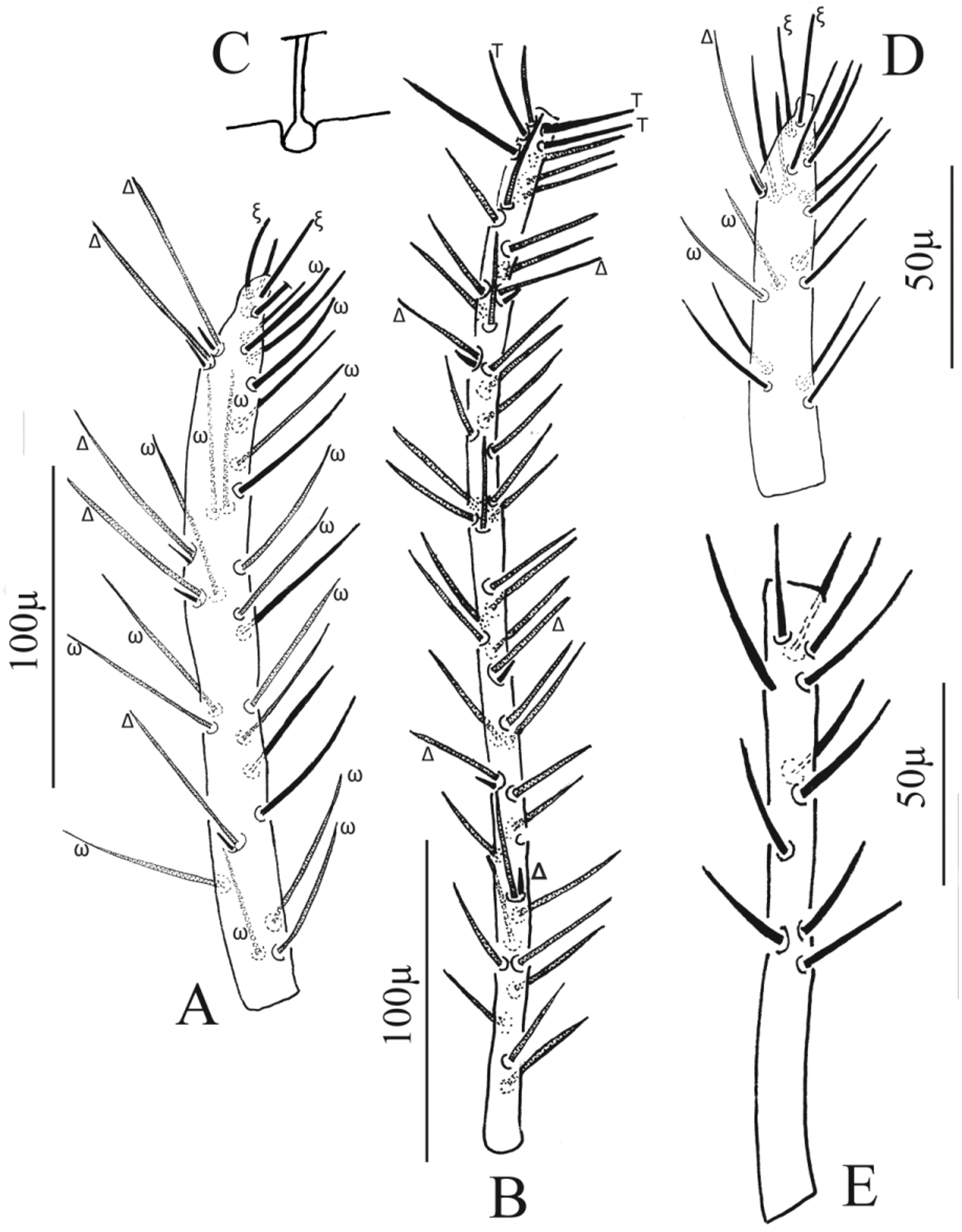
FIGURE 6. Petrobia (Tetranychina) hispaniola Sánchez & Flechtmann n. sp., Male, legs. A. Tarsus I (tactile setae unmarked; solenidia, eupathidia and duplex setae indicated by ω, ξ and Δ, respectively); B. Tibia I (tactile, duplex and trichobothrium-like setae indicated by T, Δ and b, respectively); C. Detail of seta b; D. Tarsus II (tactile setae unmarked; solenidia, eupathidia and duplex setae indicated by ω, ξ and Δ, respectively); E. Tibia II (all tactile setae).

FIGURE 7. Petrobia (Tetranychina) hispaniola Sánchez & Flechtmann n. sp., Male, legs. A. Tarsus III; B. Tibia III; C. Tarsus IV; D. Tibia IV. Tactile setae unmarked; solenidia indicated by ω.

No known copyright restrictions apply. See Agosti, D., Egloff, W., 2009. Taxonomic information exchange and copyright: the Plazi approach. BMC Research Notes 2009, 2:53 for further explanation.
|
Kingdom |
|
|
Phylum |
|
|
Class |
|
|
Order |
|
|
Family |
|
|
Genus |
1 (by plazi, 2016-04-12 13:46:59)
2 (by ImsDioSync, 2016-12-31 15:50:22)
3 (by ImsDioSync, 2016-12-31 15:51:01)
4 (by ExternalLinkService, 2019-09-26 19:42:26)
5 (by ExternalLinkService, 2022-01-30 07:48:14)
6 (by ExternalLinkService, 2022-02-18 17:53:21)
7 (by plazi, 2023-10-26 07:28:42)