
Macrostomum shenda Wang & Xin, 2019
|
publication ID |
https://doi.org/10.11646/zootaxa.4603.1.5 |
|
publication LSID |
lsid:zoobank.org:pub:0E1A44A9-C958-4869-9023-EB3086E679E7 |
|
persistent identifier |
https://treatment.plazi.org/id/03B387CD-AC67-FF98-FF4C-FE91FBA9FC4A |
|
treatment provided by |
Plazi |
|
scientific name |
Macrostomum shenda Wang & Xin |
| status |
sp. nov. |
Macrostomum shenda Wang & Xin View in CoL , n. sp.
( Figs. 2–4 View FIGURE 2 View FIGURE 3 View FIGURE 4 )
Material examined. Holotype (PLA–Ma0110): one mounted specimen . Paratypes (PLA–Ma0111–112): two serially-sectioned specimens. The type specimens were collected in OCT mangrove wetland, Shenzhen City , Guangdong Province, China ( 22°31′41″ N, 113°59′9″ E) (see Fig. 1 View FIGURE 1 for sampling location) from September to October, 2016. All specimens are deposited in IZCAS GoogleMaps .
Etymology. The name of this new species is derived from the abbreviated Chinese name of Shenzhen University.
Description. Body flattened dorsoventrally and without pigment. The body length and width of living mature individual is 1000 ± 123 µm (n=5) and 225 ± 30 µm (n=5), respectively ( Figs. 2A View FIGURE 2 , 4A View FIGURE 4 ). The thickness of the epidermis is 5 ± 1.0 µm (n=5), with cilia up to 4 ± 0.5 µm (n=5) covering the entire surface. There are tufts of sensory hairs that are 10 ± 1.3 µm (n=5) long, sparsely distributed on the body edge. The anterior end is slightly more rounded than the posterior extremity, while tufts of rigid cilia (4 ± 0.7 µm long, n=5) are located at both the anterior and posterior ends of the body. Generally, the rigid cilia are shorter and thicker than sensory hairs. The rhabdite rods, 7 ± 1.1 µm (n=5) long, are scattered in groups (mostly 3–9 rhabdites in each group) and are particularly abundant on the dorsal surface of the body. The distance between the two rounded eyes is 30 ± 4.6 µm (n=5) ( Figs. 2A View FIGURE 2 , 4A View FIGURE 4 ). The mouth is 101 ± 9.2 µm (n=5) long and the pharynx is ventrally surrounded by several pharyngeal glands ( Fig. 3A & C View FIGURE 3 ). The tail tapers gradually and has large amount of adhesive glands ( Fig. 4A View FIGURE 4 ).
The paired ovoid testes, 82 ± 9.7 µm (n=5) long, are situated ventrally between the pharynx glands and ovaries ( Figs. 2B View FIGURE 2 , 4A View FIGURE 4 ). The long oval-shaped false vesicula seminalis connects to the vesicula seminalis at its right side. The oval-shaped vesicula seminalis has a thickened muscular wall. The vesicula granulorum connects to the vesicula seminalis and is partially inserted into the proximal end of the penis stylet ( Figs. 2C View FIGURE 2 , 3B View FIGURE 3 & E–F, 4B–C). The narrow funnel-shaped penis stylet is 56 ± 3.1 µm (n=5) long. It has a slightly curved end (curvature less than 15°, the value was measured according to Ferguson 1940a) and is gradually narrowed towards the distal beveled end ( Figs. 2D View FIGURE 2 , 4D View FIGURE 4 ). The proximal end of the stylet is 14 ± 3.8 µm (n=5) in diameter, while the willow leaf shaped distal opening is 8 ± 1.0 µm (n=5) in length ( Fig. 2D View FIGURE 2 ).
The mature sperm are 28 ± 1.3 µm (n=5) long when swimming ( Figs. 2 View FIGURE 2 E–F, 4E). They have neither bristles nor brush, and are surrounded by an undulating membrane in the mid-body region ( Fig. 2F View FIGURE 2 ). The boundaries between the feeler, body and shaft of the sperm are not obvious.
The paired elongated ovoid ovaries are 163 ± 14.2 µm (n=5) long and are situated posterior to the testes on both sides of the body ( Fig. 2B View FIGURE 2 ). The big light-brown mature oocytes usually contain abundant yolk ( Fig. 2A View FIGURE 2 ). The female antrum is located posterior to the gut on ventral side. Besides, there are numerous cement glands surrounding the female gonopore ( Fig. 3A & D View FIGURE 3 ).
Remarks. A comparison of the penis stylets of Macrostomum shenda n. sp. and six similar species within the genus is shown in Table 4 View TABLE 4 . They all have a funnel-shaped stylet with an oblique and non-thickening terminal opening. However, in M. ensiferum , M. phillipsi and M. sinyaense , the ratios of the proximal diameter to the stylet length are less than 16%, while the corresponding value of M. shenda n. sp. is around 25%. Besides, in M. ensiferum , the bending angle of the stylet (> 30°) is larger than that of M. shenda n. sp. (<15°, the values were measured according to Ferguson 1940a). Another similar species, namely M. sinyaense , has a “Verschlussapparat ( Young 1976, and named “cellular valve” in Ladurner et al. 2005)”, but this structure is absent in M. shenda n. sp.. Moreover, the stylet of M. obtusum ( 80–91 µm, Luther 1960) is longer than that of M. shenda n. sp. ( 56 µm) and bends around 65° ( Papi 1951). As for M. vejdovskyi , the stylet length ( 35 µm) and proximal end diameter ( 9 µm) of the stylet are obviously shorter than those of M. shenda n. sp. ( 56 µm and 14 µm). In addition, the sperm of M. vejdovskyi and M. phillipsi have bristles, while this structure is absent in M. shenda n. sp..
In M. orthostylum Braun, 1885 View in CoL , there are a number of publications identifying this species (for example, Braun 1885; von Hofsten 1911; Meixner 1915; Ferguson & Jones 1940; Papi 1951; Beklemishev 1951; Luther 1960; Young 1976 and so on). However, the morphology of their stylets vary considerably, some specimens are up to 90 or 142 µm in their stylet length ( Meixner 1915; von Hofsten 1911). Among them, the descriptions in Ferguson & Jones (1940) and Luther (1960) are most similar with that of M. shenda View in CoL n. sp.. In Ferguson & Jones (1940), the diameter of the stylet is almost unchanged from the proximal end to the anterior 33% part, while the stylet of M. shenda View in CoL n. sp. narrows gradually. Moreover, the stylet distal opening of M. orthostylum View in CoL is oval shaped, while the corresponding opening of M. shenda View in CoL n. sp. is willow leaf shaped. As for Luther (1960), the stylet narrows gradually only in the first half. In terms of morphology of the ovaries, those of M. orthostylum View in CoL are heavily indented ( Ferguson & Jones 1940; Luther 1960; Young 1976), while those of M. shenda View in CoL n. sp. are elongated ovals with a smooth margin. In summary, it is evident that M. shenda View in CoL n. sp. is a new species within the genus Macrostomum View in CoL .
* Measurement based on images and scales from the references. NA: not available.
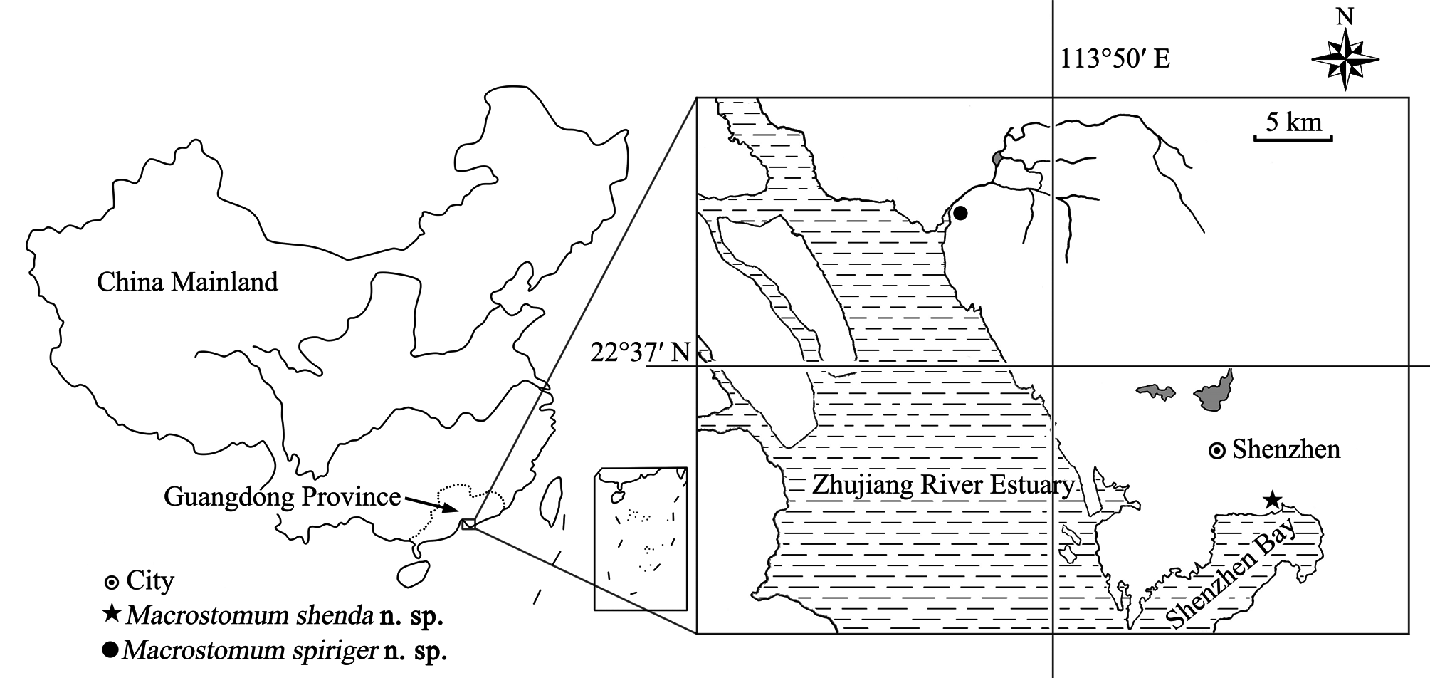
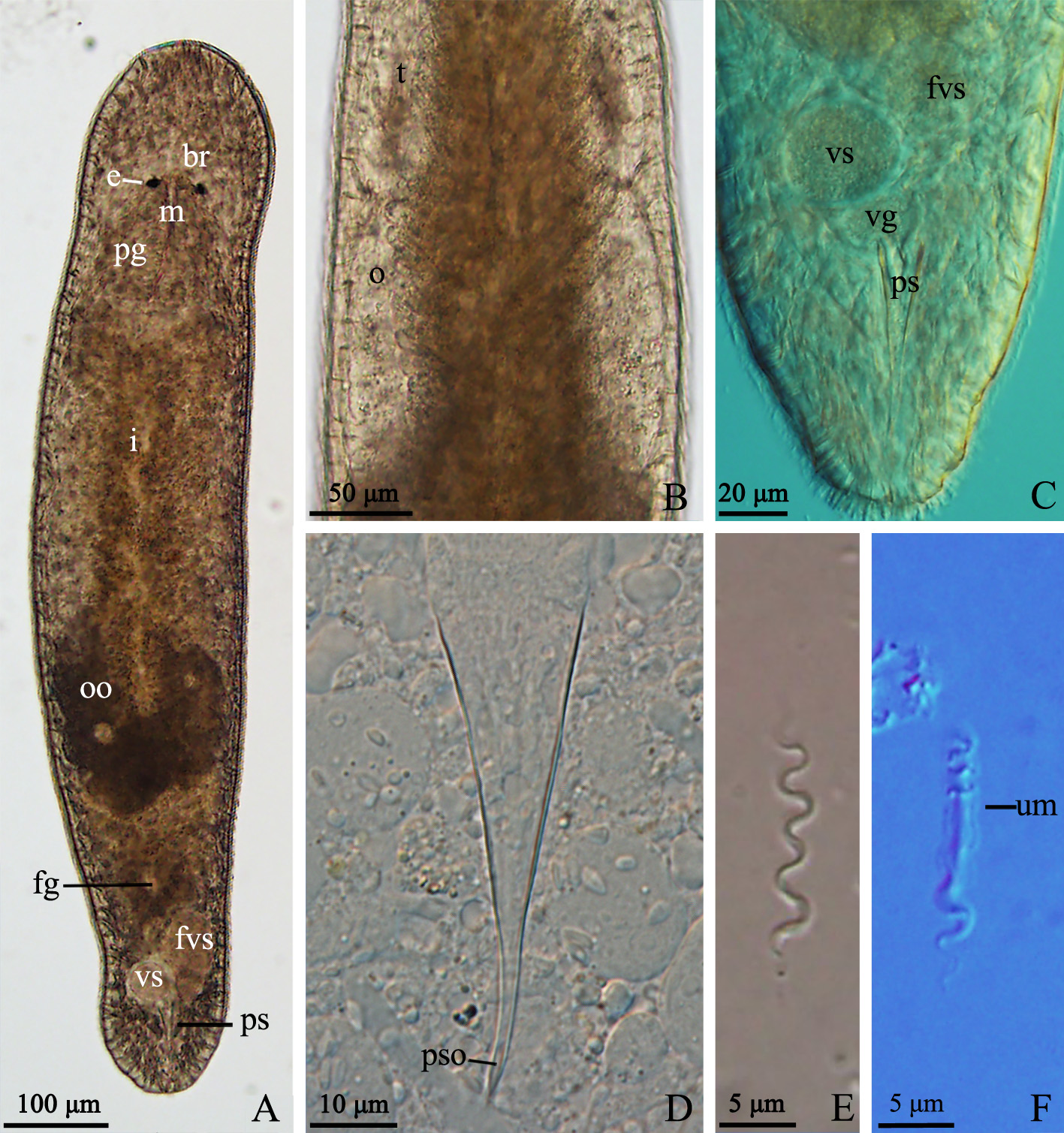
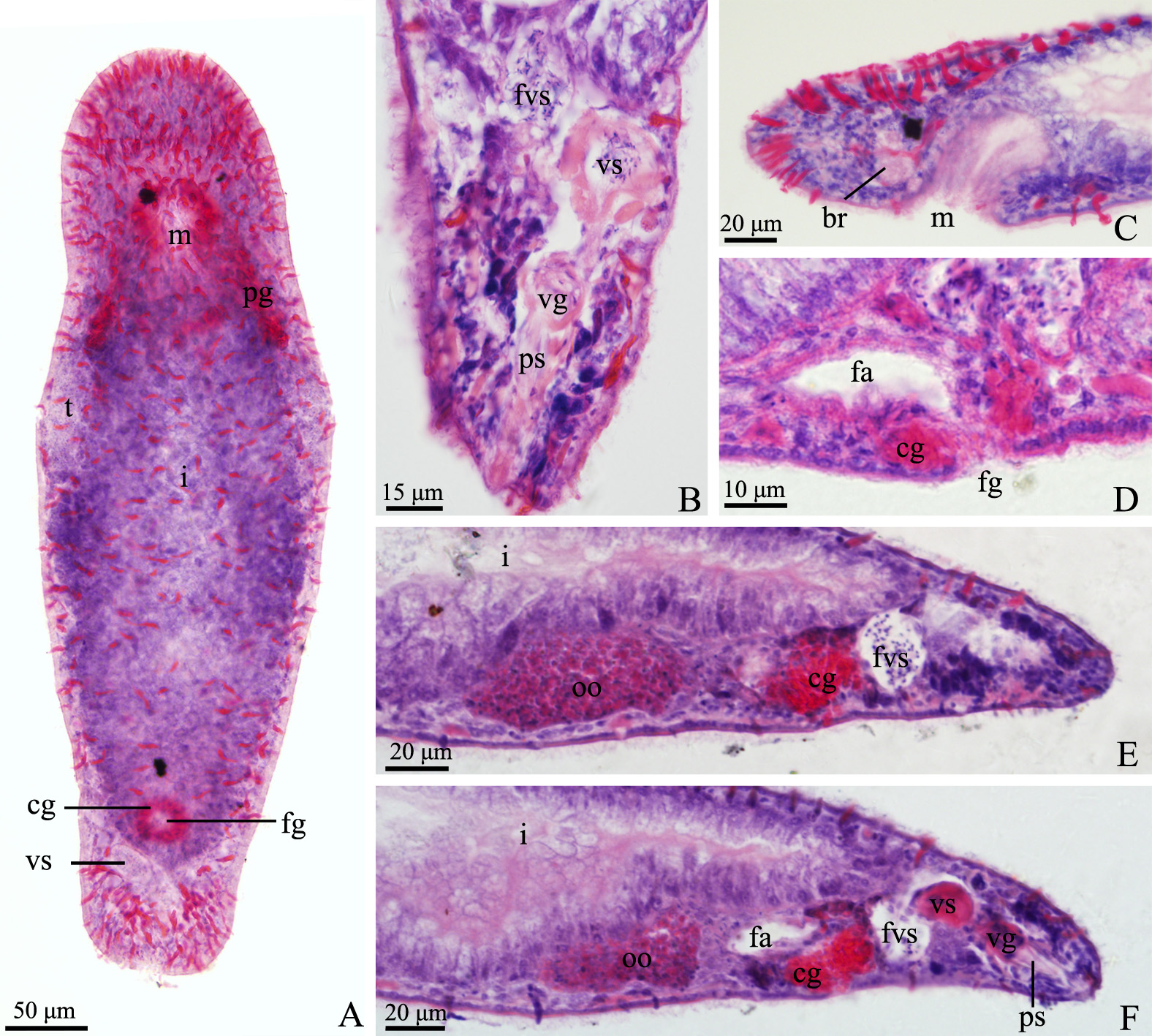
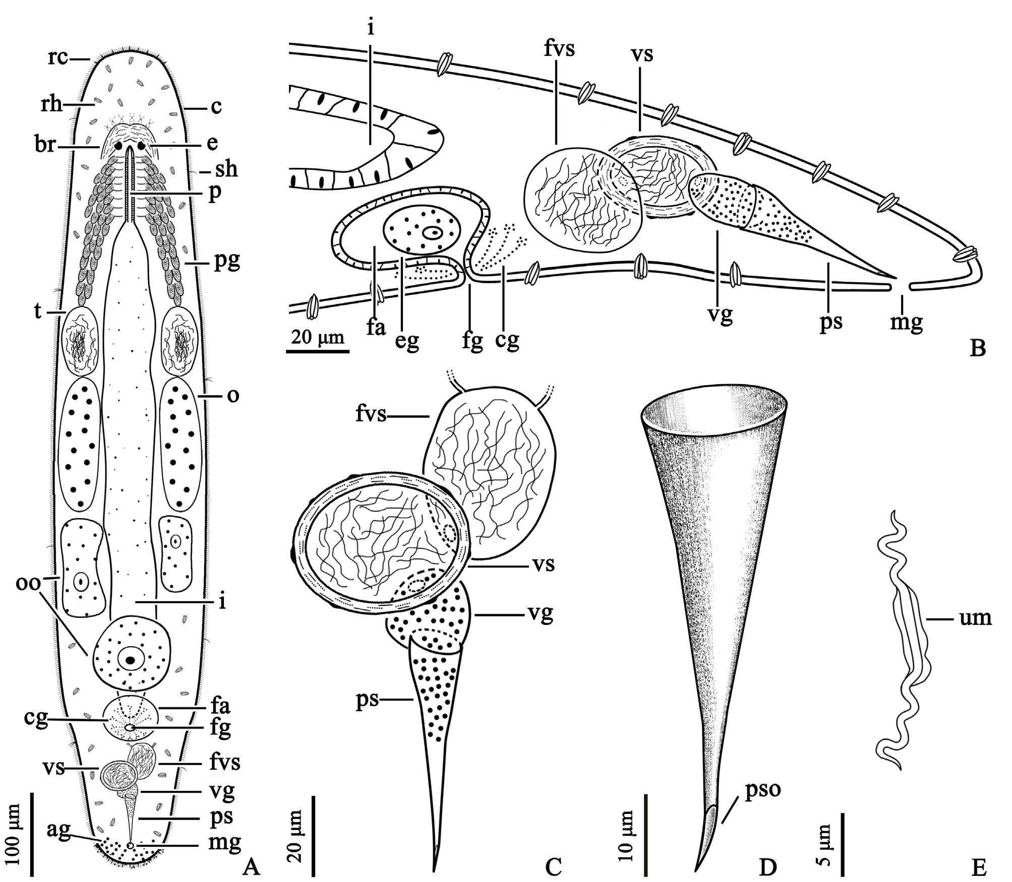
TABLE 4. Comparison on Macrostomum shenda n. sp. with six similar species.
| Species | Stylet length (µm) | Diameter of proximal opening (µm) | Diameter of distal opening (µm) | Ratio of proximal diameter to stylet length | Habitat | Distribution | References |
|---|---|---|---|---|---|---|---|
| M. shenda View in CoL n. sp. | 56 | 14 | 8 | 25.0% | Brackish | China | This study |
| M. ensiferum View in CoL | 60* | 8* | NA | 13.3% | Freshwater | Azerbaijan | Beklemishev 1951 |
| M. obtusum View in CoL | 80–91 | 18 | 5–12* | 19.8%–22.5% | Freshwater | various | Papi 1951 and Luther 1960 |
| M. orthostylum View in CoL | 57 | 10* | 11* | 17.5% | Freshwater | various | Ferguson & Jones 1940 |
| 61 | 13* | 13* | 21.3% | Luther 1960 | |||
| M. phillipsi View in CoL | 65 | 9* | NA | 13.8% | Freshwater | USA | Ferguson & Stirewalt 1938 |
| M. sinyaense View in CoL | 65–85 | 10 | 3.4 | 11.8%–15.3% | Freshwater | Kenya | Young 1976 |
| M. vejdovskyi View in CoL | 35 | 9* | 7* | 25.7% | Freshwater | USA | Ferguson 1940a |
No known copyright restrictions apply. See Agosti, D., Egloff, W., 2009. Taxonomic information exchange and copyright: the Plazi approach. BMC Research Notes 2009, 2:53 for further explanation.
|
Kingdom |
|
|
Phylum |
|
|
Class |
|
|
SuperOrder |
Macrostomorpha |
|
Order |
|
|
Family |
|
|
Genus |
Macrostomum shenda Wang & Xin
| Xin, Fan, Zhang, Si-Yu, Shi, Yong-Shi, Wang, Lei, Zhang, Yu & Wang, An-Tai 2019 |
M. shenda
| Xin & Zhang & Shi & Wang & Zhang & Wang 2019 |
M. shenda
| Xin & Zhang & Shi & Wang & Zhang & Wang 2019 |
M. shenda
| Xin & Zhang & Shi & Wang & Zhang & Wang 2019 |
M. shenda
| Xin & Zhang & Shi & Wang & Zhang & Wang 2019 |
M. shenda
| Xin & Zhang & Shi & Wang & Zhang & Wang 2019 |
Macrostomum shenda
| Xin & Zhang & Shi & Wang & Zhang & Wang 2019 |
M. shenda
| Xin & Zhang & Shi & Wang & Zhang & Wang 2019 |
M. sinyaense
| Young 1976 |
M. ensiferum
| Beklemischev 1951 |
M. phillipsi
| Ferguson & Stirewalt 1938 |
M. obtusum
| Vejdovsky 1895 |
M. orthostylum
| Braun 1885 |
M. orthostylum
| Braun 1885 |
M. orthostylum
| Braun 1885 |
M. orthostylum
| Braun 1885 |
Macrostomum
| Schmidt 1848 |